1. Tumour-Related Angiogenesis
The formation of new blood vessels from pre-existing ones—denoted as angiogenesis—is required for tumour maintenance and development as well as for metastasis formation
[1][2][1,2]. Angiogenesis is regulated by a delicate balance between a host of pro-angiogenic and antiangiogenic factors
[3]. Hence, intensive attention has been placed on the assessment of cancer-associated neovascularization
[4][5][4,5]. Producing various angiogenic factors, proteases, heparanase, digestive enzymes, and chemotactic and stimulatory factors, tumour cells exert a direct effect on capillary endothelial cells, induce the assembly of activated immune cells, and trigger the activity of stromal cells; thus, they create a pro-angiogenic niche in the tumour microenvironment
[2][6][2,6].
Given that angiogenesis/neo-angiogenesis has crucial role in tumour development, propagation, and metastatic spread, angiogenic biomarkers are recorded as promising candidates for tumour imaging
[7]. Vascular endothelial growth factor (VEGF), platelet-derived growth factor (PDGF), fibroblast growth factor-2 (FGF-2), ephrins, α
vβ
3 integrins, aminopeptidase N (APN/CD13), fibronectin, nitroimidazole, matrix metalloproteinases, gastrin-releasing peptide receptor (GRPR), and E-selectin represent the most important angiogenic molecules
[8]. The detection of angiogenesis-related markers makes timely tumour identification possible, and these biomolecules also serve as potential targets for anti-tumour treatment and patient follow-up
[1][9][1,9]. Anti-angiogenic drugs can inhibit tumour expansion and decelerate, delay, or even inhibit tumour augmentation and metastasis formation
[10][11][10,11]. Therefore, angiogenesis-directed molecular probes may lay the basis of personalised cancer diagnostics and treatment.
Non-invasive positron emission tomography (PET) or positron emission tomography/computed tomography (PET/CT) are regarded as the mainstay diagnostic tools in the detection of primary tumours and pertinent metastases. Furthermore, PET can be a useful means of angiogenesis-targeted drug development, authorization, and dose estimation
[8][12][8,12]. Several PET radionuclides have been used in angiogenesis-directed diagnostical settings, including fluoride-18 (
18F), gallium-68 (
68Ga), copper-64 (
64Cu), and even the gamma emitter indium-111 (
111In). Although scandium (Sc) radioisotopes were already recognized as valuable radiometals for isotope diagnostic use in the late 1990s, they have been set aside for almost 20 years
[13].
2. Scandium-44 (44Sc)
The following three radionuclides of Sc are well suited for either diagnostic or therapeutic applications: scandium-43 (
43Sc), scandium-44 (
44Sc), and scandium-47 (
47Sc)
[14]. Positron emitters
43Sc and
44Sc are relevant diagnostic radiometals, whereas β
−-emitting
47Sc is the therapeutic sister with accompanying γ rays for imaging purposes
[15][16][15,16].
There are two major ways to produce
44Sc. Via the proton irradiation of natural calcium or enriched
44Ca targets (through a
44Ca(p,n)
44Sc nuclear reaction), the production of
44Sc can be easily obtained by applying low-energy cyclotrons
[17][18][19][17,18,19]. Cyclotron-based production provides outstanding radiochemical yield (RCY) and radiochemical purity (RCP) for clinical applications
[18][20][18,20]. Of note,
44Sc production via applying a cyclotron has been confirmed to be economically viable
[17]. Although
44Sc can also be eluated from titanum-44/scandium-44 (
44Ti/
44Sc) generator systems, the radioactive-waste handling of long-lived
44Ti (62 ± 2 years) makes this approach challenging
[21][22][23][21,22,23]. In addition, the demanding production of
44Ti implies another obstacle regarding the routine usage of
44Ti/
44Sc generators
[17][24][17,24]. Difficulties around the production of
44Sc may underpin why this radiometal came to the focus of exhaustive investigation as a potential labelling isotope so late.
44Sc, with physical properties of T
1/2: 3.97 h (lately reported as 4.04 h), E
β+average: 632 KeV, E
β+mean: 0.63 MeV, and I:94.3%, has begun to emerge as a radiometal of particular interest in imaging, dosimetry, and treatment follow-up
[20][25][26][20,25,26]. Positron emitter
44Sc also has a gamma co-emission of 1157 keV (99.9%), which may limit the radioactive dose that can be injected into patients
[26]. However, this high-energy gamma radiation made the radiometal a precious candidate for the novel β + γ coincidence PET imaging
[27]. Since the positron energy of the applied PET radionuclide is inversely proportional to the resolution of the reconstructed image, better spatial-image resolution and better quality could be obtained with
44Sc relative to other radiometals—for example,
68Ga (
68Ga: E
β+max: 1.9 MeV and E
β+mean: 0.89 MeV vs.
44Sc: E
β+max: 1474 KeV and E
β+mean: 0.63 MeV)
[28][29][30][28,29,30]. These advantageous physical characteristics together with its decay to nontoxic Ca make
44Sc widely feasible in PET and PET/CT imaging
[13][31][13,31]. Given its small ionic radius, the chemistry of
44Sc is comparable to that of
68Ga; thus,
44Sc could be applied in several fields, including dosimetry investigations in theranostic settings and in trafficking ligands with longer pharmacokinetics and subsequent requirements for longer imaging times (even 24 h post administration)
[32][33][32,33].
Prior studies have already dealt with the investigation of the feasibility of
44Sc in pre-therapeutic dosimetry
[34][35][34,35]. Khawar et al. published a paper indicating that the pharmacokinetics of [
44Sc]Sc-PSMA-617-based (PSMA (prostate-specific membrane antigen)) PET/CT imaging serves as a useful tool for the calculation of normal organ-absorbed doses and the maximum allowable activity in prostate-cancer patients prior to lutetium-177 (
177Lu)-labelled PSMA-617 ([
177Lu]Lu-PSMA-617) radiotherapy
[36]. Pioneering clinical studies further strengthened the efficacy of
44Sc-labelled somatostatin analogue [
44Sc]Sc-DOTATOC and [
44Sc]Sc-PSMA-617 as valuable radiotracers in dosimetry settings
[35]. Delayed acquisition granted by the longer half-life of
44Sc enables better pre-therapeutic dosimetry for [
177Lu]Lu-PSMA-617 radiotherapy
[34][37][34,37]. Clinical successes in the treatment of leukaemia, non-Hodgkin lymphoma, malignant melanoma, urinary bladder cancer, glioma, neuroendocrine tumours, and prostate cancer with alpha emitter bismuth-213 (
213Bi)- and actinium-225 (
225Ac)-linked radiopharmaceuticals project that these isotopes could also benefit from dosimetry estimations based on
44Sc
[38]. Moreover, in the long run, other radionuclides, such as yttrium-90 (
90Y), ioide-131 (
131I), strontium-90 (
90Sr), and phosphorus-32 (
32P) may be potential candidates for theranostic applications. Although preclinical research findings are available on radiation-absorbed doses from
47Sc, the limited availability of the radiometal hampers its usage as a potential radiotherapeutic agent
[39].
Similar to
68Ga,
44Sc is also able to form thermodynamically stable complexes with chelator DOTA (1,4,7,10-teraazacyclododecane-N,N′,N″,N″′-teraacetic acid)
[40][41][40,41]. In addition to this,
44Sc seems superior to
68Ga in several facets. Given its longer half-life (T
1/2 44Sc: 3.97 h vs. T
1/2 68Ga: 68 min) and cyclotron-dependent extensive synthesis,
44Sc can be easily shipped to remote nuclear medical facilities. This contributes not only to the wider distribution of the radioisotope but also to the accomplishment of protein- or antibody-based PET studies with lengthened examination times
[42]. Furthermore, radiopharmaceuticals with longer pharmacokinetic characteristics can also be proposed due to the longer half-life of the radiometal
[17]. Moreover, the pharmacokinetic properties of
44Sc-appended imaging probes are largely comparable to those of the
68Ga-labelled counterparts
[43]. The nearly four-hour half-life of
44Sc could be easily fitted to the pharmacokinetics of several targeting molecules, including peptides, antibodies, or their fragments, as well as oligonuclides, which makes facile radiopharmaceutical synthesis possible
[13][17][13,17]. Additionally, long-lived
44Sc favours delayed imaging and the achievement of appropriate tumour-to-background (T/M) ratios. The time of imaging is of critical importance from the point of view of patient management and the scheduling of examinations. Therefore, the use of
44Sc-labelled radiopharmaceuticals would contribute to the establishment of a fluent workflow.
44Sc-labelled PET radiopharmaceuticals could even be appropriate for intraoperative radio-guided surgery, including the detection of lymphatic metastases at later time points post injection
[32]. Consequently,
44Sc-based PET probes could stand out as valuable imaging agents.
47Sc (T
1/2 = 3.35 days)—the therapeutic match of
44Sc—emits a β
− radiation of a maximum energy of 0.600 MeV (31.6%) and 0.439 MeV (68.4%), which could be exploited in targeted radiotherapy
[33][44][33,44]. Therefore,
44Sc/
47Sc has exquisite potential as a radiotheranostic pair in PET diagnostics and in therapeutic settings
[33][45][33,45]. Besides
44Sc and
47Sc, scandium-43 (
43Sc) is another significant member of the group of Sc isotopes. Investigating the quantitative capabilities of
43Sc/
44Sc and comparing it to
18F and
68Ga, Lima et al. published a paper indicating that the application of radiotracers labelled with the mixture of
43Sc/
44Sc may be beneficial in clinical fields
[46]. The findings of their phantom study proved that precise, quantitative PET/CT could be obtained by applying commercial PET systems
[46]. Given that
43Sc does not have high-energy gamma emission, the use of
43Sc/
44Sc could be especially important in terms of overcoming the limitation of
44Sc derived from gamma radiation. These favourable features propelled both preclinical and clinical investigations with
44Sc down new lines. Some experiments have been spawned to focus on the evaluation of the clinical feasibility of
44Sc-labelled molecules at a translational level as well as the comparison of
44Sc with other widely used radionuclides, such as
68Ga and
64Cu
[47].
3. PET Radioisotopes Other Than Scandium-44 (44Sc)
The optimal short half-life (109.8 min) and the high positron abundance (β ≥ 97%) of
18F made it the most commonly used radioisotope for the labelling of PET biomolecules
[48]. Positron emitter
18F possesses a relatively low positron energy (E
max = 0.635 MeV and E
mean = 0.250 MeV) and a short positron range within the tissue (maximum of 2.3 mm)
[49][50][49,50]. These nuclear properties ensure ideal image quality and high spatial resolution obtained with
18F-labelled tracers
[51]. The 109.8 min-long half-life is beneficial for synthesis procedures as well as for the performance of examinations of a few hours
[51]. Owing to the short longevity,
18F-labelled radiopharmaceuticals can be safely administered without an increased risk of excess radiation. Moreover, another advantage is the facile cyclotron-based production, which provides immense quantities of the isotope at high specific activity
[51]. Although a cyclotron is required to produce the radiometal, its decay characteristics make the distribution of
18F- and
18F-labelled radiotracers to distant nuclear medical laboratories without an on-site cyclotron possible. Even though
18F is well suited for the labelling of a wide range of small and medium-sized molecules, the radiolabelling of peptides with
18F is still cumbersome
[51]. Hence, different isotopes have come into focus that address some of the difficulties associated with
18F-based radiotracers.
Up to now, the favourable physical characteristics of
68Ga and
64Cu made them attractive for the radiolabelling of a broad set of molecules. Currently,
68Ga-labelled PET radiopharmaceuticals are applied most frequently for labelling purposes of radiometal-based PET tracers. The short-lived positron emitter
68Ga (T
1/2: 67.71 min ≈ 68 min, Eβ
+average: 830 KeV, maximum β
+ energy: 1.92 MeV, Iβ
+: 89%, Eγ: 1077 KeV, Iγ: 3.2%) can be obtained from a germanium-68/gallium-68 (
68Ge/
68Ga)-generator system
[52][53][52,53]. With such physical properties, decay characteristics, and easy accessibility from an on-site
68Ge/
68Ga generator,
68Ga has emerged as the mainstay PET-imaging radiometal
[32][52][54][32,52,54]. Since the radiosynthesis of peptides—complexed with different macrocyclic ligands—with
68Ga is easy to perform, the use of
68Ga is effective in imaging settings
[55].
68Ga-labelled DOTA-conjugated somatostatin analogues (SSTR), including DOTATOC, DOTATATE, and DOTANOC, meant a substantial step forward in the imaging of SSTR-positive neuroendocrine tumours
[33].
The short half-life and related transport challenges, however, may limit the widespread implementation of
68Ga into clinical and preclinical settings. Owing to the short longevity of the radiometal, radiolabelling procedures are only possible in laboratories equipped with in-house
68Ge/
68Ga-generator systems. In addition, due to its short half-life,
68Ga-labelling can be applied solely in case of small molecules and peptides featured with a rapid pharmacokinetic profile
[17][56][17,56]. Furthermore,
68Ga-labelling is only suitable for PET examinations of relatively short duration. Because of the breakthrough of
68Ge and sorbent material, the purification and concentration of
68Ga is warranted following elution, which could also restrict its applicability in routine diagnostic usage
[57]. Image noise associated with the elevated positron energy of the radionuclide constitutes another shortcoming of
68Ga imaging
[32]. These facts render
68Ga-labelled radiotracers of limited attractiveness for centralized distribution.
The application of
64Cu may bridge the limitations derived from
68Ga-associated imaging. Given its longer half-life (T
1/2: 12.7 h) and generator-independent production, the use of
64Cu (β
+ emission,
Eaverage = 278 keV, abundance: 19%) seems to be economically more viable for PET imaging
[58][59][58,59]. Due to its longer-lived nature, the easy transport to distant laboratories without an on-site cyclotron facilitates the integration of
64Cu-appended radiopharmaceuticals into diagnostics. Moreover, the 12.7 h half-life of the radiometal can be easily tailored to biomarker-targeting small and large molecules, peptides, antibodies, and nanomolecules with prolonged elimination kinetics
[60]. Additionally, its longer half-life makes
64Cu-based radiochemical procedures facile
[60]. The coordination chemistry of the radiometal enables complexation with various chelating agents, which further supports the frictionless performance of radiolabelling
[60]. Therefore,
64Cu is extremely useful in the establishment of a broad set of radiotracers for diagnostic purposes. However, the use of
64Cu is not without shortcomings. Its concomitant β
− emission (β
− = 39.0%,
E = 190.2 keV) and relatively low positron-branching ratio (17.6%) mean a meaningful extra radiation danger for patients
[58][61][62][58,61,62]. Furthermore, in case of complexation, chelators must be customized to the complex redox chemistry of
64Cu
[54].
Beyond
44Sc, the use of zirconium-89 (
89Zr) has also been exhaustively investigated in PET imaging
[63]. Given its favourable decay half-life (T
1/2 = 3.3 days; 78.4 h), appropriate radiochemistry, and the accessibility of different chelating agents for complexation with the radioisotope,
89Zr is welcomed for the radiolabelling of PET-based imaging molecules
[63]. The relatively long half-life of the radiometal could be easily adjusted to the pharmacokinetic profile of monoclonal antibodies (mABs); therefore,
89Zr seems to be well suited for the radiolabelling of mABs and for PET immunoimaging applications
[63][64][65][63,64,65]. Several mAbs, including anti-human epidermal growth factor receptor 2 (HER2) trastuzumab, anti-epidermal growth factor receptor (EGFR) mAb cetuximab, anti-PSMA mAb J591, and anti-vascular endothelial growth factor (VEGF) bevacizumab, were successfully labelled with
89Zr for the PET imaging of breast cancer, squamous-cell carcinoma, prostate tumours, and ovarian tumours, respectively, at both preclinical and clinical levels
[66][67][68][69][66,67,68,69].
Due to their decay characteristics (positron emission: E
max and E
average, 897 keV and 396.9 keV, respectively), PET images obtained with
89Zr-labelled compounds are of adequate spatial resolution
[70][71][72][70,71,72]. However, similar to
44Sc,
89Zr has spontaneous high-energy gamma radiation of 908.97 keV, which accounts for a major drawback of its usage
[70]. Although the concomitant gamma radiation does not influence either the quality or the quantification of the PET scans, it needs to be taken into account when patient doses are determined
[73][74][73,74]. Another potential disadvantage could be the limited availability of the radiometal
[63].
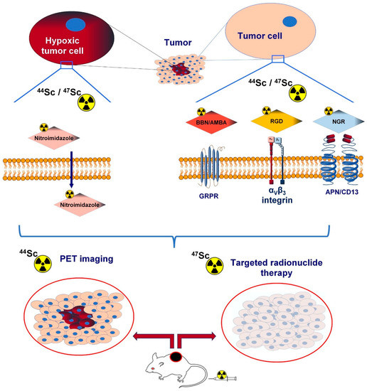
Taking the abovementioned facts into account, 44Sc may therefore be a valuable substitute for the currently applied 68Ga and 64Cu in the establishment of peptide-based targeted PET radiopharmaceuticals. RIn this researchers view, we provide a comprehensive overview of the role of 44Sc-labelled peptide-based radiopharmaceuticals in the molecular imaging of cancer-related angiogenesis (Figure 1). Table 1 summarises the preclinical studies with 44Sc-labelled PET radiotracers that selectively target angiogenic biomarkers. In Table 2 the most important physical characteristics of the discussed PET radioisotopes are displayed.
Figure 1. Sc44- and Sc47-labelled peptide-based radiopharmaceuticals in the molecular imaging and therapy of cancer-related angiogenesis.
Table 1. Preclinical studies with 44Sc-labelled PET radiotracers.
| Investigated Object |
Investigated Phenomenon |
Target Molecule |
(Radio) Labelled Vector |
Imaging Technique |
Reference |
| Zr |
|---|
| PCa PC-3 and HaCaT cell lines |
In vitro receptor-binding affinity utilizing in vitro blocking studies with BBN |
GRPR |
[ |
| Half-life (h) |
1.83 |
1.13 | 44 | Sc]Sc-NODAGA-AMBA and [ | 68 | Ga]Ga-NODAGA-AMBA, |
In vitro gamma counter measurements %ID/million cells units |
[ |
12.775] |
| 3.89 |
3.97 |
78.41 |
PCa PC-3 tumor-bearing CB17 SCID mice and healthy control |
| Decay method (%) | In vitro and in vivo biodistribution pattern, tumor-targeting capability based on blocking experiments |
EC (3)
ßGRPR |
+ | (97) |
EC (11.1)
ß | + | (88.9)[ | 44 | Sc]Sc-NODAGA-AMBA and [ | 68 | Ga]Ga-NODAGA-AMBA |
In vivo miniPET imaging, ex vivo radioactivity determination by gamma counter (%ID/g), in vivo and ex vivo blocking studies with BBN |
EC (43.9)
ß | + | (17.6)
ß | − | (38.5) |
EC (12)
ß | + | (88)[75] |
| EC (5.7) |
| ß | + | (94.3) |
EC (77.3) |
ß | + | (22.7) |
PC-3 cell line |
In vitro receptor-binding affinity with blocking studies applying [ | 125 | I-Tyr | 4 | ]-BN |
GRPR |
| β | + | endpoint energy, keV (%) | nat |
633.5 (96.7%)Sc-DOTA-BN[2-14]NH |
1899 (87.7)
822 (1.2) | 2 | and | nat | Ga-DOTA-BN[2-14]NH | 2 |
Competitive displacement cell-binding assay |
[33] |
| 653 (17.6) |
1200 (70.9) |
| 826 (17.2) |
1474 (94.3) |
902 (22.8) |
Healthy male Sprague-Dawley rats, male Copenhagen rats bearing androgen-independent Dunning R-3327-AT-1 prostate cancer tumour |
In vivo and ex vivo organ distribution, GRPR-targeting ability applying blocking studies with BBN |
GRPR |
44 | Sc-DOTA-BN[2-14]NH |
| Principal γ energies, keV (Abs.%) |
511 (194) |
511 (177.8)
1077 (3.2)
1261 (0.1)
1883 (0.1) | 2 | and | 68 | Ga-DOTA-BN[2-14]NH | 2 |
In vivo dynamic microPET imaging (in tumourous rats), ex vivo (in healthy rats) radioactivity calculations with a dose calibrator (% ID/g) |
511 (35.2)
1346 (0.5)[33] |
| 372.8 (23) |
511 (188.5) |
| 1157 (99.9) |
| 1499 (0.9) |
511 (45.5)
909 (99.0)
1713 (0.7)
1745 (0.1) |
U87MG and AR42J tumour-bearing female athymic nude mice (CD-1 nude) |
Evaluation of in vitro and in vivo behaviour, in vivo and ex vivo biodistribution |
α | v | β | 3 | integrin |
[ | 44 | Sc]Sc-DOTA-RGD, [ | 44 | Sc]Sc-NODAGA-RGD, [ | 68 | Ga]Ga-DOTA-RGD, [ | 68 | Ga]Ga-NODAGA-RGD |
In vivo PET/CT acquisition, ex vivo gamma counting (%IA/g) |
[76] |
| 4T1 tumor-bearing BALB/c mice and healthy control |
applicability of chelator AAZTA, in vivo biodistribution |
α | v | β | 3 | integrin |
44 | Sc | 3+ | , and | 44 | Sc(AAZTA) | − | and | 44 | Sc(CNAAZTA-c(RGDfK) |
in vivo PET/MRI examinations |
[77] |
| U87MG cells |
In vitro receptor-binding affinity and specificity with blocking studies using | 125 | I-Echistatin |
α | v | β | 3 | integrin |
cRGD, (cRGD) | 2 | , and DOTA-(cRGD) | 2 |
In vitro competitive cell-binding assay, gamma counter-based detection of the tracer concentration |
[17] |
| U87MG glioblastoma-bearing female athymic nude mice |
Tumour-targeting competence, specificity applying in vivo and ex vivo receptor blocking with c(RGD) | 2 | , in vivo and ex vivo biodistribution |
α | v | β | 3 | integrin |
[ | 44 | Sc]Sc-DOTA-c(RGD) | 2 |
In vivo microPET/microCT imaging, ex vivo radioactivity determination (%ID/g) |
[17] |
| CB17 SCID adult male mice bearing KB tumours and healthy control mice |
Synthesis procedure, comparison of | 44 | Sc- and | 68 | Ga-labelled derivatives, in vivo and ex vivo biodistribution |
Hypoxia |
[ | 44 | Sc]Sc-DO3AM-NI and [ | 68 | Ga]Ga- DO3AM-NI |
In vivo PET/MRI studies, ex vivo radiopharmaceutical uptake measurement %ID/g |
[78] |
| Pro-angiogenic VEGF-A | 165 | /NRP-1 complex formation |
Investigation of physicochemical properties and affinity for NRP-1 |
NRP-1 |
44 | Sc-radiocompounds ( | 44 | Sc-1, | 44 | Sc-1bis, | 44 | Sc-2, | 44 | Sc-3):Sc-DOTA-Ahx-A7R (Sc-1), Lys(DOTA-Sc)-A7R (Sc-1bis), Lys(hArg)-Dab(Ahx-DOTA-Sc)-Pro-Arg (Sc-2) and DR7A-DLys(DOTA-Sc) (Sc-3) |
Competitive ELISA test |
[79] |
Table 2. Physical characteristics of commonly used PET radionuclides.
| |
18 | F |
68 | Ga |
64 | Cu |
43 | Sc |
44 | Sc |
89 |
AMBA: aminobenzoyl–bombesin analogue; APN/CD13: aminopeptidase N; BBN: bombesin; GRPR: gastrin-releasing peptide receptor; NGR: asparagine–glycine–arginine; PET: positron emission tomography; RGD: Arg-Gly-Asp; 44Sc: scandium-44; 47Sc: scandium-47.
AAZTA: (1,4-bis(carboxymethyl)-6-[bis(carboxymethyl)]amino-6-methylperhydro-1,4-diazepine) AMBA: aminobenzoyl–bombesin analogue; AR42J: rat exocrine pancreatic tumour; BBN: bombesin; BN: bombesin; c(RGD)2: dimeric cyclic arginine–glycine–aspartic acid; c(RGDfK): cyclo(-Arg-Gly-Asp-d-Phe-Lys); DOTA: 1,4,7,10-teraazacyclododecane-N,N′,N″,N″′-teraacetic acid; ELISA: enzyme-linked immunosorbent assay; 68Ga: gallium-68; GRPR: gastrin-releasing peptide receptor; HaCaT: human immortal keratinocyte; 125I: iodine-125; KB: human epidermal carcinoma; NI: nitroimidazole; NODAGA: 1,4,7-triazacyclononane-1-glutaric acid-4,7-diacetic acid; NRP-1: neuropilin-1 co-receptor; PCa PC-3: prostate cancer; PET/CT: positron emission tomography/computed tomography; PET/MRI: positron emission tomography/magnetic resonance imaging; RGD: Arg-Gly-Asp; 44Sc: scandium-44; SCID: severe combined immunodeficient; 4T1: mouse breast cancer; U87MG: human glioblastoma; VEGF: vascular endothelial growth factor.