C-X-C Motif Chemokine Ligand 1 in Breast Cancer: Comparison
Please note this is a comparison between Version 2 by Peter Tang and Version 1 by Irena Baranowska-Bosiacka.

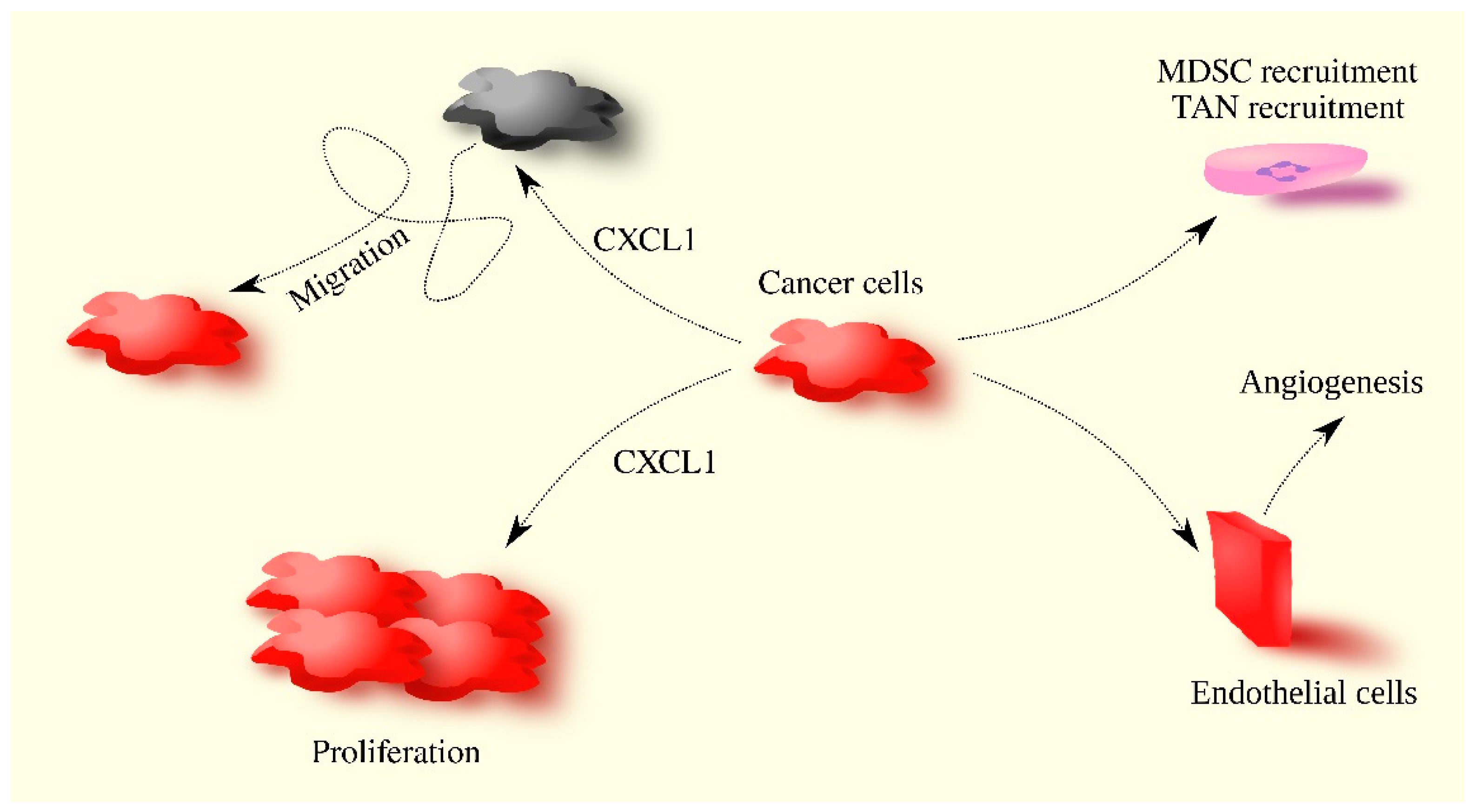
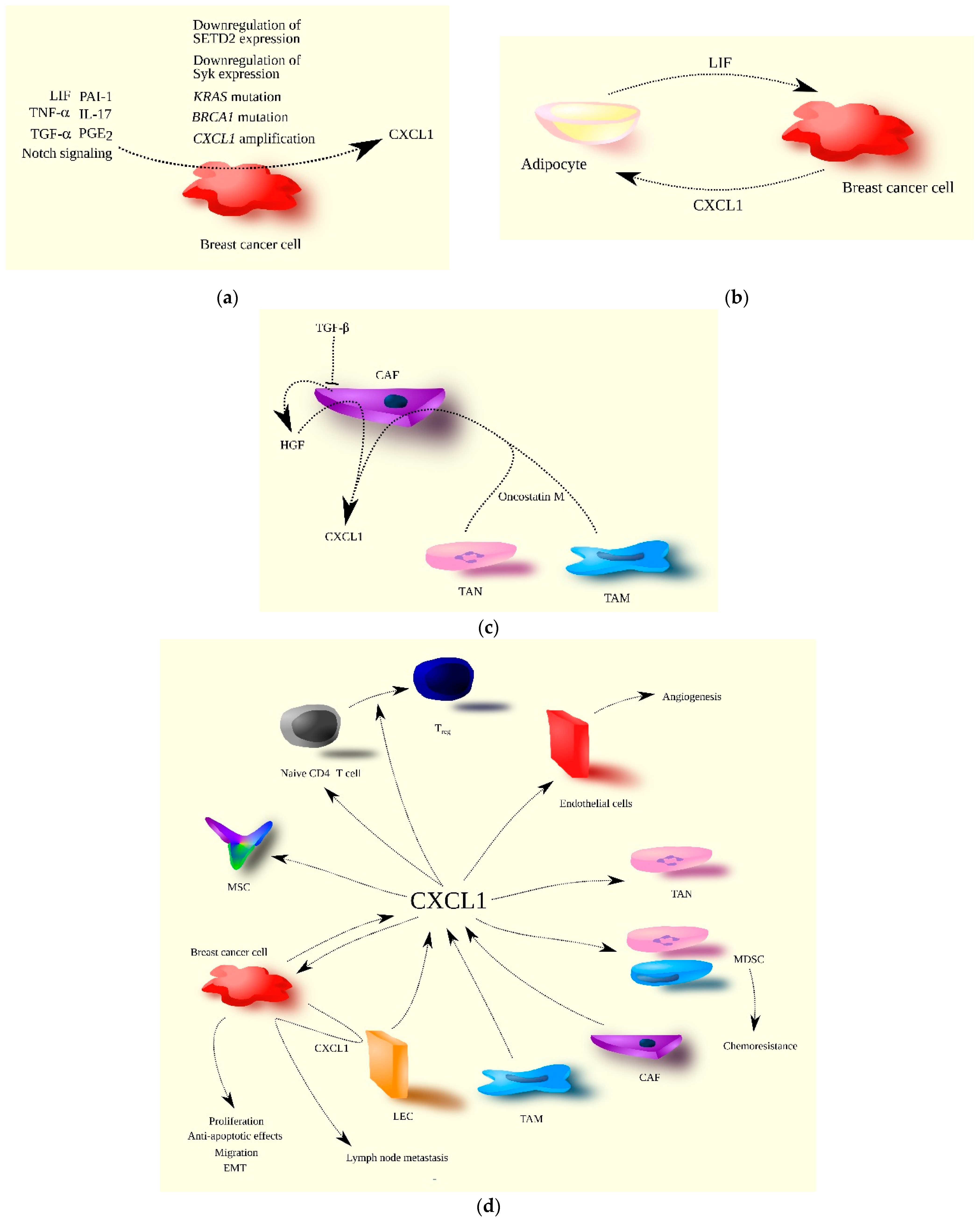
C-X-C motif chemokine ligand 1 (CXCL1) is a member of the CXC chemokine subfamily and a ligand for CXCR2. Its main function in the immune system is the chemoattraction of neutrophils. Female breast cancer is the most commonly diagnosed cancer. In 2020, 2.26 million new cases of this cancer were diagnosed, accounting for 11.7% of all cancer diagnoses

  • chemokine
  • CXCL1
  • breast cancer

1. Introduction

Chemokines are one of the components of the tumor microenvironment [4][1]. Chemokines are chemotactic cytokines that, in the immune system, are responsible for the migration and infiltration of tissues by immune cells. As immune cells constitute an important element of the tumor microenvironment [3][2], chemokines play a significant role in cancer processes [5][3].
Chemokines have been divided into four subfamilies based on the conserved cysteine motif at the N-terminus [4][1]. One of the CXC chemokines with a CXC motif at the N-terminus has 16 representatives in humans: C-X-C motif chemokine ligand 1 (CXCL1) to CXCL17, except for CXCL15. These chemokines can be further divided based on their ability to activate specific chemokine receptors. CXC chemokines that activate C-X-C motif chemokine receptor 2 (CXCR2) include CXCL1 to CXCL8, but not CXCL4. CXCL1 was first described in the 1980s [6][4]; its first described property was its autocrine stimulation of melanoma cell proliferation, hence its original name: melanoma growth-stimulatory activity (MGSA) [6][4].
CXCL1 is a chemokine whose main receptor is CXCR2 [7][5]. However, this chemokine can also activate CXCR1, but the half maximal effective concentration (EC50) is two orders of magnitude worse than that for CXCR2 [7][5]. Another receptor for CXCL1 is atypical chemokine receptor 1 (ACKR1)/Duffy antigen receptor for chemokines (DARC) [8][6]. The role of this receptor in CXCL1 function is unclear.
The activation of CXCR2 by CXCL1 leads to signal transduction via Gαi [9,10][7][8]. Multiple proteins interact with this receptor, resulting in signal transduction independent of G proteins [11][9]. CXCR2 activation causes cell migration [12][10] and can also increase proliferation [13][11]. CXCR2 expression is found in neutrophils [4][1], making CXCL1 a chemoattractant for these cells. This is its most important physiological property. In many diseases such as multiple sclerosis [14][12], viral encephalitis [15][13], periodontitis [16][14], and alcohol-induced liver injury [17][15], CXCL1 levels increase, resulting in tissue infiltration by neutrophils. These cells then participate in disease processes, including damage to healthy tissue. In chronic inflammatory states, CXCL1 also participates in the recruitment of MDSCs, leading to a decreased immune response [18][16]. CXCR2 expression is also found on endothelial cells [19][17], giving CXCL1 proangiogenic properties. These physiological and non-cancer disease-related properties reflect the role of CXCL1 in cancer processes (Figure 1).
Figure 1. CXCL1’s involvement in cancer processes. CXCL1 increases proliferation and migration, and also acts on tumor-associated cells. CXCL1 recruits neutrophils, MDSCs, and induces angiogenesis.

2. CXCL1 in Breast Cancer

Female breast cancer is the most commonly diagnosed cancer. In 2020, 2.26 million new cases of this cancer were diagnosed, accounting for 11.7% of all cancer diagnoses [2][18]. It also caused 685,000 deaths, which accounted for 6.9% of deaths caused by all cancers. Men can also have breast cancer, although it is more than 100 times less common than in women [20][19]. For this reason, this subsection will only discuss the role of CXCL1 in female breast cancer. Breast cancer is divided into subtypes based on the presence or loss of the expression of estrogen receptor (ER), progesterone receptor (PR), and human epidermal growth factor receptor 2 (HER2) [21,22,23,24,25][20][21][22][23][24]. The following main subtypes of breast cancer exist:
  • Luminal A (ER+PR+HER2);
  • Luminal B (ER+HER2 + PR or Ki-67high);
  • Luminal HER2-positive (HER2+ + ER+ or PR+);
  • Non-luminal HER2-positive (ERPRHER2+);
  • Triple-negative breast cancer (ERPRHER2).
Triple-negative breast cancer is sometimes referred to as basal-like breast cancer, which is inaccurate because 1/3 of triple-negative breast cancer cases are not basal-like breast cancers [26][25]. The median age of breast cancer incidence in women is 61 [27][26]. Risk factors include obesity and an unhealthy diet, and factors that reduce the likelihood of the disease are menopause, a large number of children, and prior breastfeeding. Genetic factors are also important risk factors, accounting for 5–10% of breast cancer cases [24,27][23][26]. These include mutations in the breast cancer type 1 and 2 susceptibility gene (BRCA1 and BRCA2); a mutation in one of these genes is associated with a 55–70% probability of developing breast cancer [27][26].
The action of sex hormones plays an important role in the development of breast cancer—no less important are cytokines, including CXCL1. A bioinformatics study indicated that CXCL1 is an important gene in breast cancer processes in young adults [28][27]. There are reports that CXCL1 expression in breast tumors is lower than in normal tissue [29[28][29][30][31],30,31,32], although some studies show that CXCL1 expression in breast tumors does not differ from healthy tissue [33][32]. This indicates that CXCL1 may not play an important role in all cases of breast cancer. Other CXCR2 ligands may play a more important role in breast cancer than CXCL1, for example, CXCL5/epithelial-cell-derived neutrophil activating factor 78 (ENA78) and CXCL8/interleukin-8 (IL-8), whose expression is increased in breast tumors [33][32]. That result was obtained in an analysis of all breast tumors without distinguishing their subtypes.
CXCL1 expression is the lowest in luminal breast cancer and at the same time lower than in normal tissue [31][30]. In contrast, CXCL1 expression is the highest in basal-like breast cancer and higher than in normal tissue [34][33]. CXCL1 expression is also increased relative to normal tissue in mesenchymal-like triple-negative breast cancer and basal-like triple-negative breast cancer [35][34]. CXCL1 expression is higher in inflammatory breast cancer than in other breast cancer subtypes [36][35]. Inflammatory breast cancer is a rare breast cancer characterized by intense inflammatory responses in the breast. CXCL1 expression in stroma breast cancer is also higher in invasive ductal carcinoma than in ductal carcinoma in situ, which indicates that CXCL1 expression increases with tumor growth [37][36].
Another study showed that expression in breast cancer decreases with increasing tumor grade [31[30][31],32], while another showed that CXCL1 expression does not differ between grade I and III breast cancers [29][28]. CXCL1 expression is higher in breast cancer metastasis (a group composed mainly of liver, lung, skin, and other metastases) than in primary tumors and higher than in normal tissue [29,36][28][35]. Circulating levels of CXCL1 are increased in breast cancer patients compared to healthy subjects [38][37].
CXCL1 expression in breast cancer cells is lineage dependent. MCF-7 cells (ER+PR+HER2) produce very small amounts of CXCL1 and there is no autocrine stimulation of proliferation by this chemokine in these cells [39,40][38][39]. The highest production of CXCL1 of all breast cancer lines occurs in triple-negative MDA-MB-231 breast cancer cells (ERPRHER2) [29][28]. This chemokine is produced in 14 to 330 times higher amounts by triple-negative MDA-MB-231 breast cancer cells than by MCF-7 cells [29,40][28][39]. On the other hand, high CXCL1 expression does not always occur in triple-negative breast cancer cell lines. An example of this is triple-negative BT-20 cells, which do not secrete CXCL1 in detectable amounts [29][28].
The expression of the receptor for CXCL1, i.e., CXCR2, is lower in triple-negative breast cancer compared to other breast cancer subtypes [41][40]. CXCR2 expression is also reduced in ER-negative and HER2-negative breast cancers, indicating a decrease in CXCL1 activity in these tumors and in tumor cells that have lost the expression of these receptors.
The frequency of CXCL1 amplification increases with the progression of breast cancer [42][41]. It is estimated that 7.5% of primary breast tumors have CXCL1 gene amplification, while in lymph node and lung metastases, the percentage is around 20% [42][41]. Furthermore, analyses conducted using the cBioPortal tool (http://www.cbioportal.org/ accessed on 30 March 2023) [43,44][42][43] based on TCGA PanCancer Atlas studies [45,46,47,48,49,50][44][45][46][47][48][49] showed that CXCL1 gene amplification was observed in 1.96% (21/1072) of breast invasive carcinoma cases. In tumors with PT53 gene mutation, the percentage of CXCL1 gene amplification is 2.88% (10/347). Additionally, in breast cancer with BRCA1 or BRCA2 gene mutations, the amplification percentage of CXCL1 gene is 3.85% (2/52). When it comes to subtypes of breast cancer, CXCL1 gene amplification was observed in 1.20% (6/499) of luminal A breast cancer cases, 3.55% (7/197) of luminal B breast cancer cases, 7.69% (6/78) of HER2-positive breast cancer cases, and 1.17% (2/171) of basal breast cancer cases.
The expression of CXCL1 is dependent on GATA-binding protein 3 (GATA3), as demonstrated by experiments on T47D cells [51][50]. Decreased GATA3 expression leads to increased CXCL1 expression, although the exact mechanism of this relationship is not yet fully understood. It is believed to be associated with the formation of a BRCA1-GATA3 complex, which decreases the expression of CXCL1 by binding to its promoter [51][50].
Another factor that increases CXCL1 expression is obesity. BMI is positively correlated with CXCL1 expression levels in breast tumors [52][51]. Due to the significant influence of CXCL1 in tumorigenesis, obesity in breast cancer patients is associated with a worse prognosis.
Other factors are also responsible for CXCL1 expression in breast cancer. The high expression of CXCL1 in triple-negative MDA-MB-231 cells is partly due to decreased expression of the nonreceptor protein tyrosine kinase Syk. This kinase inhibits nuclear factor κB (NF-κB) activation and thus downregulates CXCL1 expression in MCF-7 cells and other breast cancer cell lines [40][39]. Other proteins also affect CXCL1 expression in breast cancer. One example is the DEK protein, which downregulates CXCL1 expression [53][52], and another is SET domain-containing 2 histone lysine methyltransferase (SETD2), a histone methyltransferase that causes histone methylation in the CXCL1 gene promoter [54][53]. In breast tumors, SETD2 expression is downregulated compared to healthy tissue [55][54], and thus CXCL1 expression is upregulated [54][53]. Another factor that decreases CXCL1 expression in breast cancer cells is either decreased expression or loss of HER2 expression [56][55]. Additionally, a mutation in KRAS leads to the activation of mitogen-activated protein kinase (MAPK) cascades, which then leads to increased CXCL1 expression in breast cancer cells [57][56].
CXCL1 expression in breast tumors is also significantly influenced by intercellular signaling and secretory factors. High CXCL1 expression in triple-negative breast cancer may be due to transforming growth factor-α (TGF-α) [35][34]. It is an epidermal growth factor receptor (EGFR) ligand. At the same time, TGF-α does not affect the expression of CXCR2 ligands in non-triple-negative breast cancer cells. CXCL1 expression in breast cancer cells is also increased by secretory factors such as prostaglandin E2 (PGE2) [58][57], interleukin-17 (IL-17) derived from T helper type 17 (Th17) cells [59[58][59],60], loss of response to transforming growth factor-β (TGF-β) [61[60][61],62], tumor necrosis factor-α (TNF-α) [42][41], leukemia inhibitory factor (LIF) [63][62], plasminogen activator inhibitor-1 (PAI-1) [64][63], and Notch signaling [65][64].
CXCL1 expression in breast cancer cells is downregulated by TGF-β [61,66][60][65]. In particular, TGF-β prevents the induction of CXCL1 expression by IL-17 [61][60]. At the same time, the expression of the receptor for TGF-β, i.e., TβRII, is downregulated in breast tumors [62][61], resulting in an increase in CXCL1 expression in the tumor. This is followed by the recruitment of myeloid-derived suppressor cells (MDSC) into the tumor niche—these cells secrete TGF-β and interleukin-6 (IL-6), which results in the differentiation of CD4+ T cells into Th17 cells that secrete IL-17, a mechanism further increasing CXCL1 expression in breast cancer cells [59,60,61][58][59][60].
The effect of TNF-α on CXCL1 expression is important in resistance to chemotherapy. Anticancer drugs increase TNF-α expression, which results in the activation of the immune system and the destruction of breast cancer cells [42][41]. At the same time, TNF-α also increases the expression of CXCL1, which results in the recruitment of MDSC, cells that inhibit the antitumor immune system response—this means that CXCL1 in this situation is responsible for chemoresistance [42][41].
CXCL1 expression is also upregulated by LIF secreted by cancer-associated adipocytes [63][62]. LIF expression in these cells is increased as a result of signal transducer and activator of transcription 3 (STAT3) activation by CXCL1 from breast cancer cells. Therefore, a positive feedback loop is formed between cancer-associated adipocytes and breast cancer cells; CXCL1 is one of the components of the loop.
CXCL1 expression in breast cancer cells can also be altered by the plasminogen activation regulatory system. A study on mouse 4T1 breast cancer cells showed that urokinase-type plasminogen activator (uPA) decreases, while PAI-1 increases, the expression of CXCR2 ligands [64][63]. CXCL1 expression in breast cancer cells is also increased by Notch signaling [65][64]. The activation of this pathway is followed by the activation of hairy/enhancer-of-split related with YRPW motif-like (HEYL), a protein that attaches to the CXCL1 promoter, thus increasing the expression of CXCL1. In this way, the expression of CXCL2/growth-regulated oncogene-β (GRO-β) and CXCL3/growth-regulated oncogene-γ (GRO-γ) also increases in breast cancer cells [65][64].
In breast tumors, cancer-associated fibroblast (CAF) may be the main source of CXCL1 [37,67,68,69][36][66][67][68]. CXCL1 expression in CAF is increased under the influence of basal-like breast cancer cells [70][69], while luminal breast cancer cells do not increase CXCL1 expression in CAF. This may be the reason for the higher CXCL1 expression in basal-like triple-negative breast cancer than in other breast cancer subtypes [34,35][33][34].
CXCL1 expression in CAF is also associated with a decrease in TGF-β levels in breast tumors. This cytokine reduces CXCL1 expression in CAF [37,69][36][68]. Decreased levels of TGF-β increase the expression of hepatocyte growth factor (HGF) [68][67], a growth factor that, through its receptor c-Met, increases the expression of CXCR2 ligands in CAF, as shown by experiments on mouse cells [68][67].
Oncostatin M, which is secreted by tumor-associated neutrophils (TAN) and tumor-associated macrophages (TAM) [71[70][71],72], is also responsible for the increase in CXCL1 expression in CAF. On the other hand, oncostatin M also increases the expression of other factors in CAF, such as vascular endothelial growth factor (VEGF), CC motif chemokine ligand (CCL)2/monocyte chemoattractant protein 1 (MCP-1), CCL5/regulated on activation, normally T-cell-expressed and -secreted (RANTES), CCL19/EBI1-ligand chemokine (ELC), CCL21/secondary lymphoid tissue chemokine (SLC), and CXCL12/stromal-derived factor-1 (SDF-1) [72][71].
TAM are also responsible for CXCL1 production in breast tumors [34,73,74,75][33][72][73][74]. The amount of CXCL1 secreted by TAM may be the highest of all cytokines produced by these cells, as shown by studies of TAM isolated from lung metastasis from a mouse model [34][33].
Another source of CXCL1 in breast tumors may be adipose-derived mesenchymal stem cells (MSC) [76][75], which increase CXCL1 expression under the influence of interleukin-1β (IL-1β) from breast cancer cells [77][76]. Importantly, adipose-derived MSC also secrete CXCL8/IL-8. However, these cells are important in breast tumor development only in some models—they support the development of tumors arising from MCF-7 and ZR-75-30 lines but not from MDA-MB-231 lines [76][75].
CXCL1 is involved in tumorigenesis in breast cancer. This effect depends on whether a given cell line expresses the CXCL1 receptor, i.e., CXCR2 [29][28]. CXCL1 increases the proliferation of breast cancer cells, as has been shown in experiments on triple-negative MDA-MB-231 breast cancer lines, triple-negative HCC-1937 breast cancer cells, HER2-negative MCF-7 cells, and HER2-positive SKBR3 breast cancer cells (ERPRHER2+) [78][77]. In triple-negative breast cancer cells, there is often an autocrine increase in cell proliferation by CXCL1. This chemokine is produced and secreted by these cells, which then increases their proliferation [78][77].
CXCL1 is also important in the function of breast cancer stem cells. These cells express CXCL1 as well as the receptor for this chemokine, i.e., CXCR2 [79][78]. CXCR2 expression in breast tumors is mainly found in cancer stem cells [41][40], and for this reason, CXCR2 can be considered a marker of breast cancer stem cells. The simultaneous expression of CXCL1 and the receptor for this chemokine, CXCR2, allows breast cancer stem cells to act on themselves in an autocrine manner. This results in the proliferation of these cells, which increases their stemness and causes self-renewal [41,79][40][78], as shown by experiments on triple-negative MDA-MB-231 breast cancer [73,74][72][73].
CXCL1 has pro-survival and anti-apoptotic effects on breast cancer cells. CXCL1 increases the expression of anti-apoptotic Bcl-2 family proteins and decreases the expression of pro-apoptotic Bcl-2 family proteins [80][79], which is important in chemoresistance and radioresistance.
CXCL1 also causes the migration and epithelial-to-mesenchymal transition (EMT) of breast cancer cells, with the effect being lineage-dependent. CXCL1 causes the migration of triple-negative MDA-MB-231 breast cancer cells [40[39][72][77],73,78], MCF-7 cells (ER+PR+HER2) [78[77][80],81], Zr-75-1 cells (ER+PR+HER2+) [81][80], triple-negative HCC-1937 breast cancer cells, and HER2-positive SKBR3 breast cancer cells (ERPRHER2+) [78][77]. On the other hand, CXCL1 does not cause the migration of T47D cells (ER+PR+HER2) [81][80], even though T47D cells express CXCR2 [29][28]. CXCL1 induces breast cancer cell migration through the activation of extracellular signal-regulated kinase (ERK) MAPK, which increases the expression of matrix metalloproteinase 2 (MMP2) and matrix metalloproteinase 9 (MMP9) [82][81]. Also important in the induction of breast cancer cell migration by CXCL1 are Akt/protein kinase B (PKB), NF-κB, and STAT3 [69][68].
CXCL1 also induces the EMT of breast cancer cells, as shown by experiments on MDA-MB-231, HCC-1937, SKBR3, and MCF-7 cell lines [34,78][33][77]. This process is dependent on the CXCL1-induced activation of the NF-κB-sex-determining region Y-related high-mobility group box 4 (SOX4) pathway [34][33].
CXCL1-induced migration can be either directly via the activation of the CXCR2 receptor on breast cancer cells or indirectly. Indirectly, CXCL1 causes the recruitment of neutrophils into the tumor niche. TAN cause breast cancer cells to migrate by activating intracellular adhesion molecule-1 (ICAM-1) on these cells [83][82]. TAN also show the expression of β2-integrins, which involves the direct interaction of neutrophils with breast cancer cells and the interaction of β2-integrins with ICAM-1 on the cancer cell. CXCL1 can also indirectly induce the EMT of breast cancer cells through recruiting MDSC, which then causes the EMT of breast cancer cells via IL-6 [84][83].
CXCL1 is also important in breast cancer lymph node metastasis. Breast cancer cells cause an increase in CXCL1 expression in lymphatic endothelial cells (LEC) [85][84], which causes breast cancer cells to migrate into lymphatic vessels, resulting in lymph node metastasis.
CXCL1 may also be important in the formation of breast cancer metastasis in other organs. Blood levels of this chemokine are positively correlated with the number of circulating breast cancer cells [38][37], and the more circulating cancer cells, the greater the likelihood of metastasis.
CXCL1 has also been found to have a significant effect on the formation of metastasis of breast cancer cells to various organs. CXCL1 may be important in the bone metastasis of the cancer in question. This chemokine acts on osteoclasts precursors through the CXCR2 receptor on these cells [86,87,88,89][85][86][87][88]. This leads to osteoclast maturation, bone remodeling, and the formation of bone metastasis. At the same time, CXCL8/IL-8 appears to be more important in the formation of bone metastasis of breast cancer than CXCL1 [90][89].
CXCL1 seems to be involved in the formation of brain metastasis of breast cancer. In the formation of brain metastasis, a feedback loop is formed between breast cancer cells and astrocytes [91][90]. Breast cancer cells secrete IL-1β, in response to which astrocytes secrete HGF, which in turn increases IL-1β expression in breast cancer cells. IL-1β also increases the expression of CXCL1 and CXCL8/IL-8. These chemokines are responsible for the formation of the perivascular niche and angiogenesis in the initial stages of brain metastasis in breast cancer [91][90].
CXCL1 may also play some role in the formation of lung metastasis in breast cancer. The circulating levels of CXCL1 are the highest in patients with lung metastasis in breast cancer and higher than in patients with bone metastasis [38][37]. CXCL1 causes an increase in the adhesion of circulating breast cancer cells to human pulmonary microvascular endothelial cells, that is, to the walls of blood vessels in the lungs [92][91]. In lung fibroblasts, CXCL1, as well as other CXCR2 ligands, increases the expression of CCL2/MCP-1 and CCL7/MCP-3, both of which increase cholesterol synthesis in breast cancer cells in the lung. This leads to angiogenesis and the generation of lung metastasis in breast cancer [93][92]. Notably, this mechanism occurs in triple-negative breast cancer.
CXCL1 acts on tumor-associated cells. CXCL1 in breast tumors causes angiogenesis by acting directly on endothelial cells [58,76][57][75]. CXCL1 can also indirectly cause angiogenesis by inducing an increase in VEGF expression in breast cancer cells [78][77].
CXCL1 is involved in the recruitment of various cells to the tumor niche. It causes the recruitment of neutrophils [57[56][82][93],83,94], granulocytic-myeloid-derived suppressor cells (G-MDSC) [52,57][51][56], and breast resident adipose tissue-derived MSC [95][94]. This process may also be important for the location of the recruited cells in the tumor. Breast cancer cells secrete CXCL1 and CXCL8/IL-8, resulting in the chemotaxis of neutrophils into breast cancer cells [83][82]. Direct contact between these cells is then possible, resulting in the stimulation of breast cancer cell migration. This process is dependent on ICAM-1 on breast cancer cells and β2-integrins on neutrophils [83][82].
CXCL1 can recruit c-Kit+Ly6a/Sca1+ hematopoietic stem/progenitor cells into the tumor niche, as demonstrated by experiments in mice [96][95]. These cells differentiate into MDSC. CXCL1 also induces the recruitment of naive CD4+ T cells, which are transformed into regulatory T (Treg) cells under the influence of the tumor microenvironment [75][74]. At the same time, CXCL1 can induce the differentiation of naive CD4+ T cells into Treg. CXCL1 also acts on cancer-associated adipocytes, causing STAT3 activation in them, which leads to the production of LIF [63][62] which in turn increases CXCL1 production in breast cancer cells.
CXCL1 may be involved in chemoresistance and radioresistance. The expression of this chemokine in breast cancer cells is increased by ionizing radiation in microbeam radiotherapy [97][96], as well as by chemotherapeutics such as paclitaxel [98][97] and doxorubicin [99][98]. At least in the case of doxorubicin, this is dependent on a defect in p53 in breast cancer cells and the activation of NF-κB in these cells. The chemotherapeutic-induced increase in CXCL1 expression may be responsible for the increase in TNF-α expression [42][41]. CXCL1 also induces the recruitment of MDSC, cells that inhibit an anticancer immune response produced by radio- and chemotherapy (Figure 2). CXCL1 exerts its anti-apoptotic effect by affecting the expression of Bcl-2 family proteins [80][79], which inhibits breast cancer cell apoptosis induced by anticancer therapy.
Figure 2. The significance of CXCL1 in breast cancer progression. (a) CXCL1 expression is increased in breast cancer cells by both extracellular and intracellular factors. TNF-α, TGF-α (EGFR ligand), PGE2, IL-17, LIF, PAI-1, and Notch signaling are examples of extracellular factors, while mutations in genes such as KRAS and BRCA1, CXCL1 gene amplification, and the decreased expression of SETD2 and Syk are intracellular factors increasing CXCL1 expression in breast cancer cells. (b) The interaction of breast cancer cells with tumor-associated adipocytes results in increased CXCL1 expression in breast cancer cells due to the secretion of LIF by adipocytes. In turn, CXCL1 increases LIF expression in adipocytes, creating a positive feedback loop between these two types of cells in the breast tumor. (c) In the breast tumor, CXCL1 is secreted by CAF, and its expression in CAF is increased by oncostatin M produced by TAM and TAN. Increased CXCL1 expression in CAF is also due to their loss of responsiveness to TGF-β, resulting in increased HGF production by CAF, which further increases CXCL1 expression. (d) CXCL1 plays multiple roles in breast cancer progression, acting on various types of cells. Source of CXCL1 in breast tumor may be breast cancer cells, CAF, TAM, and LEC. CXCL1 promotes breast cancer cell proliferation, migration, EMT, and anti-apoptotic effects. Breast cancer cells also increase CXCL1 expression in LEC, promoting the migration of breast cancer cells to lymphatic vessels, resulting in lymph node metastasis. CXCL1 also acts on tumor-associated cells. It recruits immune cells such as naïve CD4+ T cells, TAN, MDSC, and MSC to the tumor niche. It can differentiate naïve CD4+ T cells into Treg and facilitate chemoresistance through the recruitment of MDSC. CXCL1 also promotes angiogenesis via its influence on endothelial cells.
High CXCL1 expression in breast tumors is positively correlated with lymph node metastasis [34,100][33][99] and TNM stage [34][33]. On the other hand, there is a study showing that stromal CXCL1 expression is not correlated with lymph node metastasis [37][36]. The level of CXCL1 expression is also not related to tumor size [34,37,100][33][36][99] or histologic grade [34][33]. The level of CXCL1 in the tumor is not related to the age of patients [37][36]. Its highest expression is found in basal-like breast cancer [34][33] and triple-negative breast cancer [35][34].
Depending on the literature source, the correlation of CXCL1 expression with negative PR and ER status may vary. CXCL1 can be correlated with negative PR [100][99] and negative ER status [82,94[81][93][99],100], and in some studies, it is not correlated [34,37][33][36]. Circulating levels of CXCL1 are not associated with PR and ER status in breast tumors [38][37]. Additionally, CXCL1 expression is not associated with HER2-negative status in breast tumors [34,37,100][33][36][99].
Considering all breast cancer cases, the association between the level of CXCL1 expression in the tumor and prognosis varies depending on the literature source (Table 1). High CXCL1 expression in breast tumors may be associated with either a worse [34,37,100][33][36][99] or better prognosis [30,32][29][31]. The level of CXCL1 in the blood may be associated with the prognosis of breast cancer patients, with a higher circulating level of CXCL1 indicating a worse prognosis [38][37]. Conversely, higher CXCL1 expression is associated with a worse prognosis for patients with basal breast cancer [52][51]. For ERα-positive breast cancer, depending on the work cited, high CXCL1 expression in the tumor may be associated with either a worse [29][28] or better prognosis [31][30].
Table 1.
Effect of CXCL1 expression level on survival of breast cancer patients.

Type of Cancer

Expression Testing Method

Impact on Survival at High CXCL1 Expression

Number of Patients in the Study

Notes

Source

Breast cancer: ERα-positive breast cancer

qRT-PCR

Worse prognosis

48

RFS

[29][28]

Breast cancer: ERα-positive breast cancer

Microarray

Kaplan–Meier Plotter database

Better prognosis

2061

RFS

Analysis based on the Kaplan–Meier Plotter database

[31][30]

Breast cancer

IHC

Worse prognosis

655

OS, nuclear CXCL1 expression,

at cytoplasmic CXCL1 expression there was only a trend (p = 0.08)

[100][99]

Breast cancer

Microarray

UALCAN/TCGA database

Better prognosis

1066

OS,

Analysis based on UALCAN

[32][31]

Breast cancer

Microarray

Worse prognosis

121

OS

[34][33]

Breast cancer

Microarray

Kaplan–Meier Plotter database

Better prognosis

3951/RFS

1402/OS

OS, RFS

analysis based on the Kaplan–Meier Plotter database

[30][29]

Breast cancer

Microarray

Kaplan–Meier Plotter database

No significant impact on prognosis

1402

OS,

trend of worse prognosis at high CXCL1 (p = 0.064),

based on the Kaplan–Meier Plotter database

[31][30]

Breast cancer

ELISA

Worse prognosis

61

OS, PFS

circulating level of CXCL1

[38][37]

Breast cancer: basal breast cancer

Microarray

Kaplan–Meier Plotter database

Worse prognosis

54

OS,

based on the Kaplan–Meier Plotter database

[52][51]

Breast cancer

Microarray

Finak microarray database

Worse prognosis

53

RFS

stromal CXCL1 expression

based on Finak microarray database

[37][36]

ELISA—enzyme-linked immunosorbent assay; IHC—immunohistochemistry; OS—overall survival; PFS—progression-free survival; RFS—relapse-free survival; qRT-PCR—quantitative real-time polymerase chain reaction. Red color—worse prognosis, Blue color—better prognosis.

References

  1. Hughes, C.E.; Nibbs, R.J.B. A guide to chemokines and their receptors. FEBS J. 2018, 285, 2944–2971.
  2. Egeblad, M.; Nakasone, E.S.; Werb, Z. Tumors as organs: Complex tissues that interface with the entire organism. Develop. Cell 2010, 18, 884–901.
  3. Do, H.T.T.; Lee, C.H.; Cho, J. Chemokines and their Receptors: Multifaceted Roles in Cancer Progression and Potential Value as Cancer Prognostic Markers. Cancers 2020, 12, 287.
  4. Richmond, A.; Lawson, D.H.; Nixon, D.W.; Chawla, R.K. Characterization of autostimulatory and transforming growth factors from human melanoma cells. Cancer Res. 1985, 45, 6390–6394.
  5. Ludwig, A.; Petersen, F.; Zahn, S.; Götze, O.; Schröder, J.M.; Flad, H.D.; Brandt, E. The CXC-chemokine neutrophil-activating peptide-2 induces two distinct optima of neutrophil chemotaxis by differential interaction with interleukin-8 receptors CXCR-1 and CXCR-2. Blood 1997, 90, 4588–4597.
  6. Szabo, M.C.; Soo, K.S.; Zlotnik, A.; Schall, T.J. Chemokine class differences in binding to the Duffy antigen-erythrocyte chemokine receptor. J. Biol. Chem. 1995, 270, 25348–25351.
  7. Damaj, B.B.; McColl, S.R.; Mahana, W.; Crouch, M.F.; Naccache, P.H. Physical association of Gi2α with interleukin-8 receptors. J. Biol. Chem. 1996, 271, 12783–12789.
  8. Kuwano, Y.; Adler, M.; Zhang, H.; Groisman, A.; Ley, K. Gαi2 and Gαi3 differentially regulate arrest from flow and chemotaxis in mouse neutrophils. J. Immunol. 2016, 196, 3828–3833.
  9. Raman, D.; Neel, N.F.; Sai, J.; Mernaugh, R.L.; Ham, A.J.; Richmond, A.J. Characterization of chemokine receptor CXCR2 interacting proteins using a proteomics approach to define the CXCR2 “chemosynapse”. Methods Enzymol. 2009, 460, 315–330.
  10. Kawanishi, H.; Matsui, Y.; Ito, M.; Watanabe, J.; Takahashi, T.; Nishizawa, K.; Nishiyama, H.; Kamoto, T.; Mikami, Y.; Tanaka, Y.; et al. Secreted CXCL1 is a potential mediator and marker of the tumor invasion of bladder cancer. Clin. Cancer Res. 2008, 14, 2579–2587.
  11. Lee, C.H.; Syu, S.H.; Liu, K.J.; Chu, P.Y.; Yang, W.C.; Lin, P.; Shieh, W.Y. Interleukin-1 beta transactivates epidermal growth factor receptor via the CXCL1-CXCR2 axis in oral cancer. Oncotarget 2015, 6, 38866–38880.
  12. Grist, J.J.; Marro, B.S.; Skinner, D.D.; Syage, A.R.; Worne, C.; Doty, D.J.; Fujinami, R.S.; Lane, T.E. Induced CNS expression of CXCL1 augments neurologic disease in a murine model of multiple sclerosis via enhanced neutrophil recruitment. Eur. J. Immunol. 2018, 48, 1199–1210.
  13. Michael, B.D.; Bricio-Moreno, L.; Sorensen, E.W.; Miyabe, Y.; Lian, J.; Solomon, T.; Kurt-Jones, E.A.; Luster, A.D. Astrocyte- and Neuron-Derived CXCL1 Drives Neutrophil Transmigration and Blood-Brain Barrier Permeability in Viral Encephalitis. Cell Rep. 2020, 32, 108150.
  14. Cortés-Vieyra, R.; Rosales, C.; Uribe-Querol, E. Neutrophil Functions in Periodontal Homeostasis. J. Immunol. Res. 2016, 2016, 1396106.
  15. Roh, Y.S.; Zhang, B.; Loomba, R.; Seki, E. TLR2 and TLR9 contribute to alcohol-mediated liver injury through induction of CXCL1 and neutrophil infiltration. Am. J. Physiol. Gastrointest. Liver Physiol. 2015, 309, G30–G41.
  16. Sander, L.E.; Sackett, S.D.; Dierssen, U.; Beraza, N.; Linke, R.P.; Müller, M.; Blander, J.M.; Tacke, F.; Trautwein, C. Hepatic acute-phase proteins control innate immune responses during infection by promoting myeloid-derived suppressor cell function. J. Exp. Med. 2010, 207, 1453–1464.
  17. Addison, C.L.; Daniel, T.O.; Burdick, M.D.; Liu, H.; Ehlert, J.E.; Xue, Y.Y.; Buechi, L.; Walz, A.; Richmond, A.; Strieter, R.M. The CXC chemokine receptor 2, CXCR2, is the putative receptor for ELR+ CXC chemokine-induced angiogenic activity. J. Immunol. 2000, 165, 5269–5277.
  18. Sung, H.; Ferlay, J.; Siegel, R.L.; Laversanne, M.; Soerjomataram, I.; Jemal, A.; Bray, F. Global Cancer Statistics 2020: GLOBOCAN Estimates of Incidence and Mortality Worldwide for 36 Cancers in 185 Countries. CA Cancer J. Clin. 2021, 71, 209–249.
  19. Lomma, C.; Chan, A.; Chih, H.; Reid, C.; Peter, W. Male Breast Cancer in Australia. Asia Pac. J. Clin. Oncol. 2021, 17, e57–e62.
  20. Onitilo, A.A.; Engel, J.M.; Greenlee, R.T.; Mukesh, B.N. Breast cancer subtypes based on ER/PR and Her2 expression: Comparison of clinicopathologic features and survival. Clin. Med. Res. 2009, 7, 4–13.
  21. Yersal, O.; Barutca, S. Biological subtypes of breast cancer: Prognostic and therapeutic implications. World J. Clin. Oncol. 2014, 5, 412–424.
  22. Johansson, A.L.V.; Trewin, C.B.; Hjerkind, K.V.; Ellingjord-Dale, M.; Johannesen, T.B.; Ursin, G. Breast cancer-specific survival by clinical subtype after 7 years follow-up of young and elderly women in a nationwide cohort. Int. J. Cancer 2019, 144, 1251–1261.
  23. Loibl, S.; Poortmans, P.; Morrow, M.; Denkert, C.; Curigliano, G. Breast cancer. Lancet 2021, 397, 1750–1769.
  24. Harbeck, N. Insights into biology of luminal HER2 vs. enriched HER2 subtypes: Therapeutic implications. Breast 2015, 24 (Suppl. 2), S44–S48.
  25. Bertucci, F.; Finetti, P.; Cervera, N.; Esterni, B.; Hermitte, F.; Viens, P.; Birnbaum, D. How basal are triple-negative breast cancers? Int. J. Cancer 2008, 123, 236–240.
  26. Rojas, K.; Stuckey, A. Breast Cancer Epidemiology and Risk Factors. Clin. Obstet. Gynecol. 2016, 59, 651–672.
  27. Yi, S.; Zhou, W. Tumorigenesis-related key genes in adolescents and young adults with HR(+)/HER2(−) breast cancer. Int. J. Clin. Exp. Pathol. 2020, 13, 2701–2709.
  28. Bièche, I.; Chavey, C.; Andrieu, C.; Busson, M.; Vacher, S.; Le Corre, L.; Guinebretière, J.M.; Burlinchon, S.; Lidereau, R.; Lazennec, G. CXC chemokines located in the 4q21 region are up-regulated in breast cancer. Endocr. Relat. Cancer 2007, 14, 1039–1052.
  29. Chen, E.; Qin, X.; Peng, K.; Xu, X.; Li, W.; Cheng, X.; Tang, C.; Cui, Y.; Wang, Z.; Liu, T. Identification of Potential Therapeutic Targets Among CXC Chemokines in Breast Tumor Microenvironment Using Integrative Bioinformatics Analysis. Cell Physiol. Biochem. 2018, 45, 1731–1746.
  30. Li, Y.; Liang, M.; Lin, Y.; Lv, J.; Chen, M.; Zhou, P.; Fu, F.; Wang, C. Transcriptional Expressions of CXCL9/10/12/13 as Prognosis Factors in Breast Cancer. J. Oncol. 2020, 2020, 4270957.
  31. Wang, F.; Yuan, C.; Wu, H.Z.; Liu, B.; Yang, Y.F. Bioinformatics, Molecular Docking and Experiments In Vitro Analyze the Prognostic Value of CXC Chemokines in Breast Cancer. Front. Oncol. 2021, 11, 665080.
  32. Abrahamsson, A.; Rzepecka, A.; Dabrosin, C. Equal Pro-inflammatory Profiles of CCLs, CXCLs, and Matrix Metalloproteinases in the Extracellular Microenvironment In Vivo in Human Dense Breast Tissue and Breast Cancer. Front. Immunol. 2018, 8, 1994.
  33. Wang, N.; Liu, W.; Zheng, Y.; Wang, S.; Yang, B.; Li, M.; Song, J.; Zhang, F.; Zhang, X.; Wang, Q.; et al. CXCL1 derived from tumor-associated macrophages promotes breast cancer metastasis via activating NF-κB/SOX4 signaling. Cell Death Dis. 2018, 9, 880.
  34. Ignacio, R.M.C.; Gibbs, C.R.; Lee, E.S.; Son, D.S. The TGFα-EGFR-Akt signaling axis plays a role in enhancing proinflammatory chemokines in triple-negative breast cancer cells. Oncotarget 2018, 9, 29286–29303.
  35. Lerebours, F.; Vacher, S.; Andrieu, C.; Espie, M.; Marty, M.; Lidereau, R.; Bieche, I. NF-kappa B genes have a major role in inflammatory breast cancer. BMC Cancer 2008, 8, 41.
  36. Zou, A.; Lambert, D.; Yeh, H.; Yasukawa, K.; Behbod, F.; Fan, F.; Cheng, N. Elevated CXCL1 expression in breast cancer stroma predicts poor prognosis and is inversely associated with expression of TGF-β signaling proteins. BMC Cancer 2014, 14, 781.
  37. Divella, R.; Daniele, A.; Savino, E.; Palma, F.; Bellizzi, A.; Giotta, F.; Simone, G.; Lioce, M.; Quaranta, M.; Paradiso, A.; et al. Circulating levels of transforming growth factor-βeta (TGF-β) and chemokine (C-X-C motif) ligand-1 (CXCL1) as predictors of distant seeding of circulating tumor cells in patients with metastatic breast cancer. Anticancer Res. 2013, 33, 1491–1497.
  38. Fujisawa, N.; Sakao, Y.; Hayashi, S.; Hadden, W.A., 3rd; Harmon, C.L.; Miller, E.J. alpha-Chemokine growth factors for adenocarcinomas; a synthetic peptide inhibitor for alpha-chemokines inhibits the growth of adenocarcinoma cell lines. J. Cancer Res. Clin. Oncol. 2000, 126, 19–26.
  39. Li, J.; Sidell, N. Growth-related oncogene produced in human breast cancer cells and regulated by Syk protein-tyrosine kinase. Int. J. Cancer 2005, 117, 14–20.
  40. Wang, Y.; Tu, L.; Du, C.; Xie, X.; Liu, Y.; Wang, J.; Li, Z.; Jiang, M.; Cao, D.; Yan, X.; et al. CXCR2 is a novel cancer stem-like cell marker for triple-negative breast cancer. OncoTargets Ther. 2018, 11, 5559–5567.
  41. Acharyya, S.; Oskarsson, T.; Vanharanta, S.; Malladi, S.; Kim, J.; Morris, P.G.; Manova-Todorova, K.; Leversha, M.; Hogg, N.; Seshan, V.E.; et al. A CXCL1 paracrine network links cancer chemoresistance and metastasis. Cell 2012, 150, 165–178.
  42. Cerami, E.; Gao, J.; Dogrusoz, U.; Gross, B.E.; Sumer, S.O.; Aksoy, B.A.; Jacobsen, A.; Byrne, C.J.; Heuer, M.L.; Larsson, E.; et al. The cBio cancer genomics portal: An open platform for exploring multidimensional cancer genomics data. Cancer Discov. 2012, 2, 401–404.
  43. Gao, J.; Aksoy, B.A.; Dogrusoz, U.; Dresdner, G.; Gross, B.; Sumer, S.O.; Sun, Y.; Jacobsen, A.; Sinha, R.; Larsson, E.; et al. Integrative analysis of complex cancer genomics and clinical profiles using the cBioPortal. Sci. Signal 2013, 6, pl1.
  44. Bonneville, R.; Krook, M.A.; Kautto, E.A.; Miya, J.; Wing, M.R.; Chen, H.Z.; Reeser, J.W.; Yu, L.; Roychowdhury, S. Landscape of Microsatellite Instability Across 39 Cancer Types. JCO Precis Oncol. 2017, 2017, PO.17.00073.
  45. Ding, L.; Bailey, M.H.; Porta-Pardo, E.; Thorsson, V.; Colaprico, A.; Bertrand, D.; Gibbs, D.L.; Weerasinghe, A.; Huang, K.L.; Tokheim, C.; et al. Perspective on Oncogenic Processes at the End of the Beginning of Cancer Genomics. Cell 2018, 173, 305–320.e10.
  46. Ellrott, K.; Bailey, M.H.; Saksena, G.; Covington, K.R.; Kandoth, C.; Stewart, C.; Hess, J.; Ma, S.; Chiotti, K.E.; McLellan, M.; et al. Scalable Open Science Approach for Mutation Calling of Tumor Exomes Using Multiple Genomic Pipelines. Cell Syst. 2018, 6, 271–281.e7.
  47. Gao, Q.; Liang, W.W.; Foltz, S.M.; Mutharasu, G.; Jayasinghe, R.G.; Cao, S.; Liao, W.W.; Reynolds, S.M.; Wyczalkowski, M.A.; Yao, L.; et al. Driver Fusions and Their Implications in the Development and Treatment of Human Cancers. Cell Rep. 2018, 23, 227–238.e3.
  48. Hoadley, K.A.; Yau, C.; Hinoue, T.; Wolf, D.M.; Lazar, A.J.; Drill, E.; Shen, R.; Taylor, A.M.; Cherniack, A.D.; Thorsson, V.; et al. Cell-of-Origin Patterns Dominate the Molecular Classification of 10,000 Tumors from 33 Types of Cancer. Cell 2018, 173, 291–304.e6.
  49. Taylor, A.M.; Shih, J.; Ha, G.; Gao, G.F.; Zhang, X.; Berger, A.C.; Schumacher, S.E.; Wang, C.; Hu, H.; Liu, J.; et al. Genomic and Functional Approaches to Understanding Cancer Aneuploidy. Cancer Cell 2018, 33, 676–689.e3.
  50. Tkocz, D.; Crawford, N.T.; Buckley, N.E.; Berry, F.B.; Kennedy, R.D.; Gorski, J.J.; Harkin, D.P.; Mullan, P.B. BRCA1 and GATA3 corepress FOXC1 to inhibit the pathogenesis of basal-like breast cancers. Oncogene 2012, 31, 3667–3678.
  51. Gibson, J.T.; Orlandella, R.M.; Turbitt, W.J.; Behring, M.; Manne, U.; Sorge, R.E.; Norian, L.A. Obesity-Associated Myeloid-Derived Suppressor Cells Promote Apoptosis of Tumor-Infiltrating CD8 T Cells and Immunotherapy Resistance in Breast Cancer. Front. Immunol. 2020, 11, 590794.
  52. Pease, N.A.; Shephard, M.S.; Sertorio, M.; Waltz, S.E.; Vinnedge, L.M.P. DEK Expression in Breast Cancer Cells Leads to the Alternative Activation of Tumor Associated Macrophages. Cancers 2020, 12, 1936.
  53. Zhou, Y.; Zheng, X.; Xu, B.; Deng, H.; Chen, L.; Jiang, J. Histone methyltransferase SETD2 inhibits tumor growth via suppressing CXCL1-mediated activation of cell cycle in lung adenocarcinoma. Aging 2020, 12, 25189–25206.
  54. Al Sarakbi, W.; Sasi, W.; Jiang, W.G.; Roberts, T.; Newbold, R.F.; Mokbel, K. The mRNA expression of SETD2 in human breast cancer: Correlation with clinico-pathological parameters. BMC Cancer 2009, 9, 290.
  55. Vazquez-Martin, A.; Colomer, R.; Menendez, J.A. Protein array technology to detect HER2 (erbB-2)-induced ‘cytokine signature’ in breast cancer. Eur. J. Cancer 2007, 43, 1117–1124.
  56. Franklin, D.A.; Sharick, J.T.; Ericsson-Gonzalez, P.I.; Sanchez, V.; Dean, P.T.; Opalenik, S.R.; Cairo, S.; Judde, J.G.; Lewis, M.T.; Chang, J.C.; et al. MEK activation modulates glycolysis and supports suppressive myeloid cells in TNBC. JCI Insight 2020, 5, e134290.
  57. Wang, D.; Wang, H.; Brown, J.; Daikoku, T.; Ning, W.; Shi, Q.; Richmond, A.; Strieter, R.; Dey, S.K.; DuBois, R.N. CXCL1 induced by prostaglandin E2 promotes angiogenesis in colorectal cancer. J. Exp. Med. 2006, 203, 941–951.
  58. Varikuti, S.; Oghumu, S.; Elbaz, M.; Volpedo, G.; Ahirwar, D.K.; Alarcon, P.C.; Sperling, R.H.; Moretti, E.; Pioso, M.S.; Kimble, J.; et al. STAT1 gene deficient mice develop accelerated breast cancer growth and metastasis which is reduced by IL-17 blockade. Oncoimmunology 2017, 6, e1361088.
  59. Ma, K.; Yang, L.; Shen, R.; Kong, B.; Chen, W.; Liang, J.; Tang, G.; Zhang, B. Th17 cells regulate the production of CXCL1 in breast cancer. Int. Immunopharmacol. 2018, 56, 320–329.
  60. Novitskiy, S.V.; Pickup, M.W.; Gorska, A.E.; Owens, P.; Chytil, A.; Aakre, M.; Wu, H.; Shyr, Y.; Moses, H.L. TGF-β receptor II loss promotes mammary carcinoma progression by Th17 dependent mechanisms. Cancer Discov. 2011, 1, 430–441.
  61. Wei, C.Y.; Tan, Q.X.; Zhu, X.; Qin, Q.H.; Zhu, F.B.; Mo, Q.G.; Yang, W.P. Expression of CDKN1A/p21 and TGFBR2 in breast cancer and their prognostic significance. Int. J. Clin. Exp. Pathol. 2015, 8, 14619–14629.
  62. Zhou, C.; He, X.; Tong, C.; Li, H.; Xie, C.; Wu, Y.; Wang, L.; Yan, X.; Luo, D.; Tang, Y.; et al. Cancer-associated adipocytes promote the invasion and metastasis in breast cancer through LIF/CXCLs positive feedback loop. Int. J. Biol. Sci. 2022, 18, 1363–1380.
  63. Jing, Y.; Kovacs, K.; Kurisetty, V.; Jiang, Z.; Tsinoremas, N.; Merchan, J.R. Role of plasminogen activator inhibitor-1 in urokinase’s paradoxical in vivo tumor suppressing or promoting effects. Mol. Cancer Res. 2012, 10, 1271–1281.
  64. Han, L.; Korangath, P.; Nguyen, N.K.; Diehl, A.; Cho, S.; Teo, W.W.; Cope, L.; Gessler, M.; Romer, L.; Sukumar, S. HEYL Regulates Neoangiogenesis Through Overexpression in Both Breast Tumor Epithelium and Endothelium. Front. Oncol. 2021, 10, 581459.
  65. Bierie, B.; Chung, C.H.; Parker, J.S.; Stover, D.G.; Cheng, N.; Chytil, A.; Aakre, M.; Shyr, Y.; Moses, H.L. Abrogation of TGF-beta signaling enhances chemokine production and correlates with prognosis in human breast cancer. J. Clin. Investig. 2009, 119, 1571–1582.
  66. Erez, N.; Glanz, S.; Raz, Y.; Avivi, C.; Barshack, I. Cancer associated fibroblasts express pro-inflammatory factors in human breast and ovarian tumors. Biochem. Biophys. Res. Commun. 2013, 437, 397–402.
  67. Fang, W.B.; Mafuvadze, B.; Yao, M.; Zou, A.; Portsche, M.; Cheng, N. TGF-β Negatively Regulates CXCL1 Chemokine Expression in Mammary Fibroblasts through Enhancement of Smad2/3 and Suppression of HGF/c-Met Signaling Mechanisms. PLoS ONE 2015, 10, e0135063.
  68. Bernard, S.; Myers, M.; Fang, W.B.; Zinda, B.; Smart, C.; Lambert, D.; Zou, A.; Fan, F.; Cheng, N. CXCL1 Derived from Mammary Fibroblasts Promotes Progression of Mammary Lesions to Invasive Carcinoma through CXCR2 Dependent Mechanisms. J. Mammary Gland Biol. Neoplasia 2018, 23, 249–267.
  69. Camp, J.T.; Elloumi, F.; Roman-Perez, E.; Rein, J.; Stewart, D.A.; Harrell, J.C.; Perou, C.M.; Troester, M.A. Interactions with fibroblasts are distinct in Basal-like and luminal breast cancers. Mol. Cancer Res. 2011, 9, 3–13.
  70. Queen, M.M.; Ryan, R.E.; Holzer, R.G.; Keller-Peck, C.R.; Jorcyk, C.L. Breast cancer cells stimulate neutrophils to produce oncostatin M: Potential implications for tumor progression. Cancer Res. 2005, 65, 8896–8904.
  71. Araujo, A.M.; Abaurrea, A.; Azcoaga, P.; López-Velazco, J.I.; Manzano, S.; Rodriguez, J.; Rezola, R.; Egia-Mendikute, L.; Valdés-Mora, F.; Flores, J.M.; et al. Stromal oncostatin M cytokine promotes breast cancer progression by reprogramming the tumor microenvironment. J. Clin. Investig. 2022, 132, e148667.
  72. Wang, N.; Zheng, Y.; Gu, J.; Cai, Y.; Wang, S.; Zhang, F.; Chen, J.; Situ, H.; Lin, Y.; Wang, Z. Network-pharmacology-based validation of TAMS/CXCL-1 as key mediator of XIAOPI formula preventing breast cancer development and metastasis. Sci. Rep. 2017, 7, 14513.
  73. Wang, S.; Liu, X.; Huang, R.; Zheng, Y.; Wang, N.; Yang, B.; Situ, H.; Lin, Y.; Wang, Z. XIAOPI Formula Inhibits Breast Cancer Stem Cells via Suppressing Tumor-Associated Macrophages/C-X-C Motif Chemokine Ligand 1 Pathway. Front. Pharmacol. 2019, 10, 1371.
  74. Li, J.; Wang, S.; Wang, N.; Zheng, Y.; Yang, B.; Wang, X.; Zhang, J.; Pan, B.; Wang, Z. Aiduqing formula inhibits breast cancer metastasis by suppressing TAM/CXCL1-induced Treg differentiation and infiltration. Cell Commun. Signal 2021, 19, 89.
  75. Wang, Y.; Liu, J.; Jiang, Q.; Deng, J.; Xu, F.; Chen, X.; Cheng, F.; Zhang, Y.; Yao, Y.; Xia, Z.; et al. Human Adipose-Derived Mesenchymal Stem Cell-Secreted CXCL1 and CXCL8 Facilitate Breast Tumor Growth by Promoting Angiogenesis. Stem Cells 2017, 35, 2060–2070.
  76. Ryan, D.; Sinha, A.; Bogan, D.; Davies, J.; Koziol, J.; ElShamy, W.M. A niche that triggers aggressiveness within BRCA1-IRIS overexpressing triple negative tumors is supported by reciprocal interactions with the microenvironment. Oncotarget 2017, 8, 103182–103206.
  77. Bhat, K.; Sarkissyan, M.; Wu, Y.; Vadgama, J.V. GROα overexpression drives cell migration and invasion in triple negative breast cancer cells. Oncol. Rep. 2017, 38, 21–30.
  78. Ciummo, S.L.; D’Antonio, L.; Sorrentino, C.; Fieni, C.; Lanuti, P.; Stassi, G.; Todaro, M.; Di Carlo, E. The C-X-C Motif Chemokine Ligand 1 Sustains Breast Cancer Stem Cell Self-Renewal and Promotes Tumor Progression and Immune Escape Programs. Front. Cell Develop. Biol. 2021, 9, 689286.
  79. Xu, H.; Lin, F.; Wang, Z.; Yang, L.; Meng, J.; Ou, Z.; Shao, Z.; Di, G.; Yang, G. CXCR2 promotes breast cancer metastasis and chemoresistance via suppression of AKT1 and activation of COX2. Cancer Lett. 2018, 412, 69–80.
  80. Youngs, S.J.; Ali, S.A.; Taub, D.D.; Rees, R.C. Chemokines induce migrational responses in human breast carcinoma cell lines. Int. J. Cancer 1997, 71, 257–266.
  81. Yang, C.; Yu, H.; Chen, R.; Tao, K.; Jian, L.; Peng, M.; Li, X.; Liu, M.; Liu, S. CXCL1 stimulates migration and invasion in ER-negative breast cancer cells via activation of the ERK/MMP2/9 signaling axis. Int. J. Oncol. 2019, 55, 684–696.
  82. Strell, C.; Lang, K.; Niggemann, B.; Zaenker, K.S.; Entschladen, F. Neutrophil granulocytes promote the migratory activity of MDA-MB-468 human breast carcinoma cells via ICAM-1. Exp. Cell Res. 2010, 316, 138–148.
  83. Zhu, H.; Gu, Y.; Xue, Y.; Yuan, M.; Cao, X.; Liu, Q. CXCR2+ MDSCs promote breast cancer progression by inducing EMT and activated T cell exhaustion. Oncotarget 2017, 8, 114554–114567.
  84. Oliveira-Ferrer, L.; Milde-Langosch, K.; Eylmann, K.; Rossberg, M.; Müller, V.; Schmalfeldt, B.; Witzel, I.; Wellbrock, J.; Fiedler, W. Mechanisms of Tumor-Lymphatic Interactions in Invasive Breast and Prostate Carcinoma. Int. J. Mol. Sci. 2020, 21, 602.
  85. Grassi, F.; Piacentini, A.; Cristino, S.; Toneguzzi, S.; Cavallo, C.; Facchini, A.; Lisignoli, G. Human osteoclasts express different CXC chemokines depending on cell culture substrate: Molecular and immunocytochemical evidence of high levels of CXCL10 and CXCL12. Histochem. Cell Biol. 2003, 120, 391–400.
  86. Hardaway, A.L.; Herroon, M.K.; Rajagurubandara, E.; Podgorski, I. Marrow adipocyte-derived CXCL1 and CXCL2 contribute to osteolysis in metastatic prostate cancer. Clin. Exp. Metastasis 2015, 32, 353–368.
  87. Lee, Y.C.; Gajdosik, M.S.; Josic, D.; Clifton, J.G.; Logothetis, C.; Yu-Lee, L.Y.; Gallick, G.E.; Maity, S.N.; Lin, S.H. Secretome analysis of an osteogenic prostate tumor identifies complex signaling networks mediating cross-talk of cancer and stromal cells within the tumor microenvironment. Mol. Cell Proteom. 2015, 14, 471–483.
  88. Sharma, B.; Nannuru, K.C.; Saxena, S.; Varney, M.L.; Singh, R.K. CXCR2: A Novel Mediator of Mammary Tumor Bone Metastasis. Int. J. Mol. Sci. 2019, 20, 1237.
  89. Bendre, M.S.; Margulies, A.G.; Walser, B.; Akel, N.S.; Bhattacharrya, S.; Skinner, R.A.; Swain, F.; Ramani, V.; Mohammad, K.S.; Wessner, L.L.; et al. Tumor-derived interleukin-8 stimulates osteolysis independent of the receptor activator of nuclear factor-kappaB ligand pathway. Cancer Res. 2005, 65, 11001–11009.
  90. Xing, F.; Liu, Y.; Sharma, S.; Wu, K.; Chan, M.D.; Lo, H.W.; Carpenter, R.L.; Metheny-Barlow, L.J.; Zhou, X.; Qasem, S.A.; et al. Activation of the c-Met Pathway Mobilizes an Inflammatory Network in the Brain Microenvironment to Promote Brain Metastasis of Breast Cancer. Cancer Res. 2016, 76, 4970–4980.
  91. Strell, C.; Niggemann, B.; Voss, M.J.; Powe, D.G.; Zänker, K.S.; Entschladen, F. Norepinephrine promotes the β1-integrin-mediated adhesion of MDA-MB-231 cells to vascular endothelium by the induction of a GROα release. Mol. Cancer Res. 2012, 10, 197–207.
  92. Han, B.; Alonso-Valenteen, F.; Wang, Z.; Deng, N.; Lee, T.Y.; Gao, B.; Zhang, Y.; Xu, Y.; Zhang, X.; Billet, S.; et al. A chemokine regulatory loop induces cholesterol synthesis in lung-colonizing triple-negative breast cancer cells to fuel metastatic growth. Mol. Ther. 2022, 30, 672–687.
  93. SenGupta, S.; Hein, L.E.; Xu, Y.; Zhang, J.; Konwerski, J.R.; Li, Y.; Johnson, C.; Cai, D.; Smith, J.L.; Parent, C.A. Triple-Negative Breast Cancer Cells Recruit Neutrophils by Secreting TGF-β and CXCR2 Ligands. Front. Immunol. 2021, 12, 659996.
  94. Senst, C.; Nazari-Shafti, T.; Kruger, S.; Höner Zu Bentrup, K.; Dupin, C.L.; Chaffin, A.E.; Srivastav, S.K.; Wörner, P.M.; Abdel-Mageed, A.B.; Alt, E.U.; et al. Prospective dual role of mesenchymal stem cells in breast tumor microenvironment. Breast Cancer Res. Treat. 2013, 137, 69–79.
  95. Zheng, Y.; Wang, N.; Wang, S.; Yang, B.; Situ, H.; Zhong, L.; Lin, Y.; Wang, Z. XIAOPI formula inhibits the pre-metastatic niche formation in breast cancer via suppressing TAMs/CXCL1 signaling. Cell Commun. Signal 2020, 18, 48.
  96. Sprung, C.N.; Yang, Y.; Forrester, H.B.; Li, J.; Zaitseva, M.; Cann, L.; Restall, T.; Anderson, R.L.; Crosbie, J.C.; Rogers, P.A. Genome-wide transcription responses to synchrotron microbeam radiotherapy. Radiat. Res. 2012, 178, 249–259.
  97. Wang, P.; Song, D.; Wan, D.; Li, L.; Mei, W.; Li, X.; Han, L.; Zhu, X.; Yang, L.; Cai, Y.; et al. Ginsenoside panaxatriol reverses TNBC paclitaxel resistance by inhibiting the IRAK1/NF-κB and ERK pathways. PeerJ 2020, 8, e9281.
  98. Dalmases, A.; González, I.; Menendez, S.; Arpí, O.; Corominas, J.M.; Servitja, S.; Tusquets, I.; Chamizo, C.; Rincón, R.; Espinosa, L.; et al. Deficiency in p53 is required for doxorubicin induced transcriptional activation of NF-κB target genes in human breast cancer. Oncotarget 2014, 5, 196–210.
  99. Kluger, H.M.; Chelouche Lev, D.; Kluger, Y.; McCarthy, M.M.; Kiriakova, G.; Camp, R.L.; Rimm, D.L.; Price, J.E. Using a xenograft model of human breast cancer metastasis to find genes associated with clinically aggressive disease. Cancer Res. 2005, 65, 5578–5587.
More
Video Production Service