Molecular Epidemiology across Cancer Types in Microsatellite Instability: Comparison
Please note this is a comparison between Version 1 by Ekaterina Belova and Version 2 by Camila Xu.

Microsatellite instability (MSI) occurs in a wide variety of tumor types and is one of the most important predictive biomarkers for immune checkpoint inhibitor therapy.

  • microsatellite instability
  • mismatch repair
  • biomarker

1. Introduction

Microsatellite instability (MSI) results from impaired DNA mismatch repair (MMR) and causes an accumulation of mutations in microsatellites (MS), also called short tandem repeats (STRs). STRs consist of repeated sequences of 1–6 nucleotides and account for 3% of the genome, both coding and noncoding regions [1][2][3][1,2,3]. The nature of MS determines its outstanding tendency to accumulate errors. It happens due to DNA slippage in the process of DNA replication, which usually leads to a change in MS length [4][5][4,5].
The MMR system is highly conserved across species. MMR is responsible for the recognition and correction of mismatched nucleotides and plays a key role in maintaining genomic stability [6][7][8][6,7,8]. Four major proteins encoded by the MLH1 (mutL homologue 1) [9][10][9,10], MSH2 (mutS homologue 2) [11], MSH6 (mutS homologue 6), and PMS2 (postmeiotic segregation increased 2) [12] genes play a central role in this process [13]. The MMR system functions through the formation of heterodimers (hMutS and hMutL). hMutS consists of MSH2 and one of the secondary proteins, either MSH6 or MSH3, and recognizes mismatched nucleotides and small indels [7][8][7,8]. The hMutL heterodimers, consisting of MLH1 and one of the secondary proteins, PMS2, PMS1, or MLH3, participate in MMR reactions [14][15][16][14,15,16] via its endonuclease activity [17], 5′ nicking [18], modulation, and termination of exonuclease 1′s (Exo1) activity [18][19][20][18,19,20]. Thus, hMutL deficiency leads to Exo1 hyperactivity and increased DNA excision [19][20][19,20]. The inactivation of at least one of the following genes: MLH1, MSH2, MSH6, or PMS2, due to germline and/or somatic mutations or epigenetic silencing, results in the MMR system deficiency (dMMR) [21][22][21,22].
MSI occurs among various tumor types, and is most common in colorectal, small bowel, endometrial, and gastric cancers [3][23][24][25][26][27][28][29][3,23,24,25,26,27,28,29]. Most cases of MSI are sporadic, arising from epigenetic inactivation of MLH1 gene expression. However, the MSI can also be caused by Lynch syndrome, a hereditary condition resulting from germline pathogenic mutations in the MMR genes, coupled with inactivation of the second allele [30][31][32][30,31,32].
MSI is the one of the major biomarkers predictive of the immune checkpoint inhibitor (ICI) benefit across cancer types, both in Lynch syndrome-related and sporadic tumors. ICI therapy aims to overcome tumor immune escape through targeting immune inhibitory molecules (e.g., PD-1, PD-L1, LAG3, and CTLA4) expressed on the surfaces of tumor and immune cells [33][34][35][33,34,35]. Correct assessment of MSI is critical for adequate therapeutic decisions. According to ESMO recommendations [30], several methods are used in clinical practice to assess MSI status. The IHC method indirectly assesses MSI by detecting loss of staining for MLH1, MSH2, PMS2, and MSH6 proteins. An advantage of using IHC to detect MMR proteins is its convenience and ability to identify the target gene for future mutational confirmation [24]. PCR-based approaches directly determine MSI status via amplification of specific microsatellite repeats. ESMO suggests PCR in case of indeterminate IHC results, including disagreement or difficulties in interpreting IHC. The five poly-A mononucleotide repeats panel (BAT-25, BAT-26, NR-21, NR-24, NR-27) is considered the current standard [30], though the Bethesda panel, comprising of two mononucleotide (BAT-25 and BAT-26) and three dinucleotide (D5S346, D2S123, and D17S250) repeats, is also widely utilized in clinical practice [36][37][36,37]. In diagnostics, MSI (previously known as MSI-H) denotes alterations in the lengths of several MS (e.g., 2 of 5 loci in a standard PCR test with five poly-A mononucleotide repeats). In contrast, if the number of unstable loci does not exceed one, it is termed as MSS (microsatellite stable). NGS-based MSI detection is considered as one of the most promising, since its advantages include higher accuracy and an expanded spectrum of microsatellites analyzed, which is relevant for non-CRC tumors that can harbor a non-standard set of unstable microsatellites [38]. NGS also allows simultaneous analysis of a comprehensive spectrum of clinically significant biomarkers [14][30][14,30].
The first FDA-approved ICI drug was ipilimumab, which was initially approved in 2011 for the treatment of melanoma and is now used in a limited range of cancers. Pembrolizumab is a second FDA-approved ICI, which has the widest range of indications, including tissue-agnostic indications [39]. To date, more than 6 ICI have been approved for site-specific or tumor-agnostic indications. Overall, the objective response rate (ORR) for tumors with MSI varies between 34% and 69% depending on the line of therapy, while the rate of pathologic complete response (pCR) may reach 100% in neoadjuvant settings, which indicates a high efficacy of checkpoint inhibitors in MSI tumors [40][41][42][40,41,42]. At the same time, about a quarter of metastatic colorectal cancers and up to half of some other types of cancer are intrinsically resistant to ICI [43][44][43,44]. Several mechanisms of such an intrinsic resistance have been proposed, including the activating mutations in the RAS/RAF signaling pathway [45], mutations in the antigen presentation machinery and interferon pathway genes [46][47][46,47], establishment of an immunosuppressive microenvironment [48], as well as the influence of the gut microbiome [49] and immunoediting theory [50].

2. MSI: Molecular Epidemiology across Cancer Types

The MSI phenotype can be found in many cancer types. In a study analyzing more than 11,000 tissue samples from patients with 39 cancer types, MSI was found in 27 tumor types (overall in 3.8% of all samples) [51][83]. The tumor types where the MSI phenotype is observed include colon, gastric, endometrial, ovarian, hepatobiliary tract, urinary tract, brain, and skin cancers (Figure 12A). Of those, the highest prevalence of MSI is in colorectal cancer (10.2%, range 6.6–14.5%) [23][24][30][52][53][54][55][56][57][58][59][23,24,30,84,85,86,87,88,89,90,91], endometrial cancer (especially endometrioid histotype) (21.9%, range 15.1–29.6%) [25][26][27][52][60][61][62][25,26,27,84,92,93,94], gastric cancer (8.5%, range 6.4–10.9%) [3][27][28][30][52][63][3,27,28,30,84,95], and small bowel cancer (14.3%, range 5.4–26.3%) [52][84]. In gastric cancer, the frequency of MSI/dMMR varies significantly within histological subtypes: from 0.9% in the mixed-type and 2.9% in the diffuse-type, to 10.7% of the intestinal-type [28][64][28,96]. In other types of cancer, the MSI rate is relatively low [52][84], specifically 2–10% in ovarian [30][65][66][67][30,60,97,98] and only 1–2% in pancreatic cancer [30][68][69][70][30,99,100,101]. The assessments of the MSI rate in urothelial carcinoma are highly contradictory, with the reported values ranging from 1% to as high as 46% [71][72][73][102,103,104]. The MSI phenotype is also reported in Lynch syndrome-unrelated cancer types—in glioblastoma, cervical cancer, small intestine, melanoma, sarcoma, and others, but in these cancer types, MSI is much rarer [30][74][75][30,105,106].
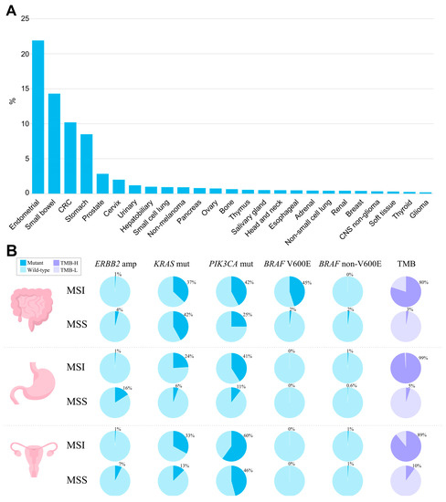
Figure 12. The landscape of MSI and genomic characteristics of MSI tumors across cancer types, according to TCGA. (A) Prevalence of MSI (%) across tumor types. (B) Prevalence of genomic alterations typically found in MSI and MSS tumors in colorectal adenocarcinoma, stomach adenocarcinoma, and uterine corpus endometrial carcinoma.
MSI is characteristic of both Lynch syndrome-associated cancer, where it is observed in nearly all cases, and sporadic cancer, where it reaches up to 10–15% of cases [59][91]. Conversely, Lynch syndrome comprises not more than 19% of MSI cancers, with the highest rate in MSI-positive colorectal cancer, followed by endometrial (5–10%), small bowel (12%), and gastric (4–15%) [64][75][76][77][78][79][80][81][96,106,107,108,109,110,111,112]. Respectively, most MSI cases (80% to 95%) arise sporadically [82][113]. The major cause of MSI is promoter hypermethylation of both MLH1 gene alleles, leading to a loss of MLH1, which is observed in ~90% of sporadic cancer [23][36][82][83][84][85][23,36,58,113,114,115]. When studying the molecular profiles of MSI tumors regardless of whether the tumor is Lynch syndrome-associated or sporadic, a number of oncogenic mutations can be found, including mutations in KRAS, NRAS, BRAF, PIK3CA, APC, TP53, etc. Specifically, KRAS is observed in MSI tumors with a frequency of 30–37% in endometrial, small bowel, and colorectal cancers, and at 15–28% in gastric cancer [29][54][64][86][87][88][29,86,96,116,117,118]. In colorectal cancer, the prevalence of KRAS mutations in MSI cases is lower than in MSS, where it reaches 46% [89][119]. Notably, MSS tumors harboring KRAS oncogenic mutations are characterized by more aggressive growth [85][90][115,120]. BRAF mutations are found with a high frequency (up to 45%) in MSI colorectal cancer, with mostly exclusive prevalence of p.V600E [91][92][121,122]. BRAF mutations have strong bias to sporadic cases, and mostly can never be observed in hereditary colorectal cancer [92][93][94][95][122,123,124,125]. According to Parsons et al.’s meta-analysis [96][126], BRAF V600E variants occur in only 1.4% of patients with Lynch syndrome. In sporadic colorectal carcinomas displaying the MSI phenotype, MLH1 hypermethylation and BRAF p.V600E mutations frequently co-occur, indicating a possible causal relationship between BRAF mutations and MLH1 loss [97][98][99][127,128,129]. However, the straight relationship between MLH1 hypermethylation and BRAF p.V600E mutation might be called into question by the fact that not all colorectal cancers with BRAF p.V600E mutations display silenced MLH1 with subsequent MSI. Such tumors remain microsatellite-stable, suggesting that other factors can influence the MSI phenotype in BRAF-positive cancer [100][130]. Indeed, only 20–30% of BRAF p.V600E-mutated metastatic colorectal cancer display MSI [101][131]. Additionally, the transition to the MSI phenotype via MLH1 hypermethylation is observed in approximately 75% of BRAF-mutated sessile serrated adenomas, with the remaining cases developing into MSS cancers. Among traditional serrated adenomas, BRAF mutations occur in two-thirds of the cases, but MLH1 silencing and the MSI phenotype are rare [102][103][132,133]. In other cancer types, the interplay between BRAF p.V600E and MSI/dMMR is not observed [3][27][62][104][105][3,27,94,134,135]. TP53 mutations are much less frequently observed in MSI as compared to MSS tumors, and their frequency in colorectal and gastric MSI cancer is 20–30%, as compared to 50–65% among MSS tumors [3][106][3,136]. Similarly, in the endometrial, pancreatic, and ovarian cancers, where TP53 mutations are common events, MSI tumors harboring TP53 can rarely be found [62][68][69][107][94,99,100,137]. This pattern of TP53 distribution among MSI and MSS tumors may indicate that TP53 mutations are unlikely to contribute to the MSI cancer tumorigenesis. The prevalence of PIK3CA mutations in colorectal and gastric tumors is higher in MSI tumors (30–45%), as compared to MSS (10–25%). However, in endometrial cancer, PIK3CA mutations are equally common among both MSI and MSS tumors, accounting for 45–60% of all cases [62][94]. Other frequently altered genes in MSI tumors include RNF43, ATM, ARID1A, BRCA2, and PTEN. Mutations in these genes have been shown to have a several-fold increased mutation frequency in MSI colorectal carcinomas compared to MSS [108][138]. Approximately one in five cases of MSI tumors have important targetable fusions in NTRK1/2/3, ALK, or RET genes [108][138], but there is no significant relationship between MSI and HER2 amplification [109][110][139,140]. In the co-occurrence of MSI and other oncogenic alterations, the role of dMMR and its onset in Lynch syndrome colorectal carcinomas is described with three models [4]. In the first (classical) model, MMR deficiency is a secondary event that occurs after adenoma development, initially driven by somatic oncogenic mutations in APC and KRAS [111][112][141,142]. Distinguishing features of adenomas developing in accordance with this model are MMR proficiency and MSS. The classical model is typically observed in patients carrying germline MSH6 or PMS2 mutations [113][143]. It is known that in case of isolated loss of MSH6, MMR activity can be retained due to overlapping functions with MSH3, which explains the relatively low risk of cancer for MSH6 mutation carriers [114][144]. Notably, MLH1 and MSH2 mutation carriers rarely develop tumors via the classical model [111][141]. The prevalence of adenomas developing via this model of carcinogenesis can be roughly estimated at 25% [115][116][117][145,146,147]. In the second and third models, biallelic inactivation of MMR genes leading to dMMR is a driver event, and therefore MSI is observed in all such tumors [111][115][141,145]. The second model is mainly observed in MLH1 and MSH2 mutation carriers and is typically characterized by inactivation of tumor suppressors involved in the WNT pathway, predominantly TGFBR2 and RNF43, due to frameshift mutations within microsatellites in the corresponding genes [108][117][138,147]. The third model accounts for about 10% of LS-associated colorectal carcinomas and is exclusively observed in patients with MLH1 mutations. Here, dMMR is accomplished with mutations in CTNNB1 and TP53 [111][141]. The spectrum of potentially actionable alterations typically observed in MSI and MSS tumors based upon data generated by the TCGA Research Network [118][148] is summarized in Figure 12B. Another important aspect worth mentioning is the relationship between MSI and tumor mutation burden (TMB). dMMR leads to a hypermutator phenotype and increased TMB, which is believed to make tumors more immunogenic. Considering all solid tumors, the simultaneous presence of MSI and TMB is quite rare and occurs only in about 3–7% of cases [30][119][30,149]. However, in tumors associated with Lynch syndrome, the overlap between TMB and MSI becomes more significant. The rate of TMB-H (≥10 mutations/megabase) patients among MSI cases is estimated at around 80–100% in colorectal cancer [52][120][121][84,150,151], 83–93% in endometrial cancer [52][122][123][124][84,152,153,154], and almost 100% in gastric and small bowel cancer [29][52][125][29,84,155]. In colorectal carcinoma, tumors with simultaneous presence of a high TMB and MSI/dMMR correspond to the CMS1 subtype [126][156], which is characterized by hypermutation, hypermethylation, enrichment in BRAF V600E mutations, as well as a strong infiltration of the tumor microenvironment with immune cells [56][127][128][88,157,158]. In this subtype, hypermethylation of the promoter regions of the MLH1 gene leads to its silencing, the accumulation of DNA mutations, and the expression of neoantigens that contribute to the high immunogenicity of the tumor [129][159]. The TMB levels in MSI tumors are likely dependent on certain MMR complex loss and tumor histology/primary site [130][131][160,161]. According to a study by Salem et al., evaluating colorectal, endometrial, and other tumors, overall, the loss of mutSα (MSH2/MSH6) leads to a more pronounced TMB than the loss of mutLα (MLH1/PMS2). However, in some types of tumor histology, secondary DNA repair pathways can better mitigate dMMR, resulting in a less pronounced TMB under the same IHC protein loss patterns. These findings support the diversity of gene- and histologically-specific heterogeneity of MSI/dMMR tumors [119][149]. Patients with MSI displaying a high TMB have a better prognosis and are also good candidates for checkpoint inhibitor therapy [129][159]. Three predictive biomarkers are currently used to select subgroups of patients eligible for ICI immunotherapy: PD-L1 expression, MSI/MMR status, and TMB. Initially, it was considered that a common mechanism of tumor immune evasion is the aberrant expression of immune inhibitory molecules, PD-L1, on the surface of cancer cells [132][162]. However, over time, evidence has emerged that even in the absence of PD-L1 expression, tumors often remain sensitive to ICI [132][133][162,163]. Additionally, TMB-H and MSI tumors respond to the immunotherapy regardless of PD-L1 expression [134][164]. To some extent, it can be explained by the focal expression of PD-L1 [135][165], which can be missed during needle biopsy, or by the dynamic and inducible nature of PD-L1 expression [136][166]. This led to the PD-L1 expression-independent indications of ICI for many types of cancers. While anti-PD-L1 therapy acts to overcome local immune resistance, CTLA-4 is expressed in T-cells and acts non-locally, often causing autoimmune reactions and lymphocyte invasion in different unaffected organs [137][138][167,168]. For that reason, prescription of anti-CTLA4 therapy does not require determination of the CTLA4 expression status. dMMR is the cause of the hypermutator phenotype and increased TMB, which likely leads to increased tumor immunogenicity. Indeed, the connection between MMR phenotype, TMB, and tumor immunogenicity is more sophisticated since some methods of TMB calculation may exclude truncating mutations in certain genes [139][169], while the dMMR phenotype is characterized by an increased frequency of frameshift mutations producing an abundance of highly immunogenic neoantigens [140][141][142][170,171,172]. Thus, dMMR/MSI status should be considered as an independent predictive biomarker of ICI effectiveness, and the presence of a TMB-H in this subgroup may be an additional factor indicating an increased immunogenicity of the tumor.
ScholarVision Creations