Inflammation, Mitochondria and Natural Compounds: Comparison
Please note this is a comparison between Version 1 by Francesca Oppedisano and Version 2 by Lindsay Dong.

Human diseases are characterized by the perpetuation of an inflammatory condition in which the levels of Reactive Oxygen Species (ROS) are quite high. Excessive ROS production leads to DNA damage, protein carbonylation and lipid peroxidation, conditions that lead to a worsening of inflammatory disorders. In particular, compromised mitochondria sustain a stressful condition in the cell, such that mitochondrial dysfunctions become pathogenic, causing human disorders related to inflammatory reactions. Indeed, the triggered inflammation loses its beneficial properties and turns harmful if dysregulation and dysfunctions are not addressed. Thus, reducing oxidative stress with ROS scavenger compounds has proven to be a successful approach to reducing inflammation. Among these, natural compounds, in particular, polyphenols, alkaloids and coenzyme Q10, thanks to their antioxidant properties, are capable of inhibiting the activation of NF-κB and the expression of target genes, including those involved in inflammation.

  • mitochondrial dysfunction
  • reactive oxygen species
  • inflammatory response
  • cytokines

1. Introduction

Complex disorders in human diseases are characterized by a cycle of inflammatory perpetuation. The “druggability” of target components of the inflammatory process represents a novel and exciting strategy for therapeutic interventions aimed at treating chronic diseases and beyond [1][2][1,2]. Reactive oxygen species (ROS) levels are typically rather high in inflammatory disorders. Increased ROS generation leads to DNA damage, protein carbonylation, and lipid peroxidation, which worsen inflammatory disorders. Impaired mitochondria are the main players in sustaining stress condition in the cell [3]. Mitochondrial dysfunction is pathogenic, causing human disorders related to inflammatory reactions [4]. The flow of energy through an organic system has led to the development of the biological complexity of living things, where energy-producing processes are established to take place in the mitochondria. As a consequence of bioenergetic failure, this perspective on human diseases provides a pathophysiological and molecular mechanism for neuromuscular and neurodegenerative disease, metabolic non-communicable and communicable diseases, autoimmune diseases, ageing, and cancer, which deeply affect the mitochondria. [5]. Alterations in cellular and organismal homeostasis are caused by a variety of chemical and physical factors of cellular damage. However, when these dysregulation and dysfunctions are not countered, the inflammation triggered ceases to be useful and becomes harmful [6].
Reducing oxidative stress with ROS scavenger compounds has proven to be a successful approach for reducing inflammation. Indeed, emerging therapies for the treatment of inflammatory illnesses include electron transfer, based on antioxidant compounds. Polyphenols, which include phenolic acids, stilbenes, flavonoids (flavonols, flavanols, anthocyanins, flavanones, flavones, and isoflavones), curcuminoids, carotenoids, capsaicinoids and capsinoids, isothiocyanates, catechins, and vitamins, are significant classes of compounds with antioxidant properties, found in plants. The ROS scavenger activities of these compounds, their ability to inhibit NF-κB (nuclear factor κ-light-chain-enhancer of activated B cells) activation, and their ability to suppress the expression of target genes, including those involved in inflammation, are determined by their antioxidant activities [7].

2. Inflammation

The cellular biological response against aggression, caused by infectious and non-infectious agents (Gram-positive and Gram-negative bacteria, viruses, parasites, toxic substances, or irradiation), is defined as inflammation [2]. Therefore, as a result of an inflammatory process, there is a disruption of or damage to homeostatic processes at the cellular level. The inflammatory process can occur with acute and/or chronic inflammatory responses, thus, involving different organs (heart, liver, kidneys, brain, pancreas, lungs, intestines, etc.). All these events can potentially lead to tissue damage or disease, such as atherosclerosis, Type 2 diabetes, obesity, neurodegenerative diseases, dysbiosis and cancer [2][8][2,8].

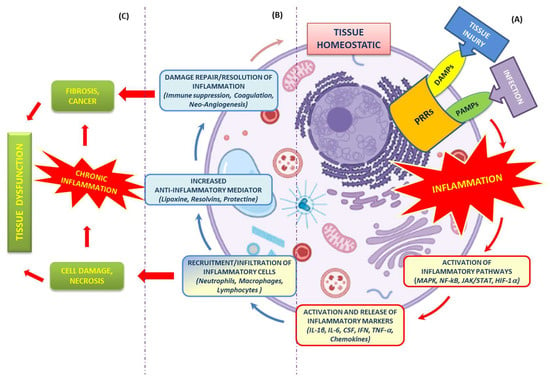
However, the inflammatory process, if it occurs in a timely and complete manner, is of paramount importance, because it ensures the survival of the organism during an infection or injury [9]. Its main goals are to neutralize harmful stimuli, allow continued homeostasis of damaged tissues, and initiate the healing process [10]. At the tissue level, inflammation is characterized by five cardinal signs: redness, swelling, heat, pain, and tissue dysfunction (rubor, tumor, calor, dolor et functio laesa). The first four signs were clinically defined in the first century Atopic Dermatitis (AD) by a Roman physician, Cornelius Celsus. The fifth was added centuries later by Rudolph Virchow in 1858 [2]. Such signs then are nothing more than the macroscopic result of the local responses that occur during the inflammatory process (changes in vascular permeability, leukocyte recruitment and accumulation, and the release of inflammatory mediators) as a result of the triggering of a complex and coordinated communication between the different cells of the immune system (neutrophils, monocytes and lymphocytes) and the blood vessels [10]. Regardless of the nature of the stimulus and location in the body, different inflammatory processes all share common mechanisms, which have been extensively discussed and explored in the literature [11], and which are summarized in the text and Figure 1.
Figure 1. Mechanisms of the inflammatory response. (A) Tissue homeostasis can be altered by external stimuli (tissue injury or infection), resulting in the activation of the innate immune system (PRRs). Such activation triggers an inflammatory response cascade (red arrows) driven by multiple pathways (MAPK, NF-kB, JAK/STAT, HIF-1α) and the release of inflammatory markers (IL-1β, IL-6, CSF, IFN, TNF-, chemokines) at the site of injury. (B) This release is aimed at the recruitment and infiltration of various immune cells (neutrophils, macrophages and lymphocytes). Their presence ensures both the continuation of “transient” inflammation, which is useful for eliminating the cause of tissue injury, as well as the secretion of anti-inflammatory and pro-regenerative cytokines that promote the resolution of inflammation, tissue repair and restoration of tissue homeostasis (blue and pink arrows). (C) Uncontrolled or chronic inflammation, promoted by cellular damage/necrosis, generates remodeling (fibrosis and cancer) and tissue dysfunction.

2.1. Recognition of Damaging Stimuli by Cell Surface Receptors: PRRs (Pattern Recognition Receptors)

PRRs are proteins expressed and present both on the plasma membrane and in the cytoplasm of macrophages, neutrophils, eosinophils, mast cells, natural killer (NK) cells, dendritic cells, and also in other cells (e.g., endothelial cells). They play a crucial role in the main functions of the innate immune system. PRRs are germline-encoded host sensors, which detect molecules that are typical for pathogens [12].
They recognize two classes of molecules/molecular patterns:
  • Microbial structures, known as Pathogen Associated Molecular Patterns (PAMPs), expressed by pathogenic microbes [13];
  • Biomolecules, known as Damage-Associated Molecular Patterns (DAMPs), expressed by host cells during cell/tissue damage or cell death [14].

2.2. Activation of Inflammatory Pathways

Inflammatory pathways primarily involve inflammatory stimuli, which are used for the activation of intracellular signaling pathways, inflammatory mediators, and all common regulatory pathways that are composed of an intricate cascade of molecular signals.
Therefore, ILs such as interleukin-1β (IL-1β), interleukin-6 (IL-6), and tumor necrosis factor-α (TNF-α), as well as microbial products, act through receptor interactions with TLRs, IL-1 receptor (IL-1R), IL-6 receptor (IL-6R), and TNF receptor (TNFR) [15][19].

2.3. Activation and Release of Inflammatory Markers

Activation by pro-inflammatory stimuli of immune and inflammatory cell origin, such as macrophages, monocytes, lymphocytes and adipocytes, induces the production of inflammatory ILs, inflammatory proteins and enzymes [16][24].
Specifically, the inflammatory cytokines involved are classified as IL (IL-1β, IL-6), CSF, IFN, TNF-α, TGF, and chemokines. These cytokines are mainly produced by cells present at the site of infection or injury, and are responsible for leukocyte recruitment [17][25].
The concerted action between pro- and anti-inflammatory cytokines (IL-4, IL-6, IL-10, IL-11, and IL-13) [18][26] ensures the proper modulation of the immune response in the event of an infection or inflammation. Excessive production of inflammatory cytokines can lead to tissue damage, hemodynamic changes, organ failure, and, ultimately, death [19][27].
Inflammation and metabolism are closely interconnected. Many studies have shown that cellular oxidative stress can also be an important pro-inflammatory stimulus, as it can increase the gene expression of growth factors, inflammatory cytokines, and chemokines. In particular, it has been shown that high levels of oxidative stress can induce the production of ROS, malondialdehyde (MDA), 8-hydroxy-2-deoxyguanosine (8-OHdG) and isoprostanes, each of which can activate various transcription factors, including NF-κB, Activator Protein-1, p53 and STAT [20][21][28,29].

2.4. Recruitment of Inflammatory Cells

All cells involved in the inflammatory process are recruited to sites of tissue injury by factors released by damaged epithelial and endothelial cells, and trigger an inflammatory cascade, along with chemokines and growth factors. The first cells attracted to an injury site are neutrophils, followed by monocytes/macrophages, dendritic cells, lymphocytes, NK cells, T cells, B cells), and mast cells [22][34].

2.5. Resolution of Inflammation

The process of resolution of inflammation is of fundamental importance, because it serves to limit both the development of further tissue damage and the onset of persistent chronic inflammation. The presence of mediators (lipoxin, resolvins, protectin, etc.) [23][36], even at this final stage of the inflammatory process, plays a key role, since their presence drives, both locally and systemically, a process of reduction in chemokine gradients over time.

3. Mitochondrial Bioenergetics and Dysfunction

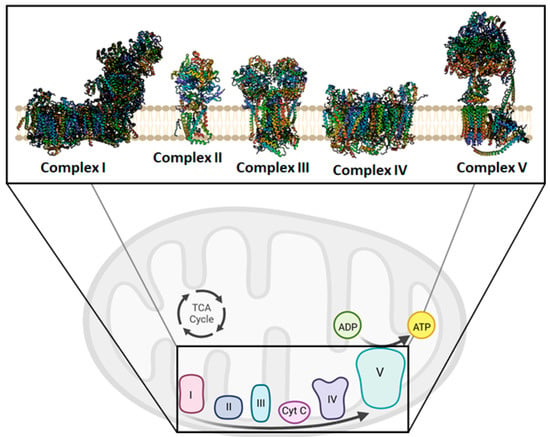
The mitochondria have always been considered the powerhouse of the cell. During aerobic metabolism, mitochondria are the primary source of ATP production. The chemiosmotic hypothesis explains the bioenergetic concept that governs the Oxidative Phosphorylation (OXPHOS) system in mitochondria [24][25][39,40] (Figure 2). OXPHOS activity relies on the inner mitochondrial membrane (IMM)-embedded enzyme (super)complexes [26][41]. As a key mechanism of energy production in cells, they are composed of respiratory machinery, which is responsible for substrate oxidation, working in concert towards the goal of producing ATP by the way of ADP phosphorylation. Mitochondrial respiration is sustained by the electron transfer chain (ETC) that drives the flow of electrons to the final acceptor, oxygen (O2), by four respiratory complexes. These are named as follows: NADH:ubiquinone oxidoreductase, or Complex I; succinate dehydrogenase, or Complex II; cytochrome bc1 oxidoreductase, or dimeric Complex III2; cytochrome c oxidase, or Complex IV. Moreover, to sustain the electron flow from NADH-dependent Complex I oxidation, or succinate-dependent Complex II oxidation, to O2-dependent Complex IV reduction, two mobile electron carriers complete the ETC, i.e., the membrane-embedded hydrophobic coenzyme Q10 (CoQ10) and the soluble cytochrome c. CoQ10 is the electron shuttle between either Complex I or Complex II and Complex III2, whereas cytochrome c facilitates the exchange of electrons from Complex III2 to Complex IV [27][42].
Figure 2. Overview of the mitochondrial oxidative phosphorylation. In the insert, Complex I, II, III, IV, and V are shown in their free form drawn as ribbon representations, obtained from modified PDB IDs: 6YJ4, 1ZOY, 6FO0, 7CP5, and 6TT7, respectively. This figure was created using BioRender (17 December 2022; https://biorender.com/).
The redox energy of electron transfer is coupled to H+ translocation during respiration, building a proton motive force (Δp) that drives ATP synthesis. Complex V, known as ATP synthase, uses the Δp created by ETC to phosphorylate ADP into ATP, the universal energy unit of cells. However, the enzyme can also work in the opposite direction when the Δp drops under phato (physio) logical conditions, and it exploits the energy of the phosphoanhydride bond of the ATP molecules to re-energize the IMM, functioning as an H+ pump [28][43]
Indeed, cristae, a typical invagination of the IMM in mitochondria, serve as the focal points for OXPHOS. To manage cristae architecture, there are two crucial curves. According to reports, the Mitochondrial Contact site and Cristae-Organizing System (MICOS) and the ATP synthase, two key cristae-shaping machines, operate in an antagonistic way concerning cristae biogenesis [29][47]. The first is placed between the concave curvature of IMM at the base of the cristae folds, and the smooth expanse of the IMM is identified as the inner boundary membrane. Contrary to MICOS, which causes a negative membrane curvature, ATP synthase creates long rows of dimers that impose a positive membrane curvature (convex when viewed from the matrix, convex when viewed from the intermembrane space) [30][48]. Monomerization of ATP synthase dimers reveals profound changes in membrane architecture, and would affect the capacity of mitochondria to provide the cell with enough ATP to maintain vital cellular processes [31][49].
Impaired bioenergetics is the result of mitochondrial dysfunction, and is closely related to ROS production [32][51]. Different conditions induce ROS production through mitochondrial polarization and cytosolic calcium (Ca2+) handling. Although the mitochondrial respiratory chain in the IMM is largely considered one of the major sources of ROS, other enzyme systems in mitochondria can also contribute significantly to ROS production [33][52]. The most significant mitochondrial superoxide generators are frequently designed as the FMN and CoQ10-binding sites of Complex I, and the QO site (at the outer or positive side of IMM) of Complex III2. In general, Complex I releases ROS into the matrix while Complex III2 is mostly contained in the intermembrane space [34][53]

4. Mitochondria Are Potentially a New Target of Natural Compounds to Counteract Inflammation

A failure of cellular homeostasis is a common accomplice of the deleterious inflammation process. Indeed, if the inflammation is not properly terminated and persists as a chronic condition it becomes pathogenic [6]. Even though the mechanisms by which dysfunctional mitochondria activate inflammatory signaling are unknown, impaired mitochondria are powerful inflammatory stimuli [4]. Nutraceuticals are bioactive compounds that promote health and have pharmaceutical potential. ROS production is a consequence of mitochondrial dysfunction and aggravates this dysfunction, triggering the inflammatory process. Nutraceuticals can counteract this, preventing abnormal inflammatory activity. The detection of and response to a wide range of pathogen- or damage-associated molecular patterns, which are generated by endogenous stress, can activate the inflammasome, a large intracellular multiprotein complex [35][55]. The physiological role of the downstream inflammatory pathways allows for the elimination of microbial infection and the repair of damaged tissues. The aberrant inflammation triggers pathological conditions, which are related to inflammatory disorders when dysregulated, such as aging-associated diseases, neurodegenerative diseases, diabetes, and atherosclerosis.
The primary immunological risk factors may encourage Nucleotide-binding Oligomerization Domain-like receptor (NOD) family pyrin domain containing 3 (NLRP3) inflammasome activation, wherein cells participate in interactions with TLRs. The following activation of Caspase (Cas)-1 and increased pro-inflammatory cytokine synthesis from this systemic inflammation pathway is finally caused by the maturation of interleukins and the proteolytic cleavage of Gasdermin D, which would be related to mitochondria dysfunction [36][56]. The latter, caused by Cas-11-dependent cleavage, localizes the N-terminal fragments to mitochondria, and forms pores, through which mtDNA is released [37][57]. In the cell, mtDNA has developed properties that allow it to function as a cellular sentinel for genotoxic stress [38][58]
The escape of mtDNA in several devastating diseases is still an obscure molecular mechanism. The critical step in the mtDNA release process is the ROS generation, which is responsible for its oxidation. Defective OXPHOS constituents or alteration of their functionality cause oxidative stress and, consequently, ROS imbalance. Antioxidants could scavenge excess ROS and reduce inflammatory responses by suppressing the inflammation phenomenon, renewing the mitochondrial bioenergetics under pathological or drug-induced imbalance conditions [39][61]. ROS-mediated oxidative modifications of biomolecules usually induces post-translational changes in proteins, i.e., phosphorylation, acetylation, ubiquitination, and SUMOylation, among others [40][62], or chemical modifications, through the oxidation of DNA bases. The most common type of DNA mutation is caused by ROS modifying guanine (G), leading to the production of 8-oxoG. This lesion causes a Hoogsteen base pair, which is a type of base pairing in nucleic acids that can result in a mismatched pairing with adenine (A) in the genome, resulting in C to A (anti) substitutions [41][63]. The enzymes of the base excision repair pathway are primarily responsible for the repair of 8-oxoG, achieved by exploiting their glycosylase activity. In mitochondria, 8-OxoG Glycosylase (OGG1) repairs major oxidative damage in mtDNA and, consequently, OGG1 deficiency results in increased levels of 8-oxoG in mtDNA [42][64]
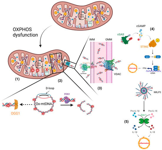
Ox-mtDNA is a mtDAMP and triggers the inflammatory response. Knowing the molecular process that underlies the inflammatory reactions could aid future therapeutic activity in response to microbial invasion or damage signals. Diets rich in bioactive compounds can modulate different stages of inflammation, but information related to their anti-inflammatory mechanisms is still not well understood [43][66]. Ox-mtDNA presence in the cytosol triggers the NLRP3 inflammasome assembly and cGAS/STING pathway-mediated type 1 interferon production [44][67]. Importantly, it was previously unknown as to how Ox-mtDNA moves into the cytoplasm to bind NLRP3 and also activate cGAS-STING signalling. The Voltage-Dependent Anion Channels (VDACs) and the PTP placed on the outer mitochondrial membrane (OMM) and the IMM, respectively, permit the escape of Ox-mtDNA from mitochondria to reach the cytosol, acting as a mtDAMP [4][45][4,59].   Conversely, mitochondrial dysfunction supports Ox-mtDNA generation, in which the mtDNA misses the opportunity for OGG1 repair, and the mutated DNA is cleaved by the Flap Endonuclease-1 (FEN1). Ox-mtDNA and ROS production are not related to FEN1 activity, but instead work to respond to the creation of a bio-signal using mitochondrial Damage-Associated Molecular Patterns (mtDAMPs). Additionally, 500–650 bp Ox-mtDNA fragments are the products of mtDNA cleavage by FEN1 that exit through the pores of the mitochondrial membranes, acting as mtDAMPs (Figure 3). In the fragments of Ox-mtDNA, it was noticed that the sequences corresponding to a region within the mitochondrial genome, the D-loop, were overrepresented and released in the cytosol [46][65].
Figure 3. Inflammatory responses elicited by impaired mitochondria. (1) Defective electron transfer in OXPHOS produces ROS, responsible for the oxidation of mtDNA. (2) Ox-mtDNA might be repaired by OGG1 or cleaved by FEN1 to fragments that are expelled from mitochondria. (3) PTP and VDAC are located on the IMM and OMM, respectively, and translocate the Ox-mtDNA into the cytosol to activate the inflammatory reactions. (4) OX-mtDNA acts as mtDAMPs and promotes cGAS/STING signalling, responsible for type I IFN production. (5) In addition, OX-mtDNA binds NRLP3 to trigger inflammasome assembly and caspase 1 (Cas-1) activation, and consequently promotes proteolytic maturation of the biologically inactive precursors of interleukin-1β (IL-1β) and interleukin-18 (IL-18), thus generating potent proinflammatory and pyrogenic activities. This figure was created using BioRender (7 March 2023; https://biorender.com/).

5. Phytosome and Pathologies

5.1. Curcumin Phytosome

It is known that human exposure to Aluminum Chloride (AlCl3) causes hepatotoxicity, which can be counteracted by the Curcumin Phytosome (CP). Curcumin is a polyphenolic compound extracted from Curcuma longa. In studies conducted in rats, Al-Kahtani et al. demonstrated that treatment with AlCl3 increases the concentrations of Aspartate Aminotransferase (AST), Alanine Aminotransferase (ALT), Alkaline Phosphatase (ALP), Lactate dehydrogenase (LDH), total bilirubin, and Lipid Peroxidation (LPO), reducing, in addition, the stores of albumin, Reduced Glutathione (GSH), Superoxide Dismutase (SOD) and Glutathione Peroxidase (GPx). Histological lesions have also been reported. All of this results in an increased expression of caspase-3 and a decreased expression of Bcl-2. On the contrary, in the presence of CP, the endogenous antioxidant status is favored; therefore, caspase-3 expression is decreased and Bcl-2 expression increases, with an improvement in liver dysfunction [47][108]. The antioxidant and anti-inflammatory properties of curcumin and its nano-phytosome were also tested in mice that exhibited acute inflammation following the administration of carrageenan. In particular, the mice were treated, for 7 days, with an oral dose equal to 15 mg/Kg of indomethacin, curcumin and its nano-phytosome. After 7 days of treatment, the mice were administered carrageenan (1%) at the level of the subplantar region of the left paw, to induce the inflammatory process. A serum antioxidant enzyme assay found that carrageenan reduced the antioxidant activities of SOD, catalase (CAT), GPx, and glutathione reductase (GRx). Conversely, these activities increased in the presence of the curcumin nano-phytosome, both separately and in combination with indomethacin, demonstrating that curcumin nano-phytosome could enhance inflammatory-related antioxidant responses [48][109].

5.2. Silybin Phytosome

Among the research related to the application of phytosomes, a very interesting study was conducted to improve the bioavailability of silybin, a natural compound known for its anticancer, antioxidant and hepatoprotective properties. Silybin is one of the main polyphenols found in silymarin, which is a complex of seven flavonolignans and polyphenols extracted from milk thistle (Silybum marianum). In particular, Chi et al. tested the phytosome-nanosuspensions formulation for silybin, defined as SPCs-NPs, in a mouse model of oxyhepatitis, induced by treatment with Carbon Tetrachloride (CCl4). CCl4 causes liver damage, following lipid peroxidation, a lower activity of antioxidant enzymes and increased generation of free radicals and ROS. The choice of silybin was dictated by the known properties of this natural compound, which is capable of protecting the liver from oxidative stress and inflammation, linked to ROS and secondary cytokine production. The CCl4-induced liver injury resulted in an increased presence of ALT, AST and ALP in the bloodstream, inducing centrilobular necrosis, ballooning degeneration and cellular infiltration. Only in the group of mice treated with SPCs-NPs was a significant reduction in ALT, AST and ALP levels recorded, while no significant changes in these parameters were reported in mice treated with silybin alone compared to the positive control.

5.3. Quercetin Phytosome

An interesting flavanol for human health is quercetin, which cannot be produced by man, but is present in abundance in foods such as fruit, especially citrus fruits, green leafy vegetables, broccoli, olive oil, cherries, and blueberries. Similar to the other flavanols, however, quercetin has a low bioavailability, due to its poor solubility in water, even if it is quite soluble in alcohol and lipids. Therefore, the quercetin-loaded phytosome nanoparticles (QP) have made it possible to overcome this difficulty, increasing the bioavailability for humans of orally administered quercetin in this formulation by about 20 times. This turns out to be very interesting, given the known antiviral, anti-atopic, pro-metabolic, and anti-inflammatory properties of quercetin. Moreover, quercetin has a protective effect on mPTP opening [49][114].

5.4. Berberine Phytosome

The efficacy of another natural product, such as berberine, was tested in women affected by Polycystic Ovary Syndrome (PCOS), an endocrine pathology characterized by hormonal imbalances, dysmetabolism and inflammation. In particular, berberine is an alkaloid used to fight infections, Type 2 diabetes and cancer, but also dyslipidemia in subjects intolerant to statins, and has been shown to enhance the expression of antioxidant enzyme activity. In the study, patients were treated with two daily oral doses of the Berberine Phytosome (BBR-PP) and evaluations were performed at baseline and after 60 days of treatment. The recorded data report a reduction in insulin resistance and acne, and an improvement in lipid metabolism and body composition, but also indicate a reduction in inflammation, with lower levels of CRP and TNF-α.

5.5. Mulberry and Ginger Phytosome

Phytosomes have also been used in the treatment of Metabolic Syndrome (MetS), characterized by visceral adiposity, insulin resistance, hypertension, high triglyceride levels, and low High-Density Lipoprotein Cholesterol (HDL-C) levels. Patients with MetS have an increased risk of developing Type 2 Diabetes Mellitus (T2DM) and Atherosclerotic Cardiovascular Disease (ASCVD). Both genetic and acquired factors generate oxidative stress, cellular dysfunction and the systemic inflammation process responsible for the pathogenesis of MetS [50][120]. In particular, the phytosome containing the combined extracts of mulberry (Morus alba Linn. Var. Chiangmai) and ginger (Zingiber officinale Roscoe) (PMG) was tested in an animal model of MetS. Specifically, male Wistar rats were fed a high-carbohydrate, high-fat diet for 16 weeks, to induce MetS. In the following 21 days, rats with MetS signs were subjected to daily oral treatment with three different doses of PMG, equal to 50, 100 and 200 mg/Kg. Data analysis demonstrated that PMG has a positive effect on body weight gain, and lipid and glucose values, as well as improving Homeostasis Model Assessment of Insulin Resistance (HOMA-IR) and Angiotensin-Converting Enzyme (ACE) levels. Even the parameters relating to the density and size of the adipocytes, to the weight of the adipose tissue, have undergone an improvement after this treatment. At the adipose tissue level, PMG also reduced inflammatory cytokines such as IL-6 and TNF-α, as well as reducing oxidative stress and Histone Deacetylase 3 (HDAC3) expression, while simultaneously increasing PPAR-γ expression. 

5.5. Eufortyn

®

Colesterolo Plus

Recently, the ANEMONE study was conducted, involving 60 healthy subjects with moderate polygenic hypercholesterolemia, treated with Eufortyn® Colesterolo Plus. This nutraceutical consists of the standardized bergamot polyphenolic fraction phytosome (Vazguard®), as well as artichoke extract (Pycrinil®), artichoke dry extract (Cynara scolymus L.), zinc and CoQ10 phytosome (Ubiqosome®).

5.6. Naringenin Phytosome

5.7. Naringenin Phytosome

Furthermore, a few years ago, Yu et al. studied the effect of Naringenin (NG) on acute lung injury. This is a respiratory pathology in which the lung undergoes an important inflammatory process. NG is a plant bioflavonoid found in citrus fruits such as bergamot, grapefruit, tangerine, known for its antioxidant, anti-inflammatory, antiproliferative and anticancer properties. In the study, rats with acute lung injury were treated with Dipalmitoylphosphatidylcholine (DPPC) phytosomes NG-loaded for dry powder inhalation (NPDPIs); in particular, NPDPIs, consisting of mannitol/DPPC/NG in a 4:2:1, w/w/w ratio, were found to be effective.

5.7. Centella Asiatica Phytosome

5.8. Centella Asiatica Phytosome

In a mouse model of phthalic anhydride-induced Atopic Dermatitis (AD), the anti-inflammatory effect of the Centella Asiatica phytosome (CA phytosome) was investigated;it is a medicinal herb used in Ayurvedic, traditional African and Chinese medicine for the treatment of venous insufficiency of legs and diabetes wounds. Following the onset of AD, lesions on the dorsal skin and ear of mice were treated by applying the CA phytosome three times a week for 4 weeks. As a result, inhibition of hyperkeratosis, mast cell proliferation and inflammatory cell infiltration has been reported. The data obtained demonstrated that the CA phytosome inhibits the NF-κB signaling pathway, the release of TNF-α, IL-1β, and IgE, as well as inhibiting the expression of iNOS and COX-2 and the production of NO, resulting in it being a good treatment for AD [51][125].

5.8. Leucoselect Phytosome

5.9. Leucoselect Phytosome

A few years ago, an open-label phase I lung cancer chemoprevention study was conducted on smokers and ex-smokers to test the chemoprotective effects of the leucoselect phytosome. This phytosome consisted of Grape Seed procyanidin Extract (GSE) complexed with soy phospholipids. At the end of the first month of treatment with leucoselect phytosome, an increase in the levels of omega-3 Polyunsaturated Fatty Acids (n-3 PUFAs) was reported, in particular of eicosapentaenoic acid (EPA) and docosahexaenoic acid (DHA), known for their anticancer properties. Three months after the start of the administration of the leucoselect phytosome, an increase in the serum levels of prostaglandin E3 (PGE3) was also recorded. The latter represents a metabolite of EPA, which, in addition to having antineoplastic properties, is also an anti-inflammatory molecule. Thus, the leucoselect phytosome represents a good chemopreventive agent for lung cancer [52][126].

5.9. CoQ10 Phytosome

5.10. CoQ10 Phytosome

Moreover, in humans, the effect of CoQ10 deficiency is known. CoQ10 is a natural compound known for its antioxidant properties, its ability to prevent damage caused by free radicals, as well as its ability to inhibit inflammation signaling pathways. The CoQ10 deficiency can be of a primary or secondary form, and determines the onset of serious diseases, such as encephalomyopathy, cerebellar ataxia and cardiovascular disease. Therefore, maintaining correct concentrations of CoQ10 is important from a therapeutic point of view, even if it is very difficult to achieve this result given its high molecular weight, high lipophilicity, light sensitivity and thermolability [53][128]. To solve these problems, over the years, studies have been conducted on various formulations of CoQ10 that could increase its oral bioavailability. One such study is by Rizzardi et al., published in 2021. In this in vitro study, the authors evaluated the effects of the CoQ10 phytosomal formulation (UBIQSOME, UBQ) on the bioenergetic and antioxidant status of human intestinal epithelial cells (I407) and rat cardiomyoblasts (H9c2). In theis formulation, CoQ10 was administered in association with lecithin.

5.10. Phytosomes and Cytotoxicity

5.11. Phytosomes and Cytotoxicity

Much evidence points to the efficacy of phytosomal formulations in increasing the cytotoxicity of certain natural compounds. Specifically, Alhakamy et al. conducted a study on treating ovarian cancer cells, OVCAR-3, with the phytosome Icariin (ICA). ICA is a flavonol glycoside found in Epimedium grandiflorum. It is best known for its efficacy in treating atherosclerosis and neurodegenerative disorders. It also possesses antioxidant, anti-inflammatory, cardioprotective and hepatoprotective activities. In addition, the antitumor activity of ICA has been demonstrated through its cytotoxicity, apoptotic activity, and regulation of cell cycle protein expression against different cell types. ICA also has anti-angiogenic, anti-metastatic, and immunomodulatory effects, explicitly enhancing the chemosensitivity of ovarian cancer cells. The limitation of ICA treatments is related to its pharmacokinetic properties; it has poor bioavailability due to its chemical structure and a short half-life when administered orally (3.15 h) and intravenously (0.56 h). Therefore, the phytosomal formulation seems to have many advantages for the cytotoxic efficacy of ICAs, particularly for ovarian cancer. This pathology represents a very serious and widespread gynecologic neoplasm worldwide, being fatal in most cases, as it has no early symptoms or adequate treatment. In the study by Alhakamy et al. an increase in the cytotoxic activity of the ICA phytosome on OVCAR-3 cells was demonstrated. Specifically, there was an increase in ICA phytosome-treated cells in the G2/M and pre-G1 phases of the cell cycle. Furthermore, Annexin V staining demonstrated an increase in early, late, and total apoptotic cells.
 
ScholarVision Creations