Chitin is a nitrogen-enriched polymer abundantly present in the exoskeletons of arthropods, cell walls of fungi, green algae, and microorganisms, radulae and beaks of molluscs and cephalopods, etc. Chitosan is a promising candidate for a wide variety of applications due to its macromolecular structure and its unique biological and physiological properties, including solubility, biocompatibility, biodegradability, and reactivity. Chitosan and its derivatives have been known to be applicable in medicine, pharmaceuticals, food, cosmetics, agriculture, the textile and paper industries, the energy industry, and industrial sustainability. More specifically, their use in drug delivery, dentistry, ophthalmology, wound dressing, cell encapsulation, bioimaging, tissue engineering, food packaging, gelling and coating, food additives and preservatives, active biopolymeric nanofilms, nutraceuticals, skin and hair care, preventing abiotic stress in flora, increasing water availability in plants, controlled release fertilizers, dye-sensitised solar cells, wastewater and sludge treatment, and metal extraction.
1. Introduction
Polysaccharides are mainly classified into two groups: homopolysaccharides and heteropolysaccharides, where the homopolysaccharides are composed of a single type of monomer while the heteropolysaccharides are originated by two or more monosaccharide units. Cellulose, chitin, amylose, amylopectin, glycogen, and dextran are some of the examples of homopolysaccharides and glycans that are present in bacterial cell walls and are made from a heteropolymer of alternating β (1-4) linked N-acetylglucosamine and N-acetylmuramic acid residues. Chitin is the most abundant natural amino polysaccharide and the second most abundant natural polysaccharide, which is comprised of N-acetyl glucosamine residues linked via β (1-4) glycosidic bonds. The structure of chitin is different from that of cellulose, the most abundant polysaccharide, where an acetylated amino group is present at the C2 position instead of the hydroxyl group. Chitin is present in green algae, the cell walls of fungi, the exoskeletons of crustaceans such as shrimps, crab, and lobster, and in the cuticles of insects and arachnids, providing structural integrity
[1]. Chitosan is a linear polysaccharide produced by deacetylation of chitin by the hydrolysis of the acetamide groups by strong alkaline treatment. It is composed of β (1-4) linked 2-amino-2-deoxy-β-D-glucopyranose with 2-acetamino-2-deoxy-β-D-glucopyranose.
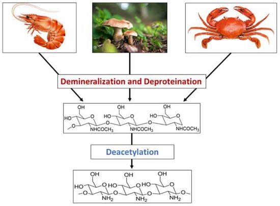
Chitosan is produced from chitin through a series of chemical reactions, as shown in
Scheme 1. The extraction of chitosan mainly involves demineralization, deproteination, and deacetylation. Few studies have reported performing decolorization as a minor step. Chitin could be taken directly from the purified synthetic substance, or it could be taken from natural sources. The most commonly used natural material for such purposes is shrimp shells, and additionally, crab, fungi, etc. have also been used. Shrimp shells are washed, dried, and pulverized to remove any dirt before subjecting them to any chemical treatment. They are comprised of chitin, protein, and minerals such as calcium carbonate and calcium phosphate, which are combined with proteins and chitin and trapped in the exoskeleton
[2]. Therefore, treating with organic or inorganic acids is essential to remove the minerals
[3][4][3,4]. HCl has been used mainly for the demineralization
[5][6][7][8][5,6,7,8], and additionally, inorganic and organic acids such as H
2SO
4, HNO
3, CH
3COOH, CH
2O
2, and HClO
3 [8][9][10][11][8,9,10,11] have also been used to remove the calcium salts. During the treatment, pH increases with the release of Ca
2+ ions, and neutralization after the treatment is essential to stop the demineralization. During the demineralization process, CaCl
2 and CO
2 are produced.
Scheme 1.
Synthesis of chitosan bio polymer using chitin presenting natural sources.
The acid concentration, extraction temperature, and time are the key factors that determine the efficiency of demineralization and the purity of the chitosan produced. Demineralized chitin is then subjected to deproteination in a diluted alkaline medium. Proteins are harder to remove as they are covalently bound to chitin-forming glycoproteins. Therefore, the deproteination process is comparatively longer and could take more than a day as well. Proteins being linked to chitin limits its applications as proteins trigger the immune response in the immune system, leading to restrictions in using chitin for biological applications. Thus, the removal of proteins is of great importance. Mainly, deproteinated chitin is treated with diluted NaOH, whose concentration varies in the range of 0.5–4 mg/L. In addition to NaOH, other basic chemicals such as KOH, Na
2CO
3, Ca(OH)
2, K
2CO
3, and NaHCO
3 have been used for deproteination
[12]. The efficiency of the process depends on the alkaline concentration, temperature, and duration. Huang et al. reported the use of a natural deep eutectic solvent made from choline chloride and malic acid for both the demineralization and deproteination of crab shells. Minerals that are deposited in the chitin-protein matrix of the crab exoskeleton are removed by malic acid, and the strong internal structure is thus weakened. Furthermore, the strong hydrogen bond network between chitin and protein is then less strengthened due to the hydrogen bonds formed between the chloride ions of the solvent and the hydroxyl groups, and hence the proteins are also removed
[13]. Deproteination could also be achieved by treating demineralized crab or shrimp shells with proteolytic enzymes, including chymotrypsin, Alkalase, Pepsin, Papain, and Trypsin
[14][15][16][14,15,16]. Demineralization should be performed before deproteination to increase tissue permeability, promote the action of the enzymes, and reduce the amount of enzyme inhibitors that may be present
[17]. Mhamdi et al. reported the use of the thermostable serine alkaline proteases from
Micromonospora chaiyaphumensis S103, with which 93% deproteination was achieved with an enzyme/substrate ratio of 20 U/mg
[18], and digestive alkaline proteases from the viscera of
Portunus segnis were used to obtain a deproteination of 85% with crab and 91% with shrimp shells
[19]. Lucas et al. reported the deproteination of chitin extracted from the cuticles of insects using Alcalase, which is a bacterial endopeptidase produced from the submerged fermentation of Bacillus licheniformis
[20]. Similarly, Valdez-Peña found the activity of Alcalase and trypsin for the deproteination of chitin
[21]. Furthermore, protease secreted by several bacterial strains during fermentation in the presence of shrimp waste have been reported in many studies. Lee et al. reported the activity of
Paenibacillus elgii TKU051 in deproteination once fermented on shrimp waste, where a maximum of 96.86% deproteination was obtained after seven days of fermentation
[22]. Younes et al. showed the activity of crude protease resulted from the
Bacillus mojavensis A21,
Balistes capriscus Bacillus licheniformis NH1,
Bacillus licheniformis MP1,
Vibrio metschnikovii J1,
Aspergillus clavatus ES1, where A21, A26, J1, and MP1 reported to have an activity of efficiency of deproteination of about 76 ± 4% and that of NH1 and ES1 were significantly lower, resulting in 65 ± 3% and 59 ± 3%, respectively
[23][24][23,24]. Therefore, it is evident that deproteination of chitin could be achieved by treating with alkaline solutions, proteolytic enzymes, and proteases secreted during the fermentation of some microorganisms. Deproteinated chitin is then subjected to N-deacetylation, where the acetyl group is removed to produce chitosan. Deacetylation is commonly performed when deproteinated chitin is subjected to a treatment with a highly alkaline (40–50%) NaOH solution. The dielectric constant of NaOH is greater than that of KOH, making NaOH more suitable for the removal of the acetyl group. Chitin is composed of crystalline and amorphous regions, where the crystalline regions are reluctant to the deacetylation and complete amorphization increases the degree of deacetylation
[25]. Deacetylation depends on the concentration of NaOH, reaction time, temperature, and the properties of the alkaline solution used. Furthermore, deacetylation of chitin can be achieved enzymatically when chitin deacetylase is used to remove the acetyl groups
[26][27][28][26,27,28]; however, enzymatic deacetylation possesses disadvantages including high cost and low productivity, which limit the products to low molecular weight and amorphous chitin
[8][28][8,28]. In addition to the main steps in converting chitin to chitosan, decoloration has also been used in many studies to remove the pigments such as astaxanthin and β-carotene associated with chitin using organic solvents including acetone, sodium hypochlorite, and hydrogen peroxide
[16][29][16,29].
2. Synthesis of Chitosan Nanoparticles
Chitosan nanoparticles have been synthesized by many methods, including ionic gelation
[30][31][32][30,31,32], microemulsion
[33][34][35][33,34,35], spray drying
[36][37][38][36,37,38], and the reverse micellar method
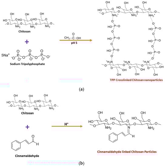
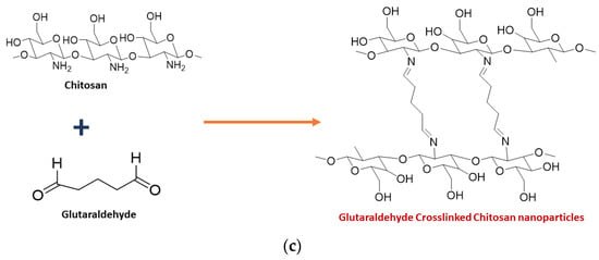
[39][40][39,40], of which the ionic gelation method has been found to be more promising due to the use of a cross linker. Low molecular weight chitosan was dissolved in 1.0% acetic acid, and the pH was then raised to 5.0 by adding 1 M NaOH solution. Sodium tripolyphosphate (TPP), the cross linker, was added, which forms electrostatic interactions between negatively charged TPP and positively charged chitosan. The obtained precipitate was centrifuged and washed to yield pure chitosan nanoparticles
[31]. The synthesis of CNPs from the ionic gelation method using TPP is summarised in
Figure 1a. The green synthesis of chitosan nanoparticles was also reported in several studies. El-Naggar et al., reported the biosynthesis of CNPs using
Pelargonium graveolens leaf extract, which was effective in inhibiting the growth of the phytopathogenic fungi
Botrytis cinerea [41]. Gadkari et al. studied the effect of cinnamaldehyde-cross-linked CNPs on inhibiting the growth of
Staphylococcus aureus and
E. coli where cinnamaldehyde was used as the cross-linker instead of TPP, as shown in
Figure 1b
[42]. Galan et al., reported the use of chitosan crosslinked with glutaraldehyde for the removal of reactive blue 4 dye
[43]. Hence, it is evident that glutaraldehyde could also be used as a cross-linker to join chitosan molecules, as shown in
Figure 1c.
Figure 1.
Synthesis of chitosan nanoparticles using (
a
) TPP (
b
) cinnamaldehyde (
c
) Glutaraldehyde as the crosslinker.
Duraisamy et al., found the antibacterial properties of CNPs prepared in the presence of an ethanol extract of
Martynia annua against
Bacteroides fragilis,
Streptococcus oralis MTCC 2696,
Propionibacterium acnes MTCC 1951,
Pseudomonas aeruginosa MTCC 424,
Staphylococcus aureus MTCC 2940,
E. coli MTCC 443,
Bacillus cereus MTCC 441,
Streptococcus mutans MTCC 890,
Aeromonas hydrophila MTCC 12301, and
Streptococcus faecalis [44]. The green synthesis of CNPs is further reported in other studies, which will be elaborated on with the applications in this re
svie
arch [45][46]w paper [45,46].
35. Toxicity of Chitosan
Chitosan is a biodegradable polymer whose biodegradability could occur through chemical or enzyme catalysis. The degradability depends on the degree of deacetylation and the availability of amino groups. The toxicity of chitosan depends on the charge density and degree of deacetylation. Kazemi et al. evaluated the cytotoxicity of the thiolated chitosan-lauric acid as a new chitosan derivative by MTT assay. The cytotoxicity of the new polymer and the normal chitosan were not significantly different, as indicated by the cell viability studies performed against normal gingiva human cells (HGF1-PI 1). Chitosan derivative, thiolated chitosan-lauric acid, shows low toxicity due to the intramolecular H bonds, which lead to low flexibility where the rigid structure limits the interactions between the cell membrane and the positive charge of the polymer, rendering it less toxic. On the other hand, lauric acid conjugation to chitosan increases its viability and biocompatibility
[47][330]. Chitosan nanogels were found to be free of cytotoxicity when tested against the HEK-293 normal cell line by MTT assay
[48][331]. Asghar et al. evaluated the cytotoxicity of green synthesized chitosan coated silver nanoparticles using the HeLa cell line using the MTT assay method and found that 93.2% cell viability was achieved with 100 µg/mL but with higher doses (300 and 400 µg/mL), the viability reduced to 54.5% and 35.2%, respectively. However, the chitosan-coated Ag nanoparticles showed less toxicity compared with Ag nanoparticles, and they exhibit more antibacterial, anticoagulant, antiplatelet, and thrombolytic activities compared with Ag nanoparticles
[49][332]. Furthermore, Frigaard et al. reported that among the 25 articles they have referred to except one, all others reported less toxicity of chitosan nanoparticles with >80% cell viability when studied both in vitro and in vivo
[50][333], and the only study where low cell viability was reported was the use of dry powder of chitosan nanoparticles (250 and 500 µg/mL) with Calu-6 cells for 24 h
[51][334]. Therefore, it is evident that chitosan and its derivatives are less toxic to human cells.
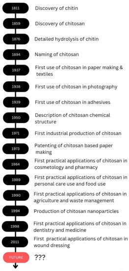
As revealed by the examples above throughout the review, it is evident that chitosan is effectively applicable in several fields, and chitosan would be a promising candidate in future applications as well. The discovery and progress of chitosan and its applications are exhibited in
Scheme 2.
Scheme 2.
The discovery and the progress of chitosan and its applications.