Nanoparticles can be synthesized through physical, chemical, and biological routes, where biologically synthesized nanoparticles are also referred to as biogenic-synthesized nanoparticles or bionanoparticles. Bionanoparticles exploit the inherent reducing property of biological entities to develop cost-effective, non-toxic, time-efficient, sustainable, and stable nanosized particles. There is a wide array biomedical focus on metallic nanoparticles, especially silver nanoparticles, due to their distinctive physiochemical properties making them a suitable therapeutic molecule carrier. This research aims to provide a broad insight into the various classes of living organisms that can be exploited for the development of silver nanoparticles, and elaboratively review the interdisciplinary biomedical applications of biogenically synthesized silver nanoparticles in health and life sciences domains.
- nanotechnology
- bionanoparticles
- silver nanoparticles
- biological entities
- biomedical applications
1. Introduction
In the past decade, the field of nanoscience and nanotechnology has revolutionized science and technology. The term nanoparticle is defined differently by ISO [1], ASTM [1], and IUPAC [2]. The definitions by different organizations consider a variety of attributes such as particle number, concentration of particles, particle size distribution, aggregation/agglomeration of nanoparticles, etc. [1]. However, an internationally recognized definition of nanoparticle has not been established. Apart from the nomenclature, other challenges in the field of nanotechnology include a lack of (a) validated analytical methods and test protocols, (b) reliable exposure and toxicity data, and (c) accurate analytical techniques to precisely characterize nanoparticle morphology [3]. All these developments and challenges suggest robust research and development in nano-research and the importance of nanotechnology in the ever-changing scientific scenario. In nanoscience and nanotechnology, there is a focus on metallic nanoparticles, especially silver nanoparticles (AgNPs), for their distinctive catalytic activity [4], electrical and thermal conductivity [5], non-linear optical properties [6], and surface-enhanced Raman scattering properties [7]. Moreover, studies suggest that AgNPs have excellent market value in comparison to other nanoparticles from the consumer’s perspective [8]. In medicine and biomedical applications, therapeutic effects largely depend on the pharmacokinetics and pharmacodynamics of AgNPs [9]. Due to their biocompatibility and viability, AgNPs act as suitable therapeutic molecule carriers of anticancer [10][11], antimicrobial [12][13], antioxidant [14], and anti-inflammatory [15][16] agents. Moreover, the intrinsic properties of AgNPs, such as binding affinity for various organic molecules and strong absorption, make them a potential candidate for vaccine development and as drug carriers for specific and selective tissue targeting [17]. Even after extended research, there lies a vast gap in the investigation of AgNPs for biomedical applications. The major hindrance is that the state of AgNPs depends on the medium in which it interacts [18]. For example, AgNPs undergo agglomeration, aggregation, and dissolution on exposure to biological media and biomolecules due to various factors such as organic matter, pH, ionic strength, etc. [18][19]. Another issue is the unavailability of a systemic pattern of comparative analysis of AgNPs for their effects in currently published papers [20][21]. The current studies are focused on synthesizing AgNPs while minimizing these limitations. AgNPs can be synthesized through three alternative methods, namely physical, chemical, and biogenic routes. The chemical and biogenic routes of AgNPs are comparable, as reducing and stabilizing agents help convert silver ions to AgNPs and further coat particles to maintain size in the nano-size range and anisotropic shape. However, in biogenic routes, biological species act as reducing and/or stabilizing agents in contrast to specific chemicals used in the chemical route. The role of biological species to reduce and/or stabilize and further coat AgNPs makes them an attractive candidate to investigate some prominent concerns such as agglomeration and aggregation. The biogenic methods of synthesizing AgNPs were initiated nearly two decades ago, and currently, more than 1000 biological species are employed in synthesizing AgNPs. It represents a massive success in the biogenic synthesis of AgNPs. This research is specifically focused on the biological route of AgNPs synthesis, where biological species act as a catalyst by reducing, stabilizing, and/or capping Ag+ ions. Various reviews on silver nanoparticles and their biomedical applications are availabl [22][23][24][25][26]. However, a single elaborative review on the use of all biological species for silver nanoparticle synthesis is not available. This research aims to review various employed biological species such as microorganisms, plants, viruses, and human cell lines involved in the synthesis of AgNPs. The research also throws insight into the biomedical applications of biogenic AgNPs.2. Biosynthesis of AgNPs
2.1. Methods of Biosynthesis: Physical, Chemical, and Biological
The synthesis of AgNPs can follow a top-down (physical route) or bottom-up approach (chemical and biological route), as depicted in Figure 1. The top-down approach does not require reducing or stabilizing agents but follows specific techniques reviewed elsewhere [27]. On the other hand, the bottom-up approach requires reducing or stabilizing agents reviewed elsewhere [27]. The reducing and stabilizing agents in the bottom-up approach may be chemical or biological entities classifying this route into chemical and biological methods of AgNPs synthesis, respectively, as depicted in Figure 1. The chemical route of AgNPs synthesis is commonly a three-component system consisting of a metal precursor, reducing agent, and stabilizing agent, where the initial concentration of the metal precursor, the concentration of the stabilizing agent, the potential of the reducing agent, and the molar ratio of metal precursor to reducing agent determine AgNPs size [28]. For example, strong reducing agents (e.g., borohydride) form small-sized AgNPs in contrast to weak reducing agents (e.g., sodium citrate) that produce large-sized AgNPs [28]. The major shortcoming in chemically synthesized AgNPs is the adsorption of certain chemicals on AgNPs surfaces leading to health hazards and toxicity and obstructing their advancement to biomedical use. In contrast to the chemical route, which generally is a three-component based system, the biological route is a two-component system where biological entities both act as reducing and stabilizing agents to reduce metal precursors and stabilize the formed nanoparticles. The use of biological entities both as reducing and stabilizing agents increases their possibility to improve stability, reduce aggregation, increase the reaction rate, and provide efficient purification of AgNPs. Moreover, the adsorption of organic molecules from biogenic species on AgNPs increases their potential for biomedical activity. For example, coating metallic nanoparticles with phytochemicals is suggested to improve the stability of nanoparticles in the external environment and prevent colloidal aggregation [29][30]. In support of this, Mousavi-Khattat et al. stated that though chemically synthesized AgNPs had higher stability after synthesis, their stability decreased with time in comparison to biogenic-synthesized AgNPs [31]. They further suggested that the synergistic effect of phytochemicals and their coating on AgNPs improve the antibacterial efficacy of biogenic-synthesized AgNPs [31]. A similar study on the functionalization of organic groups on AgNPs surfaces leading to their better anticancer activity in contrast to chemically synthesized AgNPs has been carried out by Kummara et al. [32]. In contrast, Spagnoletti et al. suggested similar bactericidal activity of chemical and biogenic-mediated AgNPs with lower toxicity by the latter [33]. Sreelekha et al. also carried out a comparative study on chemically and biogenically synthesized AgNPs, suggesting that the water-soluble biomolecules adsorbed on AgNPs surfaces provide higher stability to green-synthesized AgNPs and support their higher antioxidant activity [34]. A similar comparative analysis to differentiate the properties of AgNPs produced by the biogenic route from that of the chemical route has been carried out by Veeragoni et al. [35].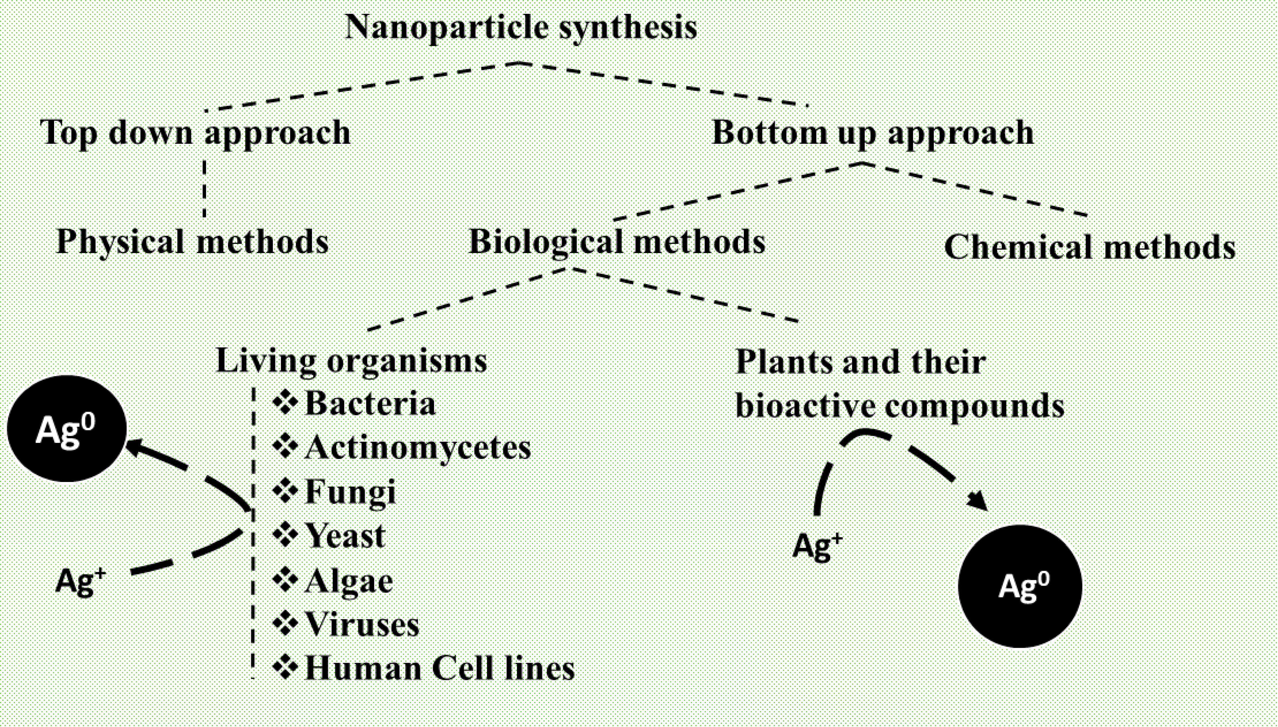
2.2. Biological Species for Nanoparticles Synthesis
2.2.1. Bacteria
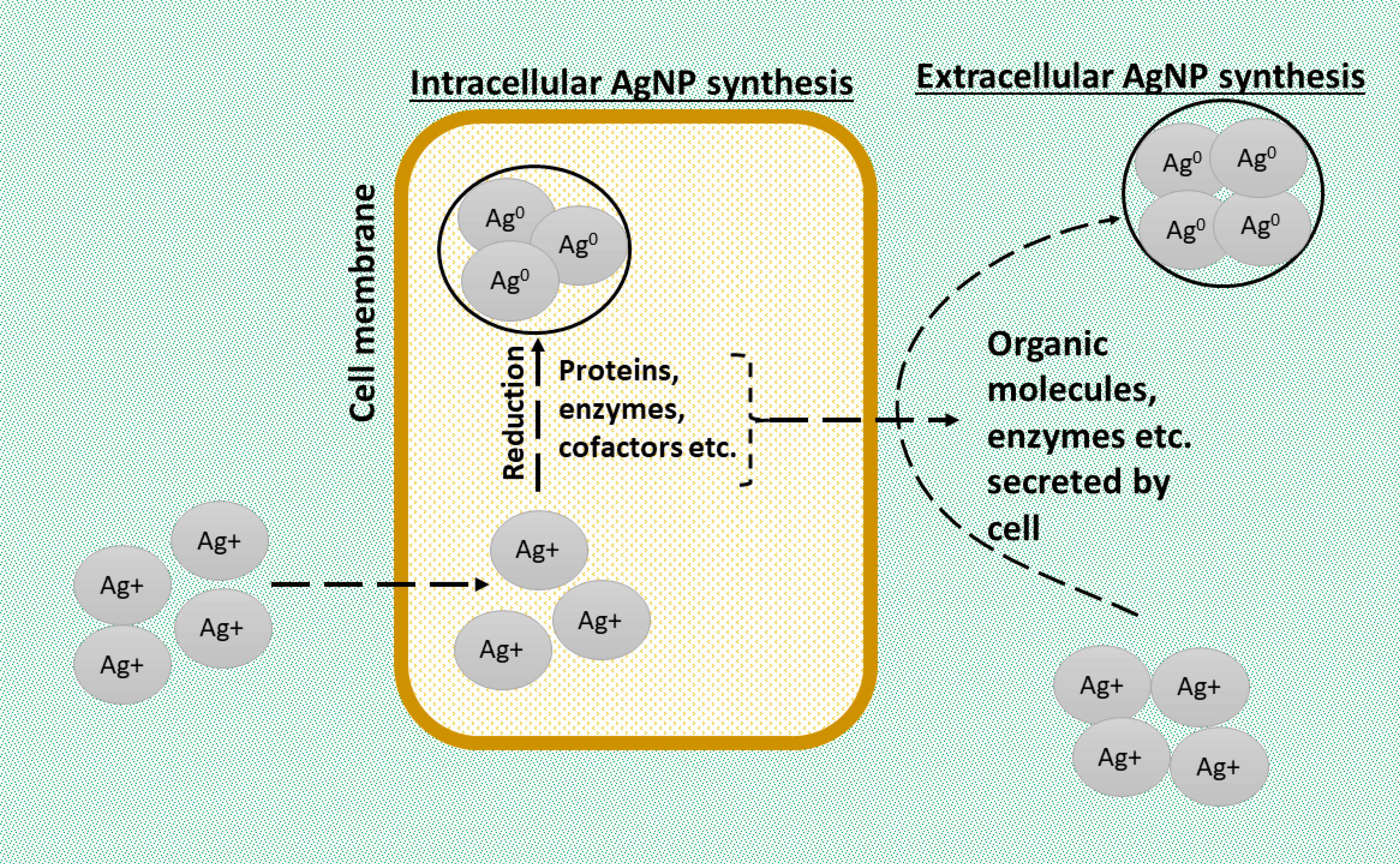
Bacteria are prokaryotes that have developed a natural defense mechanism to survive continual exposure to toxic metals and environmental conditions [51]. The resistance of bacteria to metals such as silver causes the accumulation of Ag+ in their cell wall, aiding in AgNPs synthesis. Therefore, bacteria’s natural mechanisms are exploited for nanoparticle synthesis. The bacterial biomass [52] or supernatant [53] helps in intra- or extracellular AgNPs synthesis. The bacteria’s major functional groups involved in synthesis are carboxylic, hydroxylic, and primary and secondary amides. The major advantages are a high growth rate and easy handling, amongst others. [54][55]. In bacteria-mediated AgNPs synthesis, nanoparticles can be produced from bacterial genomic DNA, culture broth, cell-free supernatants, or protein extracts. Chumpol et al. synthesized AgNPs using both ssDNA and dsDNA and stated that bacterial ssDNA-mediated AgNPs were more stable than dsDNA, as silver ions interacted more efficiently with the nitrogenous bases of ssDNA (produced from denaturing dsDNA) [56]. However, this preparation method involves a three-component system where glucose was additionally required for the conversion of silver ions [56]. Furthermore, their time-dependent synthesis studies suggested that longer time reactions resulted in aggregation, which was confirmed by the shift in SPR peak to a longer wavelength in proportion to time [56]. Saravanan et al. synthesized AgNPs using a culture broth of Bacillus brevis (NCIM 2533) and stated that the coating of proteins (from cultural extract) on AgNPs surfaces results in minimal agglomeration [57]. These results are in parallel to studies of Yurtluk et al., who synthesized AgNPs from Bacillus sp. SBT8 and obtained similar SPR peaks [58]. However, Yurtluk et al. also studied the effects of pH and temperature and stated that particle size increases with pH and efficient yield occurs at 33–37 °C [58]. To further analyze the effects of pH, Arzoo et al. isolated 155 strains of Pseudomonas spp. from the rhizosphere, of which three strains, namely SMS13, SMS100, and SMS124, were most efficient in AgNPs synthesis [59]. Arzoo et al. stated that AgNPs (from Pseudomonas aeruginosa) were synthesized at the beginning or end of the log phase when most bacterial metabolites are produced and in turn fluctuated the environmental pH [59]. Similar to this, Sable et al. demonstrated the role of nitrate reductase and other enzymes from Bacillus subtilis spizizenii in AgNPs synthesis and further suggested that the media components might alter the particle size and optical properties of AgNPs [60]. In contrast to the study conducted by Yurtluk et al., Saleem et al. synthesized AgNPs using bacterial strains (wus1, wus2, and wus5) and suggested that high pH negatively impacts AgNPs synthesis and affects SPR peak [61]. However, the optimum temperature for AgNPs synthesis reported in both studies was parallel to that reported by Singh et al. [62]. Furthermore, Singh et al. suggested the role of water-soluble biomolecules and active enzymes as reducing and capping agents, which were parallel to the results from the above-discussed studies [62]. An interesting study to confirm the role of bacterial proteins was conducted by Li et al., where they used protein extracts of Deinococcus radiodurans and suggested that bacterial protein extracts produced monodispersed and spherical AgNPs due to the interactions (reduction and capping) of silver ions with hydroxyl, amine, carboxyl, phosphate, or sulfhydryl groups of proteins of Deinococcus radiodurans [63].2.2.2. Actinomycetes
Actinomycetes are Gram-positive, aerobic entities [64] with bacteria-like cell wall composition [65] and fungus-like branched filamentous growth [66][67] found both in soil and aquatic conditions. There is comparatively a limited number of studies synthesizing AgNPs via actinomycetes. Actinomycetes are involved in the intra- and extracellular synthesis of nanoparticles [47]. In AgNPs synthesis, the reduction and consequent formation of nanoparticles occur on the mycelial surface and in the cytoplasm [47][68]. Ag+ ions get trapped on the surface, which then interacts with the functional groups of biomolecules present in mycelia, leading to Ag+ reduction [66]. Streptomyces species, the largest genus of actinobacteria, are involved in the synthesis of AgNPs with numerous biomedical applications. The actinomycetes mediating the synthesis of AgNPs have multiple advantages, such as monodispersity over polydispersity [69], small-sized particles that increase stability and biocompatibility, and biocidal features [70]. Monodispersed nanoparticles have an added advantage, as they suggest better sample-wide uniformity, lower aggregation, and higher stability. In actinomycetes-mediated AgNPs synthesis, nanoparticles are majorly produced from the culture broth or cell-free supernatant of actinomycetes. Wypij et al. synthesized small-sized AgNPs (that increased stability and biocompatibility) from the Streptomyces xinghaiensis OF1 strain, capped with organic compounds such as amino bonds [71]. Similarly, S. et al. synthesized AgNPs from the Streptomyces hirsutus strain SNPGA8, where the presence of FTIR-intense bands suggested the role of functional groups, namely alcohols, bromide, iodide, chlorides, and sulfates, in the reduction, stabilization, and capping of AgNPs [72]. However, the above two studies reported the formation of polydispersed AgNPs. To understand the dispersity phenomenon, Mabrouk et al. synthesized AgNPs from Streptomyces spiralis and Streptomyces rochei and found that the organism’s strain played a critical role in the size homogeneity of nanoparticles [73]. For example, they found that Streptomyces rochei could produce monodispersed AgNPs, as a single low-molecular-weight protein was involved in reduction, capping, and stabilization in comparison to the involvement of numerous varied molecular-weight proteins in the production of polydispersed AgNP from Streptomyces spiralis [73]. Furthermore, they stated that bactericidal activity is higher for smaller-sized nanoparticles, as they have a higher surface area than large-sized nanoparticles [73].2.2.3. Fungi
Fungi are eukaryotic, unicellular, or multicellular heterotrophs that obtain food from dead or living organisms. Fungi have a higher preference than other microorganisms for nanoparticle synthesis [74], as they are easy to grow, handle, and resist agitation amongst other extreme processing conditions. They secrete many proteins, enzymes, and polysaccharides, which play a vital role in synthesizing a diverse range of nanoparticles [75]. The major functional groups involved are carbonyl, amide, hydroxyl, etc. [76][77]. Nanoparticles can be synthesized intracellularly and extracellularly. Intracellular synthesis provides better control over size, while extracellular synthesis is hassle-free due to easy downstream processing steps. Fungi also have an appreciable binding capacity, tolerance, bioaccumulation, and intracellular uptake for silver ions under various experimental conditions [78]. Moreover, the size and structure of fungal-mediated AgNPs can be manipulated by altering pH, temperature, time, and other culture conditions. Soleimani et al. studied the effect of different pH (5.0, 6.0, 7.0, and 8.0) and temperatures (40 °C and 60 °C) for fungal strains, namely Beauveria bassiana (JS1, JS2, and KA75) and Metarhizium anisopliae, and stated that 60 °C and pH 7.0 were optimum conditions for the production of small-sized AgNPs in high concentrations [79]. Furthermore, they suggested that isolates KA75 and JS1 produced the most desirable AgNPs [79]. Koli et al. synthesized AgNPs from Monascus red pigments, where sunlight catalyzed the reaction with a reaction time of 5 min [80]. Spagnoletti et al. conducted a comparative study between chemical and biogenic (via Macrophomina phaseolina) synthesis of AgNPs and stated that both modes of nanoparticles synthesis had similar bactericidal activity. However, the biogenic-mediated nanoparticles represented lower toxicity in the model organism [33]. Ansari et al. synthesized AgNPs from various fungal species, suggesting the role of carbohydrates (from exopolysaccharides) and not proteins in nanoparticle synthesis [81]. Furthermore, they suggested the highest reduction capacity for Aspergillus niger KIBGE-IB36, followed by Aspergillus terreus KIBGE-IB35, Aspergillus flavus KIBGE-IB34, and Aspergillus fumigatus KIBGE-IB33 [81]. Though FTIR analysis was not conducted to confirm the adsorption of functional groups on the nanoparticles surface, a strong SPR peak suggested the formation of stable AgNPs with no aggregation for 3 months [81]. In support of Ansari et al., Li et al. suggested the role of polysaccharides in AgNPs synthesis from Aspergillus japonicus PJ01 and stated that reducing sugars helped in reduction, and polysaccharides and proteins supported nanoparticle stabilization [82]. They further studied the role of silver nitrate concentration, pH, and temperature to suggest that the size of AgNPs is proportional to silver nitrate’s concentration. Nanoparticle synthesis decreases in extreme alkaline conditions, with the optimum temperature for synthesis being 30 °C [82]. However, the work of Wang et al. was contrary to Li et al., which suggested different optimum pH and temperatures for AgNPs synthesis from Aspergillus sydowii [83]. These contrasting results suggested the role of the strain in deciding the optimum conditions of pH and temperature.2.2.4. Yeast
Yeasts are eukaryotic, single-celled organisms, chemoorganotrophs (produce energy from organic matter), widely used in bakery and fermentation processes, that can accumulate a variety of metals. Like fungi, yeasts have a rapid growth process that can be easily manipulated in the laboratory using specific nutrient conditions [84][85]. Yeasts can synthesize nanoparticles intracellularly and extracellularly [86]. The metal ions trapped by yeast undergo oxidation, reduction, sorption, chelation, cell membrane transport, or efflux [84][87]. These processes, by different yeast genera, lead to size and shape-dependent changes in AgNPs. The role of yeast extract as a capping agent helps produce monodispersed nanoparticles that can be easily preserved without precipitation for more than a year [86][88]. Cunha et al. synthesized AgNPs from Rhodotorula glutinis and Rhodotorula mucilaginosa and suggested that the time required for AgNPs synthesis was proportional to the constituents of the extract that reduced and stabilized the nanoparticles [89]. They further stated that the adsorption of proteins on AgNPs prepared from yeast extract prevented aggregation and sedimentation and enhanced colloidal stability for nearly 15 months [89]. Their study also explained that proteins, ions, and water molecules adsorbed on AgNPs surfaces (in suspended form) caused light scattering, which explained the larger size of nanoparticles when analyzed by DLS in comparison to other techniques [89]. Supporting the work of Cunha et al., Shu et al. synthesized AgNPs from Saccharomyces cerevisiae and suggested that biomolecules, namely amino acids, alpha-linolenic acid, and aminobutyric acid, favored controllable size distribution, monodispersity, and stability for nearly a year without precipitation.2.2.5. Algae
Algae are photosynthetic, unicellular or multicellular eukaryotes, found in water [90] and soil [91]. They can be differentiated into micro- and macroalgae based on their size. Brown algae, green algae, and cyanobacteria are significant varieties of algae that help synthesize nanoparticles [91][92][93]. Algae are excellent and inexpensive sources of AgNPs production in bulk quantities. The property of algae to develop a specific charge on their surface [91][94] and reduce metals inside and outside the cell makes it a robust biological entity for nanoparticle synthesis. Algae biomass, cell-free extracts, supernatants, and filtrate of broth are used in AgNPs synthesis [93]. The major disadvantages of algae-mediated AgNPs synthesis are the difficulties in the separation of synthesized nanoparticles from the other components involved in the reaction and low production [95][96]. In algae-mediated AgNPs synthesis, the nanoparticles are majorly synthesized from aqueous extract or cell-free supernatant of algae. Rao et al. synthesized AgNPs from fucoidan solution by a microwave irradiation technique and confirmed the adsorption of fucoidan on AgNPs surfaces through a characteristic sulfate group peak, as reflected in the FTIR analysis [97]. They further suggested that the percentage of nanoparticles and fucoidan was 87% and 13%, respectively, and confirmed it through inductively coupled plasma mass spectrometry [97]. In contrast, Bao et al. synthesized AgNPs using Neochloris oleoabundans and found the adsorption of no functional groups on the nanoparticles surface, particularly due to the low concentration of cellular materials [98]. However, the reactions under low concentrations suggested the efficiency of reaction by the organism and ease in AgNPs separation. They also stated the relevance of the optimum concentration of AgNO3 (nearly 0.4 mM), optimum pH (between 5 and 7), and extraction time (0.5–10.0 h) for maximum nanoparticle yield [98]. Furthermore, as the reaction was carried out under light conditions, they suggested the dependency on light for the reaction [98]. Husain et al. synthesized AgNPs from Microchaete and suggested that intrinsic capping and stabilization by functional groups of Microchaete prevented the need for further downstream processing [99]. Veeragoni et al. carried out a comparative study analyzing the differences between chemically and Padina tetrastromatica mediated AgNPs synthesis, suggesting the role of alcohol, alkane, and nitro groups in chemical synthesis and ketones, aldehydes, and phenol in biogenic method [35]. However, they stated that biogenic-mediated AgNPs were more negatively charged contributing to low aggregation and high stability at different pH conditions and 10% serum biological media [35]. Furthermore, they suggested that the concentration of AgNPs synthesis was pH dependent in biogenic synthesis but not in chemical synthesis, probably due to the impact of ions on bioactive compounds [35].2.2.6. Virus
Viruses are linear, circular, single, or double-stranded nucleic acids with capsid (outermost layer). They hold the capacity to synthesize monodispersed, polyvalent, or symmetric nanoparticles of appreciable surface area and a high aspect ratio [100]. The outer proteinaceous coating of a virus called capsid is majorly involved in binding with metal ions [101]. Some significant viruses employed are brome mosaic virus, cowpea mosaic virus, cowpea chlorotic mottle virus, hibiscus chlorotic ringspot virus, red clover necrotic mosaic virus, tobacco mosaic virus, and turnip yellow mosaic virus [102]. AgNPs can be synthesized with the help of biological substances of virus-like viroid capsules, DNA, multicellular superstructures, and lipid cylinders. Interestingly, different combinations of viral particles can interact with plant extract for the synthesis of bionanoparticles with reduced size in more prominent numbers [103]. Comparatively, virus-mediated synthesis of AgNPs is less common.2.2.7. Plants
Amongst all the biological classes, plants hold the maximum potential for bionanoparticles synthesis, as they are natural sources that can help remove heavy metals from soil and water [104]. This characteristic property of plants is employed for the synthesis of AgNPs. Different chemical components of plants involving various phytochemicals (e.g., catechins, flavones, terpenoids, polyphenols, etc.) support nanoparticles synthesis [105][106][107]. These phytochemicals are soluble in water and act as reducing and capping agents [108]. The nanoparticles synthesized by plants could be through living plants (intracellular route), plant extracts (extracellular pathway), or phytochemicals (extracellular pathway) [109]. Some methods of plant-mediated AgNPs synthesis are elaborated in Table 6. In the intracellular route, the plant or its biomass interacts and reduces metal in the aqueous metal salt solution [110][111]. A single plant species can produce polydispersed nanoparticles with variable morphological structures. This diversity is attributed to stabilizing and reducing agents and complex nanoparticle separation and purification procedures [112][113]. In the extracellular route, plant extracts are obtained via hot or cold extraction methods or the Soxhlet apparatus. Phytoconstituents of plants play a vital role in bionanomaterials synthesis. As extracts are first separated and then used for nanoparticle synthesis, the process is called an extracellular method. The process helps in the production of nanoparticles of specific shape and structure, which have a negative potential and are stable in water. Due to variations in phytochemical composition, the process cannot produce monodispersed particles [114]. Phytochemical-mediated synthesis of nanoparticles extends the extracellular route in which specific phytochemical is isolated, quantified, and then used for nanoparticle synthesis. This method helps predict the nanoparticle synthesis mechanism [114]. The process majorly depends on flavonoids and polyphenols and helps control nanoparticle shape and size.2.2.8. Human Cell Line
Human cells are heterotrophic in nature and require an external source of energy for survival. Recent research conducted on certain human cell lines suggests that they can be utilized in nanoparticle synthesis. The epithelial cells of a healthy individual were used for the synthesis of nanoparticles, in vivo and in situ, without the use of external chemicals as reducing agents. Similarly, cancerous and non-cancerous cells such as HeLa (Homo sapiens, human), SiHa, and human embryonic kidney-293 cell lines serve as a source of AgNPs synthesis. Human cell-line-mediated AgNPs synthesis is simple, effective, and inexpensive, but less commonly explored [115][116].2.3. Characterization Techniques
Characterization of a material is the analysis of its structure, composition, and physical-chemical properties by determining properties such as size, shape, structure, and surface area. Microscopic and spectroscopic methods can help in nanomaterial characterization, as depicted in Figure 3. Spectroscopic methods include X-ray diffraction (XRD), ultraviolet spectroscopy (UV–Vis), and Fourier transform infrared spectroscopy (FTIR), while microscopic techniques include scanning electron microscopy (SEM) and transmission electron microscopy (TEM). However, these techniques are always used in combination for proper assessment and confirmation of results. The choice of characterization techniques employed depends on the applications specific to the prepared nanoparticles. In comparative studies of biogenic and chemically synthesized AgNPs, the deviation or variability of peaks and intensity has been observed [31][32][34]. The deviation or variability is specifically observed in UV–Vis, FTIR, and XRD results and is attributed to the functionalization of the nanoparticle surface.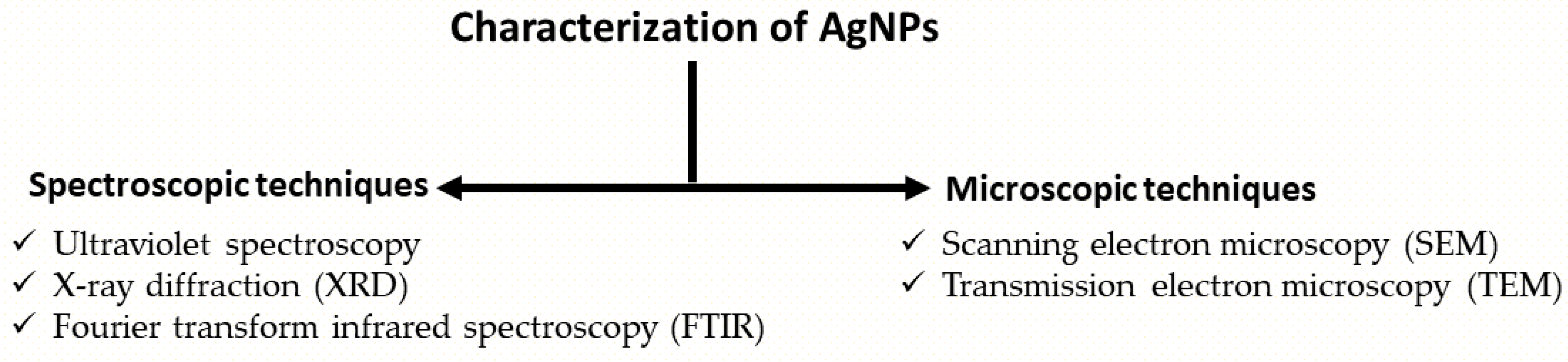
3. Biomedical Applications
3.1. Antimicrobial Activity and Associated Applications
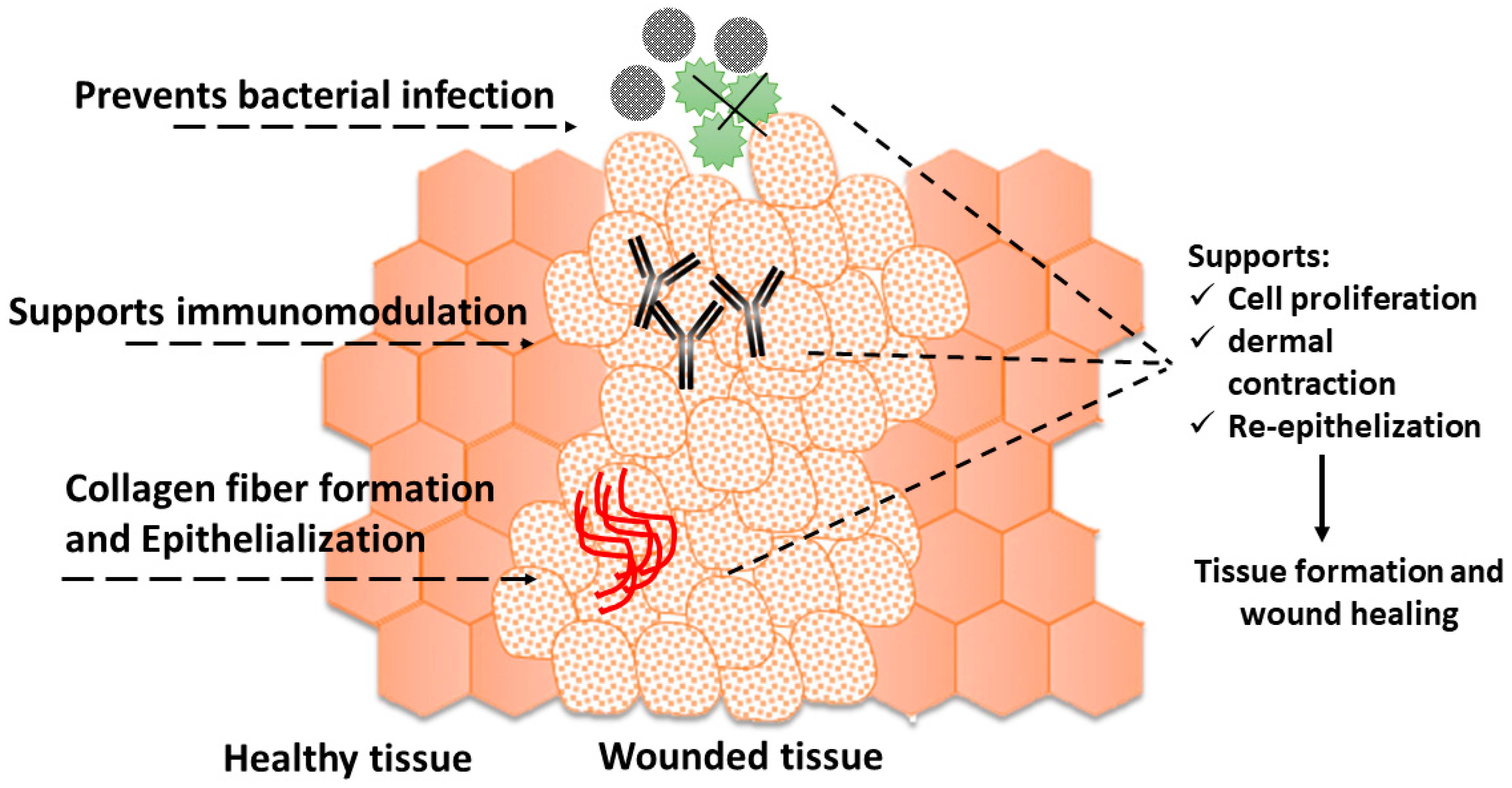
Antibiotic resistance occurs due to mutations in target microorganisms, efflux pumps, and biofilm formation [129], which is a significant problem [130][131][132], causing the emergence of multidrug-resistant pathogens. The hypothesis that drug-bound AgNPs act as carriers for antibiotics and disrupt bacterial cell walls enabling antibiotic entry is explored to overcome antibiotic resistance. AgNPs are found effective against Gram-positive and Gram-negative bacteria [133], as they work by (a) damaging the cell membrane and its components and (b) inducing cell ROS production that affects the DNA, RNA, and proteins of a cell, as depicted in Figure 4. However, AgNPs have different efficacy due to the presence of thick peptidoglycan in Gram-positive bacteria [134].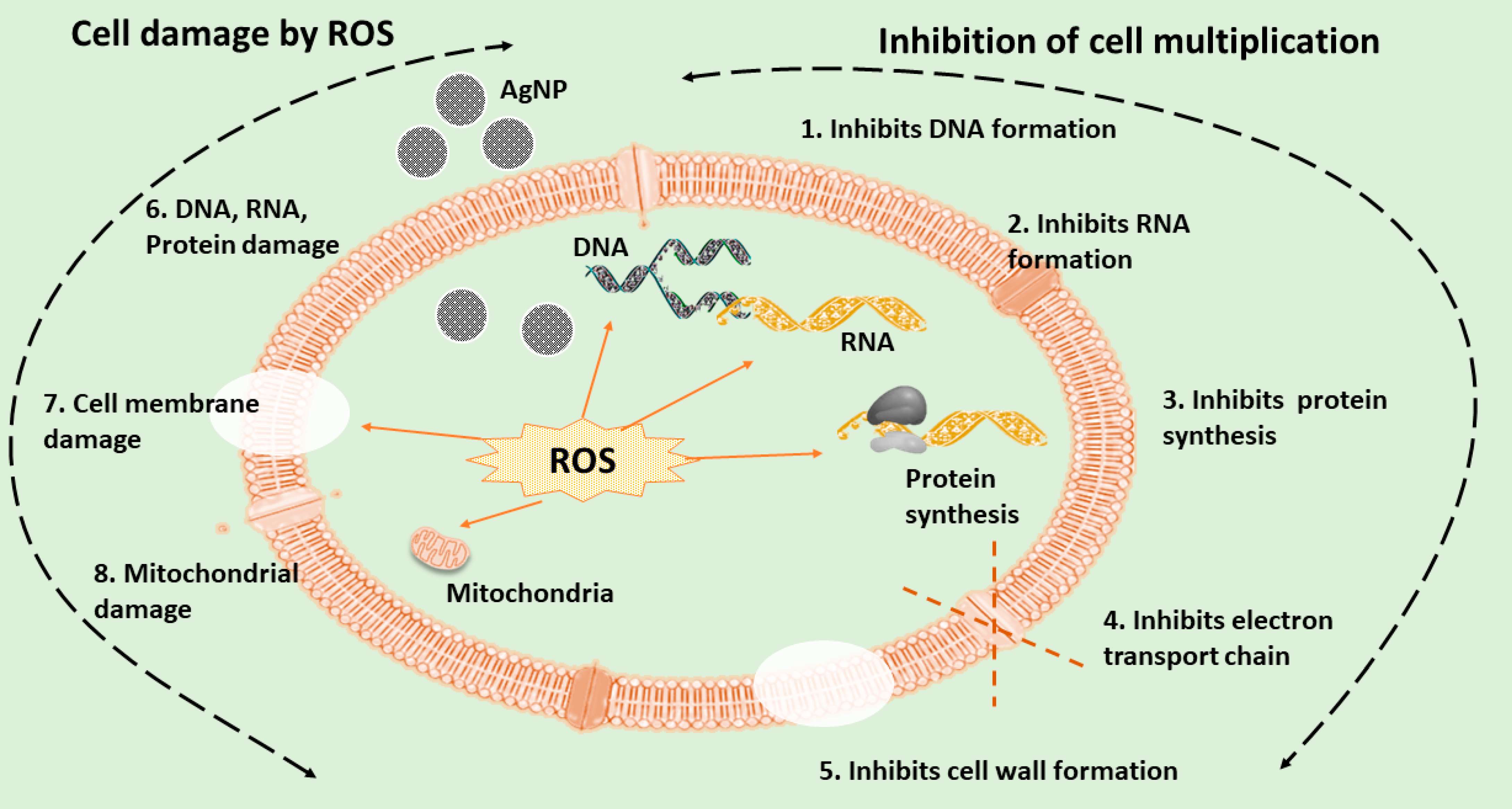
3.2. Antiviral Agents
Viruses pose a significant challenge for life sciences with their remarkable adaptability to the host [147], causing life-threatening diseases. AgNPs act as potential antiviral agents and carriers of antiviral therapies [148] by interacting with viral surface components and blocking viral entry. AgNPs are also believed to prevent viral replication and change the host cell pH, making the environment unfavorable for viruses. Various AgNPs-mediated antiviral mechanisms are depicted in Figure 6.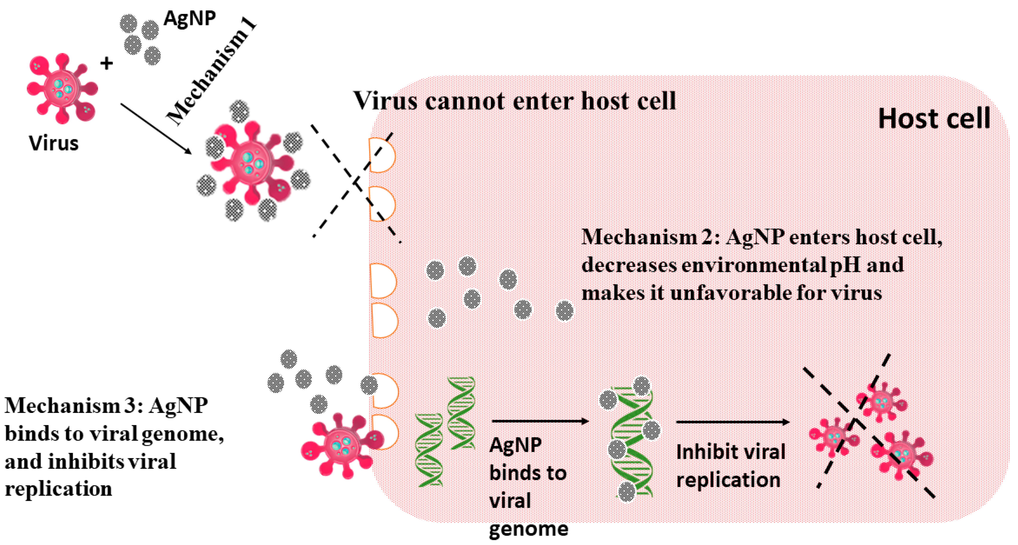
3.3. Other Biological Applications of AgNPs
Biosensors, analytical devices to detect an analyte or to measure physiological signals [149], are vital tools in improving therapeutic and diagnostic efficacy. AgNPs have been developed to improve the sensitivity and efficacy of biosensors and are employed in magnetic resonance imaging (MRI), computed tomography (CT) imaging, and photothermal therapy (PTT) [150]. AgNPs have been shown to act as antidiabetic agents, which act by inhibiting the activities of certain enzymes, such as α-amylase and α-glucosidase (vital for carbohydrate metabolism) [151]. AgNPs have also been shown to act as effective anti-inflammatory agents that suppress vascular endothelial growth factor (VEGF), hypoxia-inducible factor-1α (HIF-1α), cytokine production (IL-12, TNFα), and COX-2 expression [152], as depicted in Figure 7. VEGF is an inflammatory agent that enhances antigen sensitization, T-helper mediated inflammatory cytokines such as IL-4, IL-5, IL-9, and IL-13 [153][154], and HIF-1α-mediated bacterial cytotoxicity; and release of proinflammatory factors such as IL-1α, IL-6, and TNF-α [155][156]. Thus, the inhibition of these inflammatory factors by AgNPs helps in their anti-inflammatory activity.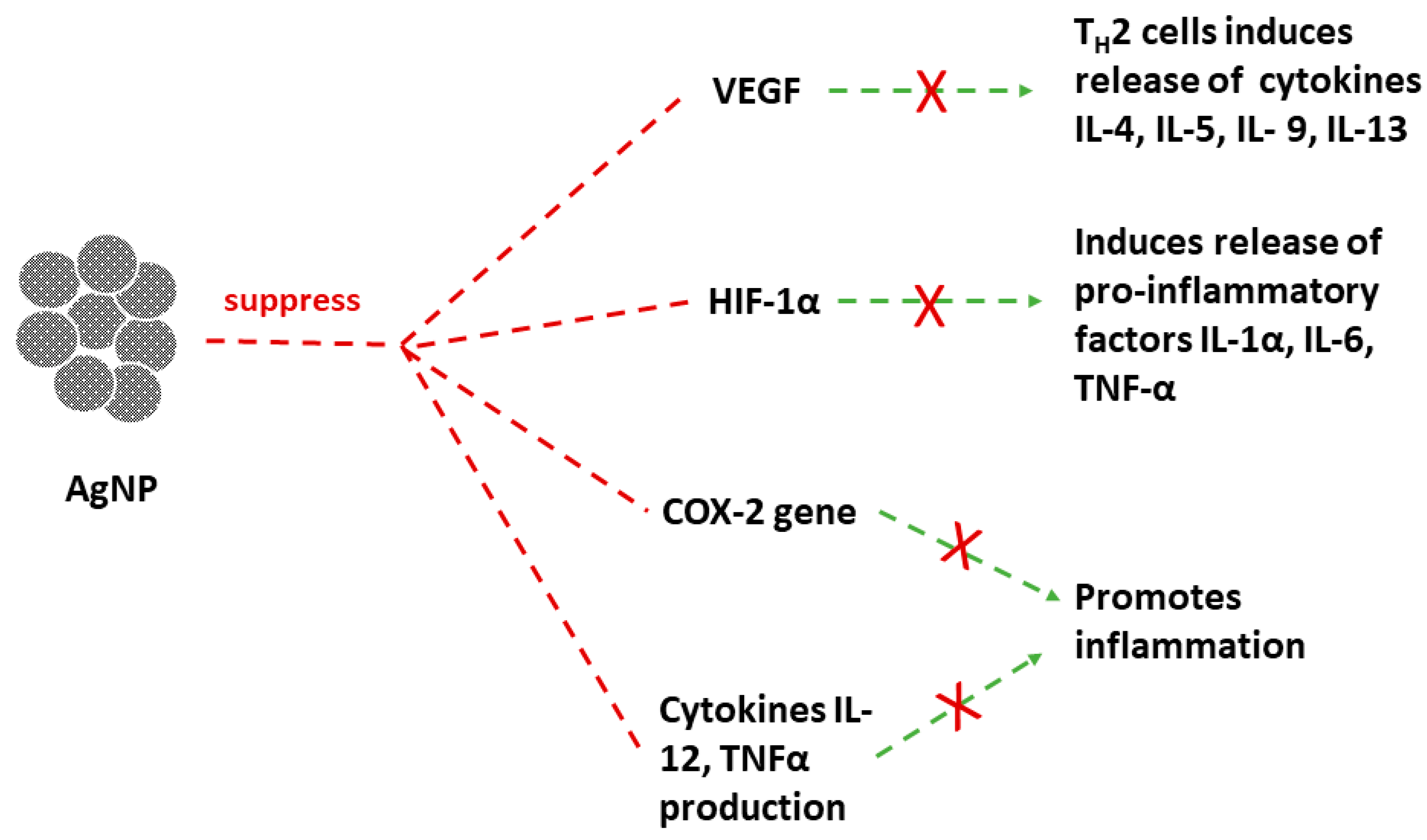
4. Toxicity Associated with AgNPs
AgNPs are highly exploited commercial products for their biomedical advantages [169]. The increasing applications of AgNPs have set an alarming concern about their uptake and toxicity. Previous studies have suggested that AgNPs exhibited higher oral or inhalation exposure as compared to their uptake by the skin. One such study has indicated that the AgNPs accumulated in various organs [170]. Research on various animal models has suggested differential accumulation of AgNPs in different organs, i.e., higher AgNPs accumulation in females than in males [171][172]. Nanomaterial toxicity depends on the physicochemical properties and local barriers in the organs. For example, small-sized silver nanoparticles have higher toxicity than their large-sized counterparts [173]. AgNPs toxicity studies have suggested that it leads to pathological changes in various organs, causing damage to certain organs such as the kidney [174][175] and the spleen [175][176][177]. The results from earlier studies have suggested that AgNPs are linked to inflammation of the blood–brain barrier [178][179] and disruption of the synaptic machinery of neurons [180], thereby affecting neurodevelopment and causing neurotoxicity [181]. These studies further contradict the anti-inflammatory activity of AgNPs. Similarly, the antimicrobial activity of AgNPs also has toxic effects on human cells [182]. In contrast to AgNPs applications for wound healing, it inhibits keratinocyte proliferation [183], and also leads to dermal cytotoxicity. AgNPs are reported to cause blood diseases due to their direct interaction with red blood cells contradicting AgNPs use as an anticoagulant [184][185]. Though AgNPs are studied as anticancer agents, there are numerous reported studies regarding their cytotoxicity related to colon cancer [186] and lung cancer [187][188]. Some elaborated reviews on the toxicity of AgNPs, their effects at the cellular level, their mechanism of cellular effects, and their physicochemical properties leading to their toxicity are out of the scope of this research and are reviewed elsewhere [189][190]. The reported toxicological studies of AgNPs are comparatively fewer than their applications. This, in turn, implies further extensive research on the associated nanotoxicity. Proper models to study AgNPs toxicity with high-throughput analysis and efficient techniques may help in critical evaluation to reach a conclusive remark of the safe and efficient applications of AgNPs. Above all, relevant measures and precautions should be opted to minimize AgNPs toxicity.5. Outlook
Several methods have been reported for the synthesis of silver nanoparticles (AgNPs), using available chemical agents or biological species, investigating the scope and role of the different organic substances in the synthetic process. The knowledge acquired from these interdisciplinary studies has helped to overcome some problems, such as poor stability, aggregation, and agglomeration of the synthesized AgNPs. The primary difference between chemically synthesized and biogenic production of AgNPs is that the first methodology deals with the functionalization of nanoparticles surfaces with organic molecules, causing deviations in the characteristic properties of AgNPs and possibly hindering a biomedical application, whereas the biogenic methods produce biocompatible materials. On the other hand, this functionalization helps to improve the stability and reduce the aggregation, overcoming some important limitations of AgNPs preparation methodologies. Therefore, the advancement in the synthesis methodologies of AgNPs is held back in various ways, preventing its translation to biomedical applications. The advancement in different methods of AgNPs synthesis is held back in various ways preventing its translation to biomedical applications. Foremost, there are various methods of synthesis, purification, characterization, and validation of data for AgNPs without an established systemic pattern to compare the characteristic properties of AgNPs. It is believed that if research follows a standard protocol such as (a) characterization with all the concerned techniques, (b) finding AgNPs size and morphology by each technique, and (c) comparing results of each technique, etc., it may provide more meaningful analysis. Secondly, AgNPs are merely prepared and studied for their applications with less focus on investigating the primary phytochemicals or organic groups responsible for their applications. Higher attention to studying the role of the organic group that causes deviations during characterization and improves efficacy for their therapeutic applications may minimize the gap in the investigation of AgNPs for biomedical applications. Thirdly, there are numerous AgNPs toxicology studies in contrast to the investigated biomedical applications that require further attention before reaching a conclusive remark on the prospective therapeutic usage of AgNPs. Lastly, the computational advances have proved to be very helpful in the assessment of AgNPs properties, as they help to predict, understand, and validate the data related to biomedical applications. However, compared to the number of reported studies of biogenically synthesized AgNPs, bare minimum articles are available that employ in silico docking and molecular dynamics simulation (MDS) techniques, requiring attention. Altogether, AgNPs may prove to be highly promising in the management of health and diseases and may contribute significantly to the advancement of life sciences research.References
- Calderón-Jiménez, B.; Johnson, M.E.; Montoro Bustos, A.R.; Murphy, K.E.; Winchester, M.R.; Vega Baudrit, J.R. Silver Nanoparticles: Technological Advances, Societal Impacts, and Metrological Challenges. Front. Chem. 2017, 5, 6.
- Alemán, J.V.; Chadwick, A.V.; He, J.; Hess, M.; Horie, K.; Jones, R.G.; Kratochvíl, P.; Meisel, I.; Mita, I.; Moad, G.; et al. Definitions of terms relating to the structure and processing of sols, gels, networks, and inorganic-organic hybrid materials (IUPAC Recommendations 2007). Pure Appl. Chem. 2007, 79, 1801–1829.
- Hofmann-Amtenbrink, M.; Grainger, D.W.; Hofmann, H. Nanoparticles in medicine: Current challenges facing inorganic nanoparticle toxicity assessments and standardizations. Nanomed. Nanotechnol. Biol. Med. 2015, 11, 1689–1694.
- Xu, R.; Wang, D.; Zhang, J.; Li, Y. Shape-dependent catalytic activity of silver nanoparticles for the oxidation of styrene. Chem. Asian J. 2006, 1, 888–893.
- Alshehri, A.H.; Jakubowska, M.; Młożniak, A.; Horaczek, M.; Rudka, D.; Free, C.; Carey, J.D. Enhanced Electrical Conductivity of Silver Nanoparticles for High Frequency Electronic Applications. ACS Appl. Mater. Interfaces 2012, 4, 7007–7010.
- Kelly, K.L.; Coronado, E.; Zhao, L.L.; Schatz, G.C. The Optical Properties of Metal Nanoparticles: The Influence of Size, Shape, and Dielectric Environment. J. Phys. Chem. B 2003, 107, 668–677.
- Nie, S.; Emory, S.R. Probing Single Molecules and Single Nanoparticles by Surface-Enhanced Raman Scattering. Science 1997, 275, 1102–1106.
- Vance, M.E.; Kuiken, T.; Vejerano, E.P.; McGinnis, S.P.; Hochella, M.F., Jr.; Rejeski, D.; Hull, M.S. Nanotechnology in the real world: Redeveloping the nanomaterial consumer products inventory. Beilstein J. Nanotechnol. 2015, 6, 1769–1780.
- Ramezanpour, M.; Leung, S.S.W.; Delgado-Magnero, K.H.; Bashe, B.Y.M.; Thewalt, J.; Tieleman, D.P. Computational and experimental approaches for investigating nanoparticle-based drug delivery systems. Biochim. Biophys. Acta 2016, 1858, 1688–1709.
- Muhammad, Z.; Raza, A.; Ghafoor, S.; Naeem, A.; Naz, S.S.; Riaz, S.; Ahmed, W.; Rana, N.F. PEG capped methotrexate silver nanoparticles for efficient anticancer activity and biocompatibility. Eur. J. Pharm. Sci. Off. J. Eur. Fed. Pharm. Sci. 2016, 91, 251–255.
- Petrov, P.D.; Yoncheva, K.; Gancheva, V.; Konstantinov, S.; Trzebicka, B. Multifunctional block copolymer nanocarriers for co-delivery of silver nanoparticles and curcumin: Synthesis and enhanced efficacy against tumor cells. Eur. Polym. J. 2016, 81, 24–33.
- Al-Obaidi, H.; Kalgudi, R.; Zariwala, M.G. Fabrication of inhaled hybrid silver/ciprofloxacin nanoparticles with synergetic effect against Pseudomonas aeruginosa. Eur. J. Pharm. Biopharm. Off. J. Arb. Pharm. Verfahr. EV 2018, 128, 27–35.
- Kaur, A.; Goyal, D.; Kumar, R. Surfactant mediated interaction of vancomycin with silver nanoparticles. Appl. Surf. Sci. 2018, 449, 23.
- Arumai Selvan, D.; Mahendiran, D.; Senthil Kumar, R.; Kalilur Rahiman, A. Garlic, green tea and turmeric extracts-mediated green synthesis of silver nanoparticles: Phytochemical, antioxidant and in vitro cytotoxicity studies. J. Photochem. Photobiol. B 2018, 180, 243–252.
- Jiang, Q.; Yu, S.; Li, X.; Ma, C.; Li, A. Evaluation of local anesthetic effects of Lidocaine-Ibuprofen ionic liquid stabilized silver nanoparticles in Male Swiss mice. J. Photochem. Photobiol. B 2018, 178, 367–370.
- Karthik, C.S.; Manukumar, H.M.; Ananda, A.P.; Nagashree, S.; Rakesh, K.P.; Mallesha, L.; Qin, H.-L.; Umesha, S.; Mallu, P.; Krishnamurthy, N.B. Synthesis of novel benzodioxane midst piperazine moiety decorated chitosan silver nanoparticle against biohazard pathogens and as potential anti-inflammatory candidate: A molecular docking studies. Int. J. Biol. Macromol. 2018, 108, 489–502.
- Rai, M.; Ingle, A.P.; Gupta, I.; Brandelli, A. Bioactivity of noble metal nanoparticles decorated with biopolymers and their application in drug delivery. Int. J. Pharm. 2015, 496, 159–172.
- Stebounova, L.V.; Guio, E.; Grassian, V.H. Silver nanoparticles in simulated biological media: A study of aggregation, sedimentation, and dissolution. J. Nanoparticle Res. 2011, 13, 233–244.
- Argentiere, S.; Cella, C.; Cesaria, M.; Milani, P.; Lenardi, C. Silver nanoparticles in complex biological media: Assessment of colloidal stability and protein corona formation. J. Nanoparticle Res. 2016, 8, 1–13.
- Gliga, A.R.; Skoglund, S.; Odnevall Wallinder, I.; Fadeel, B.; Karlsson, H.L. Size-dependent cytotoxicity of silver nanoparticles in human lung cells: The role of cellular uptake, agglomeration and Ag release. Part. Fibre Toxicol. 2014, 11, 11.
- Gorham, J.M.; Rohlfing, A.B.; Lippa, K.A.; MacCuspie, R.I.; Hemmati, A.; David Holbrook, R. Storage Wars: How citrate-capped silver nanoparticle suspensions are affected by not-so-trivial decisions. J. Nanoparticle Res. 2014, 16, 2339.
- Kanti Das, T.; Ganguly, S.; Remanan, S.; Das, N.C. Temperature-Dependent Study of Catalytic Ag Nanoparticles Entrapped Resin Nanocomposite towards Reduction of 4-Nitrophenol. ChemistrySelect 2019, 4, 3665–3671.
- Rafique, M.; Sadaf, I.; Rafique, M.S.; Tahir, M.B. A review on green synthesis of silver nanoparticles and their applications. Artif. Cells Nanomed. Biotechnol. 2017, 45, 1272–1291.
- Prasher, P.; Singh, M.; Mudila, H. Silver nanoparticles as antimicrobial therapeutics: Current perspectives and future challenges. 3 Biotech 2018, 8, 411.
- Mustapha, T.; Misni, N.; Ithnin, N.R.; Daskum, A.M.; Unyah, N.Z. A Review on Plants and Microorganisms Mediated Synthesis of Silver Nanoparticles, Role of Plants Metabolites and Applications. Int. J. Environ. Res. Public. Health 2022, 19, 674.
- Ali, S.; Chen, X.; Ajmal Shah, M.; Ali, M.; Zareef, M.; Arslan, M.; Ahmad, S.; Jiao, T.; Li, H.; Chen, Q. The avenue of fruit wastes to worth for synthesis of silver and gold nanoparticles and their antimicrobial application against foodborne pathogens: A review. Food Chem. 2021, 359, 129912.
- Kaabipour, S.; Hemmati, S. A review on the green and sustainable synthesis of silver nanoparticles and one-dimensional silver nanostructures. Beilstein J. Nanotechnol. 2021, 12, 102–136.
- Gudikandula, K.; Charya Maringanti, S. Synthesis of silver nanoparticles by chemical and biological methods and their antimicrobial properties. J. Exp. Nanosci. 2016, 11, 714–721.
- Lee, J.; Park, E.Y.; Lee, J. Non-toxic nanoparticles from phytochemicals: Preparation and biomedical application. Bioprocess Biosyst. Eng. 2014, 37, 983–989.
- Saifuddin, N.; Wong, C.W.; Yasumira, A.A.N. Rapid Biosynthesis of Silver Nanoparticles Using Culture Supernatant of Bacteria with Microwave Irradiation. J. Chem. 2009, 6, 61–70.
- Mousavi-Khattat, M.; Keyhanfar, M.; Razmjou, A. A comparative study of stability, antioxidant, DNA cleavage and antibacterial activities of green and chemically synthesized silver nanoparticles. Artif. Cells Nanomed. Biotechnol. 2018, 46, S1022–S1031.
- Kummara, S.; Patil, M.B.; Uriah, T. Synthesis, characterization, biocompatible and anticancer activity of green and chemically synthesized silver nanoparticles—A comparative study. Biomed. Pharmacother. Biomed. Pharmacother. 2016, 84, 10–21.
- Spagnoletti, F.N.; Kronberg, F.; Spedalieri, C.; Munarriz, E.; Giacometti, R. Protein corona on biogenic silver nanoparticles provides higher stability and protects cells from toxicity in comparison to chemical nanoparticles. J. Environ. Manag. 2021, 297, 113434.
- Sreelekha, E.; George, B.; Shyam, A.; Sajina, N.; Mathew, B. A Comparative Study on the Synthesis, Characterization, and Antioxidant Activity of Green and Chemically Synthesized Silver Nanoparticles. BioNanoScience 2021, 11, 489–496.
- Veeragoni, D.; Deshpande, S.; Rachamalla, H.K.; Ande, A.; Misra, S.; Mutheneni, S.R. In Vitro and In Vivo Anticancer and Genotoxicity Profiles of Green Synthesized and Chemically Synthesized Silver Nanoparticles. ACS Appl. Bio Mater. 2022, 5, 2324–2339.
- Durán, N.; Marcato, P.D.; Durán, M.; Yadav, A.; Gade, A.; Rai, M. Mechanistic aspects in the biogenic synthesis of extracellular metal nanoparticles by peptides, bacteria, fungi, and plants. Appl. Microbiol. Biotechnol. 2011, 90, 1609–1624.
- Sharma, V.K.; Yngard, R.A.; Lin, Y. Silver nanoparticles: Green synthesis and their antimicrobial activities. Adv. Colloid Interface Sci. 2009, 145, 83–96.
- Long, D.; Wu, G.; Chen, S. Preparation of oligochitosan stabilized silver nanoparticles by gamma irradiation. Radiat. Phys. Chem. 2007, 76, 1126–1131.
- Chen, J.; Wang, J.; Zhang, X.; Jin, Y. Microwave-assisted green synthesis of silver nanoparticles by carboxymethyl cellulose sodium and silver nitrate. Mater. Chem. Phys. 2008, 108, 421–424.
- Francis, S.; Joseph, S.; Koshy, E.P.; Mathew, B. Microwave assisted green synthesis of silver nanoparticles using leaf extract of elephantopus scaber and its environmental and biological applications. Artif. Cells Nanomed. Biotechnol. 2018, 46, 795–804.
- Srikar, S.K.; Giri, D.D.; Pal, D.B.; Mishra, P.K.; Upadhyay, S.N. Green Synthesis of Silver Nanoparticles: A Review. Green Sustain. Chem. 2016, 06, 34–56.
- Iravani, S.; Korbekandi, H.; Mirmohammadi, S.V.; Zolfaghari, B. Synthesis of silver nanoparticles: Chemical, physical and biological methods. Res. Pharm. Sci. 2014, 9, 385–406.
- Iravani, S. Green synthesis of metal nanoparticles using plants. Green Chem. 2011, 13, 2638.
- Mittal, A.K.; Chisti, Y.; Banerjee, U.C. Synthesis of metallic nanoparticles using plant extracts. Biotechnol. Adv. 2013, 31, 346–356.
- Naghdi, M.; Taheran, M.; Brar, S.K.; Verma, M.; Surampalli, R.Y.; Valero, J.R. Green and energy-efficient methods for the production of metallic nanoparticles. Beilstein J. Nanotechnol. 2015, 6, 2354–2376.
- Manivasagan, P.; Nam, S.Y.; Oh, J. Marine microorganisms as potential biofactories for synthesis of metallic nanoparticles. Crit. Rev. Microbiol. 2016, 42, 1007–1019.
- Golinska, P.; Wypij, M.; Ingle, A.P.; Gupta, I.; Dahm, H.; Rai, M. Biogenic synthesis of metal nanoparticles from actinomycetes: Biomedical applications and cytotoxicity. Appl. Microbiol. Biotechnol. 2014, 98, 8083–8097.
- Hulkoti, N.I.; Taranath, T.C. Biosynthesis of nanoparticles using microbes- a review. Colloids Surf. B Biointerfaces 2014, 121, 474–483.
- Patil, M.P.; Kim, G.-D. Marine microorganisms for synthesis of metallic nanoparticles and their biomedical applications. Colloids Surf. B Biointerfaces 2018, 172, 487–495.
- Ammar, H.A.; El Aty, A.A.A.; El Awdan, S.A. Extracellular myco-synthesis of nano-silver using the fermentable yeasts Pichia kudriavzeviiHA-NY2 and Saccharomyces uvarumHA-NY3, and their effective biomedical applications. Bioprocess Biosyst. Eng. 2021, 44, 841–854.
- Fredrickson, J.K.; Zachara, J.M.; Balkwill, D.L.; Kennedy, D.; Li, S.W.; Kostandarithes, H.M.; Daly, M.J.; Romine, M.F.; Brockman, F.J. Geomicrobiology of high-level nuclear waste-contaminated vadose sediments at the hanford site, washington state. Appl. Environ. Microbiol. 2004, 70, 4230–4241.
- Kalimuthu, K.; Suresh Babu, R.; Venkataraman, D.; Bilal, M.; Gurunathan, S. Biosynthesis of silver nanocrystals by Bacillus licheniformis. Colloids Surf. B Biointerfaces 2008, 65, 150–153.
- Shivaji, S.; Madhu, S.; Singh, S. Extracellular synthesis of antibacterial silver nanoparticles using psychrophilic bacteria. Process Biochem. 2011, 46, 1800–1807.
- Liu, J.; Qiao, S.Z.; Hu, Q.H.; Lu, G.Q.M. Magnetic nanocomposites with mesoporous structures: Synthesis and applications. Small Weinh. Bergstr. Ger. 2011, 7, 425–443.
- Vaseghi, Z.; Nematollahzadeh, A.; Tavakoli, O. Green methods for the synthesis of metal nanoparticles using biogenic reducing agents: A review. Rev. Chem. Eng. 2018, 34, 529–559.
- Chumpol, J.; Siri, S. Simple green production of silver nanoparticles facilitated by bacterial genomic DNA and their antibacterial activity. Artif. Cells Nanomed. Biotechnol. 2018, 46, 619–625.
- Saravanan, M.; Barik, S.K.; MubarakAli, D.; Prakash, P.; Pugazhendhi, A. Synthesis of silver nanoparticles from Bacillus brevis (NCIM 2533) and their antibacterial activity against pathogenic bacteria. Microb. Pathog. 2018, 116, 221–226.
- Yurtluk, T.; Akçay, F.A.; Avcı, A. Biosynthesis of silver nanoparticles using novel Bacillus sp. SBT8. Prep. Biochem. Biotechnol. 2018, 48, 151–159.
- Arzoo, S.; Naqvi, Z.; Hussain, M.; Shamim, S.; Zeb, T.F.; Ali, S. Production and antimicrobial activity of silver nanoparticles synthesized from indigenously isolated Pseudomonas aeruginosa from Rhizosphere. Pak. J. Pharm. Sci. 2020, 33, 2815–2822.
- Sable, S.V.; Kawade, S.; Ranade, S.; Joshi, S. Bioreduction mechanism of silver nanoparticles. Mater. Sci. Eng. C 2020, 107, 110299.
- Saleem, S.; Iqbal, A.; Hasnain, S. Bacterial mediated silver nanoparticles and their efficacy against MRSA. Trop. Biomed. 2020, 37, 482–488.
- Singh, P.; Pandit, S.; Mokkapati, V.; Garnæs, J.; Mijakovic, I. A Sustainable Approach for the Green Synthesis of Silver Nanoparticles from Solibacillus isronensis sp. and Their Application in Biofilm Inhibition. Molecules 2020, 25, 2783.
- Li, J.; Tian, B.; Li, T.; Dai, S.; Weng, Y.; Lu, J.; Xu, X.; Jin, Y.; Pang, R.; Hua, Y. Biosynthesis of Au, Ag and Au-Ag bimetallic nanoparticles using protein extracts of Deinococcus radiodurans and evaluation of their cytotoxicity. Int. J. Nanomed. 2018, 13, 1411–1424.
- Gupta, A.; Singh, D.; Singh, S.K.; Singh, V.K.; Singh, A.V.; Kumar, A. Role of actinomycetes in bioactive and nanoparticle synthesis. In Role of Plant Growth Promoting Microorganisms in Sustainable Agriculture and Nanotechnology; Woodhead Publishing: Amsterdam, The Netherlands; Elsevier: Amsterdam, The Netherlands, 2019; pp. 163–182. ISBN 978-0-12-817004-5.
- Das, S.; Lyla, P.S.; Khan, S.A. Distribution and generic composition of culturable marine actinomycetes from the sediments of Indian continental slope of Bay of Bengal. Chin. J. Oceanol. Limnol. 2008, 26, 166–177.
- Abdeen, S.; Geo, S.; Praseetha, P.K.; Dhanya, R.P. Biosynthesis of silver nanoparticles from Actinomycetes for therapeutic applications. Int. J. Nano Dimens. 2014, 5, 155–162.
- Bhatti, A.A.; Haq, S.; Bhat, R.A. Actinomycetes benefaction role in soil and plant health. Microb. Pathog. 2017, 111, 458–467.
- Ahmad, A.; Senapati, S.; Khan, M.I.; Kumar, R.; Ramani, R.; Srinivas, V.; Sastry, M. Intracellular synthesis of gold nanoparticles by a novel alkalotolerant actinomycete, Rhodococcus species. Nanotechnology 2003, 14, 824–828.
- Ahmad, A.; Senapati, S.; Khan, M.I.; Kumar, R.; Sastry, M. Extracellular Biosynthesis of Monodisperse Gold Nanoparticles by a Novel Extremophilic Actinomycete, Thermomonospora sp. Langmuir 2003, 19, 3550–3553.
- Manivasagan, P.; Venkatesan, J.; Sivakumar, K.; Kim, S.-K. Actinobacteria mediated synthesis of nanoparticles and their biological properties: A review. Crit. Rev. Microbiol. 2016, 42, 209–221.
- Wypij, M.; Czarnecka, J.; Świecimska, M.; Dahm, H.; Rai, M.; Golinska, P. Synthesis, characterization and evaluation of antimicrobial and cytotoxic activities of biogenic silver nanoparticles synthesized from Streptomyces xinghaiensis OF1 strain. World J. Microbiol. Biotechnol. 2018, 34, 23.
- Pallavi, S.S.; Rudayni, H.A.; Bepari, A.; Niazi, S.K.; Nayaka, S. Green synthesis of Silver nanoparticles using Streptomyces hirsutus strain SNPGA-8 and their characterization, antimicrobial activity, and anticancer activity against human lung carcinoma cell line A549. Saudi J. Biol. Sci. 2022, 29, 228–238.
- Mabrouk, M.; Elkhooly, T.A.; Amer, S.K. Actinomycete strain type determines the monodispersity and antibacterial properties of biogenically synthesized silver nanoparticles. J. Genet. Eng. Biotechnol. 2021, 19, 57.
- Dhillon, G.S.; Brar, S.K.; Kaur, S.; Verma, M. Green approach for nanoparticle biosynthesis by fungi: Current trends and applications. Crit. Rev. Biotechnol. 2012, 32, 49–73.
- Guilger-Casagrande, M.; Lima, R. de Synthesis of Silver Nanoparticles Mediated by Fungi: A Review. Front. Bioeng. Biotechnol. 2019, 7.
- Korbekandi, H.; Asghari, G.; Chitsazi, M.R.; Bahri Najafi, R.; Badii, A.; Iravani, S. Green biosynthesis of silver nanoparticles using Althaea officinalis radix hydroalcoholic extract. Artif. Cells Nanomed. Biotechnol. 2016, 44, 209–215.
- Fernández, J.G.; Fernández-Baldo, M.A.; Berni, E.; Camí, G.; Durán, N.; Raba, J.; Sanz, M.I. Production of silver nanoparticles using yeasts and evaluation of their antifungal activity against phytopathogenic fungi. Process Biochem. 2016, 51, 1306–1313.
- Yadav, A.; Kon, K.; Kratosova, G.; Duran, N.; Ingle, A.P.; Rai, M. Fungi as an efficient mycosystem for the synthesis of metal nanoparticles: Progress and key aspects of research. Biotechnol. Lett. 2015, 37, 2099–2120.
- Soleimani, P.; Mehrvar, A.; Michaud, J.P.; Vaez, N. Optimization of silver nanoparticle biosynthesis by entomopathogenic fungi and assays of their antimicrobial and antifungal properties. J. Invertebr. Pathol. 2022, 190, 107749.
- Koli, S.H.; Mohite, B.V.; Suryawanshi, R.K.; Borase, H.P.; Patil, S.V. Extracellular red Monascus pigment-mediated rapid one-step synthesis of silver nanoparticles and its application in biomedical and environment. Bioprocess Biosyst. Eng. 2018, 41, 715–727.
- Ansari, A.; Pervez, S.; Javed, U.; Abro, M.I.; Nawaz, M.A.; Qader, S.A.U.; Aman, A. Characterization and interplay of bacteriocin and exopolysaccharide-mediated silver nanoparticles as an antibacterial agent. Int. J. Biol. Macromol. 2018, 115, 643–650.
- Li, P.-J.; Pan, J.-J.; Tao, L.-J.; Li, X.; Su, D.-L.; Shan, Y.; Li, H.-Y. Green Synthesis of Silver Nanoparticles by Extracellular Extracts from Aspergillus japonicus PJ01. Molecules 2021, 26, 4479.
- Wang, D.; Xue, B.; Wang, L.; Zhang, Y.; Liu, L.; Zhou, Y. Fungus-mediated green synthesis of nano-silver using Aspergillus sydowii and its antifungal/antiproliferative activities. Sci. Rep. 2021, 11, 10356.
- Yeast—Industrial Applications; Morata, A.; Loira, I. (Eds.) InTech: Rijeka, Croatia, 2017; ISBN 978-953-51-3599-9.
- Jha, A.K.; Prasad, K.; Kulkarni, A.A.R. Yeast Mediated Synthesis of Silver Nanoparticles. Int. J. Nanosci. Nanotechnol. 2008, 4, 17–22.
- Boroumand Moghaddam, A.; Namvar, F.; Moniri, M.; Tahir, P.; Azizi, S.; Mohamad, R. Nanoparticles Biosynthesized by Fungi and Yeast: A Review of Their Preparation, Properties, and Medical Applications. Molecules 2015, 20, 16540–16565.
- Breierová, E.; Vajcziková, I.; Sasinková, V.; Stratilová, E.; Fišera, M.; Gregor, T.; Šajbidor, J. Biosorption of Cadmium Ions by Different Yeast Species. Z. Für Nat. C 2002, 57, 634–639.
- Skalickova, S.; Baron, M.; Sochor, J. Nanoparticles Biosynthesized by Yeast: A Review of their application. Kvas. Prum. 2017, 63, 290–292.
- Cunha, F.A.; da, C.S.O. Cunha, M.; da Frota, S.M.; Mallmann, E.J.J.; Freire, T.M.; Costa, L.S.; Paula, A.J.; Menezes, E.A.; Fechine, P.B.A. Biogenic synthesis of multifunctional silver nanoparticles from Rhodotorula glutinis and Rhodotorula mucilaginosa: Antifungal, catalytic and cytotoxicity activities. World J. Microbiol. Biotechnol. 2018, 34, 127.
- Sharma, P.; Sharma, N. Industrial and Biotechnological Applications of Algae: A Review. J. Adv. Plant Biol. 2017, 1, 1–25.
- Sharma, A.; Sharma, S.; Sharma, K.; Chetri, S.P.K.; Vashishtha, A.; Singh, P.; Kumar, R.; Rathi, B.; Agrawal, V. Algae as crucial organisms in advancing nanotechnology: A systematic review. J. Appl. Phycol. 2016, 28, 1759–1774.
- Schröfel, A.; Kratošová, G.; Bohunická, M.; Dobrocka, E.; Vávra, I. Biosynthesis of gold nanoparticles using diatoms—Silica-gold and EPS-gold bionanocomposite formation. J. Nanoparticle Res. 2011, 13, 3207–3216.
- Dahoumane, S.A.; Mechouet, M.; Wijesekera, K.; Filipe, C.D.M.; Sicard, C.; Bazylinski, D.A.; Jeffryes, C. Algae-mediated biosynthesis of inorganic nanomaterials as a promising route in nanobiotechnology—A review. Green Chem. 2017, 19, 552–587.
- Sharma, D.; Kanchi, S.; Bisetty, K. Biogenic synthesis of nanoparticles: A review. Arab. J. Chem. 2019, 12, 3576–3600.
- Sathishkumar, R.S.; Sundaramanickam, A.; Srinath, R.; Ramesh, T.; Saranya, K.; Meena, M.; Surya, P. Green synthesis of silver nanoparticles by bloom forming marine microalgae Trichodesmium erythraeum and its applications in antioxidant, drug-resistant bacteria, and cytotoxicity activity. J. Saudi Chem. Soc. 2019, 23, 1180–1191.
- Dahoumane, S.A.; Wujcik, E.K.; Jeffryes, C. Noble metal, oxide and chalcogenide-based nanomaterials from scalable phototrophic culture systems. Enzym. Microb. Technol. 2016, 95, 13–27.
- Rao, S.S.; Saptami, K.; Venkatesan, J.; Rekha, P.D. Microwave-assisted rapid synthesis of silver nanoparticles using fucoidan: Characterization with assessment of biocompatibility and antimicrobial activity. Int. J. Biol. Macromol. 2020, 163, 745–755.
- Bao, Z.; Cao, J.; Kang, G.; Lan, C.Q. Effects of reaction conditions on light-dependent silver nanoparticle biosynthesis mediated by cell extract of green alga Neochloris oleoabundans. Environ. Sci. Pollut. Res. Int. 2019, 26, 2873–2881.
- Husain, S.; Verma, S.K.; Yasin, D.; Hemlata, N.A.N.; Rizvi, M.M.; Fatma, T. Facile green bio-fabricated silver nanoparticles from Microchaete infer dose-dependent antioxidant and anti-proliferative activity to mediate cellular apoptosis. Bioorganic Chem. 2021, 107, 104535.
- Narayanan, K.B.; Han, S.S. Helical plant viral nanoparticles-bioinspired synthesis of nanomaterials and nanostructures. Bioinspir. Biomim. 2017, 12, 031001.
- Kobayashi, M.; Tomita, S.; Sawada, K.; Shiba, K.; Yanagi, H.; Yamashita, I.; Uraoka, Y. Chiral meta-molecules consisting of gold nanoparticles and genetically engineered tobacco mosaic virus. Opt. Express 2012, 20, 24856–24863.
- Young, M.; Debbie, W.; Uchida, M.; Douglas, T. Plant Viruses as Biotemplates for Materials and Their Use in Nanotechnology. Annu. Rev. Phytopathol. 2008, 46, 361–384.
- Gahlawat, G.; Choudhury, A.R. A review on the biosynthesis of metal and metal salt nanoparticles by microbes. RSC Adv. 2019, 9, 12944–12967.
- Tangahu, B.V.; Sheikh Abdullah, S.R.; Basri, H.; Idris, M.; Anuar, N.; Mukhlisin, M. A Review on Heavy Metals (As, Pb, and Hg) Uptake by Plants through Phytoremediation. Int. J. Chem. Eng. 2011, 2011, 939161.
- Marchiol, L. Synthesis of metal nanoparticles in living plants. Ital. J. Agron. 2012, 7, 37.
- Park, Y.; Hong, Y.N.; Weyers, A.; Kim, Y.S.; Linhardt, R.J. Polysaccharides and phytochemicals: A natural reservoir for the green synthesis of gold and silver nanoparticles. IET Nanobiotechnol. 2011, 5, 69.
- Rajeshkumar, S.; Bharath, L.V. Mechanism of plant-mediated synthesis of silver nanoparticles—A review on biomolecules involved, characterisation and antibacterial activity. Chem. Biol. Interact. 2017, 273, 219–227.
- Rice-evans, C.A.; Miller, N.J.; Bolwell, P.G.; Bramley, P.M.; Pridham, J.B. The Relative Antioxidant Activities of Plant-Derived Polyphenolic Flavonoids. Free Radic. Res. 1995, 22, 375–383.
- Tamuly, C.; Hazarika, M.; Bordoloi, M.; Bhattacharyya, P.K.; Kar, R. Biosynthesis of Ag nanoparticles using pedicellamide and its photocatalytic activity: An eco-friendly approach. Spectrochim. Acta. A. Mol. Biomol. Spectrosc. 2014, 132, 687–691.
- Yang, X.; Feng, Y.; He, Z.; Stoffella, P.J. Molecular mechanisms of heavy metal hyperaccumulation and phytoremediation. J. Trace Elem. Med. Biol. Organ Soc. Miner. Trace Elem. GMS 2005, 18, 339–353.
- Milner, M.J.; Kochian, L.V. Investigating heavy-metal hyperaccumulation using Thlaspi caerulescens as a model system. Ann. Bot. 2008, 102, 3–13.
- Kumar, V.; Yadav, S.K. Plant-mediated synthesis of silver and gold nanoparticles and their applications. J. Chem. Technol. Biotechnol. 2009, 84, 151–157.
- Mukunthan, K.S.; Balaji, S. Cashew Apple Juice ( Anacardium occidentale L.) Speeds Up the Synthesis of Silver Nanoparticles. Int. J. Green Nanotechnol. 2012, 4, 71–79.
- Ahmed, K.B.A.; Subramaniam, S.; Veerappan, G.; Hari, N.; Sivasubramanian, A.; Veerappan, A. β-Sitosterol-D-glucopyranoside isolated from Desmostachya bipinnata mediates photoinduced rapid green synthesis of silver nanoparticles. RSC Adv. 2014, 4, 59130–59136.
- Subramanian, P.; Shanmugam, K. Extracellular and intracellular synthesis of silver nanoparticles. Asian J. Pharm. Clin. Res. 2016, 9, 133–139.
- Rabia Kanwar, R.F.; Khalid, A. 2. Biological, physical and chemical synthesis of silver nanoparticles and their non-toxic bio-chemical application: A brief review. Pure Appl. Biol. PAB 2021, 11, 421–438.
- Sharma, N.K.; Vishwakarma, J.; Rai, S.; Alomar, T.S.; AlMasoud, N.; Bhattarai, A. Green Route Synthesis and Characterization Techniques of Silver Nanoparticles and Their Biological Adeptness. ACS Omega 2022, 7, 27004–27020.
- Tomaszewska, E.; Ranoszek-Soliwoda, K.; Kadziola, K.; Tkacz-Szczesna, B.; Celichowski, G.; Cichomski, M.; Szmaja, W.; Grobelny, J. Detection Limits of DLS and UV-Vis Spectroscopy in Characterization of Polydisperse Nanoparticles Colloids. J. Nanomater. 2013, 2013, 313081.
- Link, S.; El-Sayed, M.A. Optical Properties and Ultrafast Dynamics of Metallic Nanocrystals. Annu. Rev. Phys. Chem. 2003, 54, 331–366.
- Noginov, M.A.; Zhu, G.; Bahoura, M.; Adegoke, J.; Small, C.; Ritzo, B.A.; Drachev, V.P.; Shalaev, V.M. The effect of gain and absorption on surface plasmons in metal nanoparticles. Appl. Phys. B 2007, 86, 455–460.
- He, R.; Qian, X.; Yin, J.; Zhu, Z. Preparation of polychrome silver nanoparticles in different solvents. J. Mater. Chem. 2002, 12, 3783–3786.
- Rani, P.; Kumar, V.; Singh, P.P.; Matharu, A.S.; Zhang, W.; Kim, K.-H.; Singh, J.; Rawat, M. Highly stable AgNPs prepared via a novel green approach for catalytic and photocatalytic removal of biological and non-biological pollutants. Environ. Int. 2020, 143, 105924.
- Majeed Khan, M.A.; Kumar, S.; Ahamed, M.; Alrokayan, S.A.; AlSalhi, M.S. Structural and thermal studies of silver nanoparticles and electrical transport study of their thin films. Nanoscale Res. Lett. 2011, 6, 434.
- Parit, S.B.; Karade, V.C.; Patil, R.B.; Pawar, N.V.; Dhavale, R.P.; Tawre, M.; Pardesi, K.; Jadhav, U.U.; Dawkar, V.V.; Tanpure, R.S.; et al. Bioinspired synthesis of multifunctional silver nanoparticles for enhanced antimicrobial and catalytic applications with tailored SPR properties. Mater. Today Chem. 2020, 17, 100285.
- Mehta, B.K.; Chhajlani, M.; Shrivastava, B.D. Green synthesis of silver nanoparticles and their characterization by XRD. J. Phys. Conf. Ser. 2017, 836, 012050.
- Fissan, H.; Ristig, S.; Kaminski, H.; Asbach, C.; Epple, M. Comparison of different characterization methods for nanoparticle dispersions before and after aerosolization. Anal. Methods 2014, 6, 7324–7334.
- Hall, J.B.; Dobrovolskaia, M.A.; Patri, A.K.; McNeil, S.E. Characterization of nanoparticles for therapeutics. Nanomed. 2007, 2, 789–803.
- Reymond-Laruinaz, S.; Saviot, L.; Potin, V.; Marco de Lucas, M. del C. Protein–nanoparticle interaction in bioconjugated silver nanoparticles: A transmission electron microscopy and surface enhanced Raman spectroscopy study. Appl. Surf. Sci. 2016, 389, 17–24.
- Qayyum, S.; Khan, A.U. Biofabrication of broad range antibacterial and antibiofilm silver nanoparticles. IET Nanobiotechnol. 2016, 10, 349–357.
- Lateef, A. The microbiology of a pharmaceutical effluent and its public health implications. World J. Microbiol. Biotechnol. 2004, 20, 167–171.
- Lateef, A.; Oloke, J.K.; Gueguim-Kana, E.B. Antimicrobial resistance of bacterial strains isolated from orange juice products. Afr. J. Biotechnol. 2004, 3, 334–338.
- Adewoye, S.O.; Lateef, A. Assessment of the Microbiological Quality of Clarias gariepinus Exposed to an Industrial Effluent in Nigeria. Environmentalist 2004, 24, 249–254.
- Eltarahony, M.; Zaki, S.; ElKady, M.; Abd-El-Haleem, D. Biosynthesis, Characterization of Some Combined Nanoparticles, and Its Biocide Potency against a Broad Spectrum of Pathogens. J. Nanomater. 2018, 2018, 5263814.
- Chandrasekaran, R.; Seetharaman, P.; Krishnan, M.; Gnanasekar, S.; Sivaperumal, S. Carica papaya (Papaya) latex: A new paradigm to combat against dengue and filariasis vectors Aedes aegypti and Culex quinquefasciatus (Diptera: Culicidae). 3 Biotech 2018, 8, 83.
- Arunasri, K.; Mohan, S.V. Biofilms. In Microbial Electrochemical Technology; Elsevier: Amsterdam, The Netherlands, 2019; pp. 295–313. ISBN 978-0-444-64052-9.
- Rabin, N.; Zheng, Y.; Opoku-Temeng, C.; Du, Y.; Bonsu, E.; Sintim, H.O. Biofilm formation mechanisms and targets for developing antibiofilm agents. Future Med. Chem. 2015, 7, 493–512.
- Malheiro, J.; Simões, M. Antimicrobial resistance of biofilms in medical devices. In Biofilms and Implantable Medical Devices; Elsevier: Amsterdam, The Netherlands, 2017; pp. 97–113. ISBN 978-0-08-100382-4.
- Almaguer-Flores, A. Biofilms in the oral environment. In Bio-Tribocorrosion in Biomaterials and Medical Implants; Woodhead Publishing Limited: Sawston, UK; Elsevier: Sawston, UK, 2013; pp. 169–186. ISBN 978-0-85709-540-4.
- Loza-Correa, M.; Ramírez-Arcos, S. Detection of bacterial adherence and biofilm formation on medical surfaces. In Biofilms and Implantable Medical Devices; Woodhead Publishing: Duxford, UK; Woodhead Publishing: Cambridge, MA, USA; Woodhead Publishing: Kidlington, UK; Elsevier: Duxford, UK; Elsevier: Cambridge, MA, USA; Elsevier: Kidlington, UK, 2017; pp. 181–193. ISBN 978-0-08-100382-4.
- Alt, V.; Bechert, T.; Steinrücke, P.; Wagener, M.; Seidel, P.; Dingeldein, E.; Domann, E.; Schnettler, R. An in vitro assessment of the antibacterial properties and cytotoxicity of nanoparticulate silver bone cement. Biomaterials 2004, 25, 4383–4391.
- van de Belt, H.; Neut, D.; Schenk, W.; van Horn, J.R.; van der Mei, H.C.; Busscher, H.J. Infection of orthopedic implants and the use of antibiotic-loaded bone cements. A review. Acta Orthop. Scand. 2001, 72, 557–571.
- Liu, Y.; Zheng, Z.; Zara, J.N.; Hsu, C.; Soofer, D.E.; Lee, K.S.; Siu, R.K.; Miller, L.S.; Zhang, X.; Carpenter, D.; et al. The antimicrobial and osteoinductive properties of silver nanoparticle/poly (DL-lactic-co-glycolic acid)-coated stainless steel. Biomaterials 2012, 33, 8745–8756.
- Zheng, Z.; Yin, W.; Zara, J.N.; Li, W.; Kwak, J.; Mamidi, R.; Lee, M.; Siu, R.K.; Ngo, R.; Wang, J.; et al. The use of BMP-2 coupled—Nanosilver-PLGA composite grafts to induce bone repair in grossly infected segmental defects. Biomaterials 2010, 31, 9293–9300.
- Magalhães, A.P.R.; Santos, L.B.; Lopes, L.G.; Estrela, C.R.D.A.; Estrela, C.; Torres, M.; Bakuzis, A.F.; Cardoso, P.C.; Carrião, M.S. Nanosilver Application in Dental Cements. Int. Sch. Res. Not. 2012, 2012, e365438.
- Akhavan, A.; Sodagar, A.; Mojtahedzadeh, F.; Sodagar, K. Investigating the effect of incorporating nanosilver/nanohydroxyapatite particles on the shear bond strength of orthodontic adhesives. Acta Odontol. Scand. 2013, 71, 1038–1042.
- Hamdan, S.; Pastar, I.; Drakulich, S.; Dikici, E.; Tomic-Canic, M.; Deo, S.; Daunert, S. Nanotechnology-Driven Therapeutic Interventions in Wound Healing: Potential Uses and Applications. ACS Cent. Sci. 2017, 3, 163–175.
- Strasser, P.; Koh, S.; Anniyev, T.; Greeley, J.; More, K.; Yu, C.; Liu, Z.; Kaya, S.; Nordlund, D.; Ogasawara, H.; et al. Lattice-strain control of the activity in dealloyed core-shell fuel cell catalysts. Nat. Chem. 2010, 2, 454–460.
- Franci, G.; Falanga, A.; Galdiero, S.; Palomba, L.; Rai, M.; Morelli, G.; Galdiero, M. Silver Nanoparticles as Potential Antibacterial Agents. Molecules 2015, 20, 8856–8874.
- Singh, R. Prospects of Nanobiomaterials for Biosensing. Int. J. Electrochem. 2011, 2011, 125487.
- Ehlerding, E.B.; Grodzinski, P.; Cai, W.; Liu, C.H. Big Potential from Small Agents: Nanoparticles for Imaging-Based Companion Diagnostics. ACS Nano 2018, 12, 2106–2121.
- Liu, Y.; Zeng, S.; Liu, Y.; Wu, W.; Shen, Y.; Zhang, L.; Li, C.; Chen, H.; Liu, A.; Shen, L.; et al. Synthesis and antidiabetic activity of selenium nanoparticles in the presence of polysaccharides from Catathelasma ventricosum. Int. J. Biol. Macromol. 2018, 114, 632–639.
- Franková, J.; Pivodová, V.; Vágnerová, H.; Juránová, J.; Ulrichová, J. Effects of silver nanoparticles on primary cell cultures of fibroblasts and keratinocytes in a wound-healing model. J. Appl. Biomater. Funct. Mater. 2016, 14, 137–142.
- Lee, C.G.; Link, H.; Baluk, P.; Homer, R.J.; Chapoval, S.; Bhandari, V.; Kang, M.J.; Cohn, L.; Kim, Y.K.; McDonald, D.M.; et al. Vascular endothelial growth factor (VEGF) induces remodeling and enhances TH2-mediated sensitization and inflammation in the lung. Nat. Med. 2004, 10, 1095–1103.
- Barnes, P.J. Th2 cytokines and asthma: An introduction. Respir. Res. 2001, 2, 64.
- Imtiyaz, H.Z.; Simon, M.C. Hypoxia-Inducible Factors as Essential Regulators of Inflammation. In Diverse Effects of Hypoxia on Tumor Progression; Simon, M.C., Ed.; Current Topics in Microbiology and Immunology; Springer: Berlin/Heidelberg, Germany, 2010; Volume 345, pp. 105–120. ISBN 978-3-642-13328-2.
- Lin, N.; Simon, M.C. Hypoxia-inducible factors: Key regulators of myeloid cells during inflammation. J. Clin. Investig. 2016, 126, 3661–3671.
- Kanipandian, N.; Kannan, S.; Ramesh, R.; Subramanian, P.; Thirumurugan, R. Characterization, antioxidant and cytotoxicity evaluation of green synthesized silver nanoparticles using Cleistanthus collinus extract as surface modifier. Mater. Res. Bull. 2014, 49, 494–502.
- Reddy, N.J.; Nagoor Vali, D.; Rani, M.; Rani, S.S. Evaluation of antioxidant, antibacterial and cytotoxic effects of green synthesized silver nanoparticles by Piper longum fruit. Mater. Sci. Eng. C Mater. Biol. Appl. 2014, 34, 115–122.
- Shanmugasundaram, T.; Radhakrishnan, M.; Gopikrishnan, V.; Pazhanimurugan, R.; Balagurunathan, R. A study of the bactericidal, anti-biofouling, cytotoxic and antioxidant properties of actinobacterially synthesised silver nanoparticles. Colloids Surf. B Biointerfaces 2013, 111, 680–687.
- Karami Mehrian, S.; Heidari, R.; Rahmani, F. Effect of silver nanoparticles on free amino acids content and antioxidant defense system of tomato plants. Indian J. Plant Physiol. 2015, 20, 257–263.
- Alavi, M.; Karimi, N.; Valadbeigi, T. Antibacterial, Antibiofilm, Antiquorum Sensing, Antimotility, and Antioxidant Activities of Green Fabricated Ag, Cu, TiO2, ZnO, and Fe3O4 NPs via Protoparmeliopsis muralis Lichen Aqueous Extract against Multi-Drug-Resistant Bacteria. ACS Biomater. Sci. Eng. 2019, 5, 4228–4243.
- Jones, A.-A.D.; Mi, G.; Webster, T.J. A Status Report on FDA Approval of Medical Devices Containing Nanostructured Materials. Trends Biotechnol. 2019, 37, 117–120.
- El Moussaoui, A.; Jawhari, F.Z.; Almehdi, A.M.; Elmsellem, H.; Fikri Benbrahim, K.; Bousta, D.; Bari, A. Antibacterial, antifungal and antioxidant activity of total polyphenols of Withania frutescens L. Bioorganic Chem. 2019, 93, 103337.
- Sharma, H.; Mishra, P.K.; Talegaonkar, S.; Vaidya, B. Metal nanoparticles: A theranostic nanotool against cancer. Drug Discov. Today 2015, 20, 1143–1151.
- Patra, S.; Mukherjee, S.; Barui, A.K.; Ganguly, A.; Sreedhar, B.; Patra, C.R. Green synthesis, characterization of gold and silver nanoparticles and their potential application for cancer therapeutics. Mater. Sci. Eng. C Mater. Biol. Appl. 2015, 53, 298–309.
- Mukherjee, S.; Chowdhury, D.; Kotcherlakota, R.; Patra, S.; B, V.; Bhadra, M.P.; Sreedhar, B.; Patra, C.R. Potential theranostics application of bio-synthesized silver nanoparticles (4-in-1 system). Theranostics 2014, 4, 316–335.
- Davalos, D.; Akassoglou, K. Fibrinogen as a key regulator of inflammation in disease. Semin. Immunopathol. 2012, 34, 43–62.
- Levi, M.; Schultz, M.; van der Poll, T. Disseminated intravascular coagulation in infectious disease. Semin. Thromb. Hemost. 2010, 36, 367–377.
- Wang, L.; Zhang, T.; Li, P.; Huang, W.; Tang, J.; Wang, P.; Liu, J.; Yuan, Q.; Bai, R.; Li, B.; et al. Use of Synchrotron Radiation-Analytical Techniques To Reveal Chemical Origin of Silver-Nanoparticle Cytotoxicity. ACS Nano 2015, 9, 6532–6547.
- Tolaymat, T.M.; El Badawy, A.M.; Genaidy, A.; Scheckel, K.G.; Luxton, T.P.; Suidan, M. An evidence-based environmental perspective of manufactured silver nanoparticle in syntheses and applications: A systematic review and critical appraisal of peer-reviewed scientific papers. Sci. Total Environ. 2010, 408, 999–1006.
- Park, J.; Lim, D.-H.; Lim, H.-J.; Kwon, T.; Choi, J.; Jeong, S.; Choi, I.-H.; Cheon, J. Size dependent macrophage responses and toxicological effects of Ag nanoparticles. Chem. Commun. Camb. Engl. 2011, 47, 4382–4384.
- Sung, J.H.; Ji, J.H.; Song, K.S.; Lee, J.H.; Choi, K.H.; Lee, S.H.; Yu, I.J. Acute inhalation toxicity of silver nanoparticles. Toxicol. Ind. Health 2011, 27, 149–154.
- Cho, Y.-M.; Mizuta, Y.; Akagi, J.; Toyoda, T.; Sone, M.; Ogawa, K. Size-dependent acute toxicity of silver nanoparticles in mice. J. Toxicol. Pathol. 2018, 31, 73–80.
- Moradi-Sardareh, H.; Basir, H.R.G.; Hassan, Z.M.; Davoudi, M.; Amidi, F.; Paknejad, M. Toxicity of silver nanoparticles on different tissues of Balb/C mice. Life Sci. 2018, 211, 81–90.
- Roshan, R.; Ranjbar, S. Toxicological effects of silver nanoparticles in rats. Afr. J. Microbiol. Res. 2012, 6, 5587–5593.
- Wen, H.; Dan, M.; Yang, Y.; Lyu, J.; Shao, A.; Cheng, X.; Chen, L.; Xu, L. Acute toxicity and genotoxicity of silver nanoparticle in rats. PLoS ONE 2017, 12, e0185554.
- Mazen, N.F.; Saleh, E.Z.; Mahmoud, A.A.; Shaalan, A.A. Histological and immunohistochemical study on the potential toxicity of sliver nanoparticles on the structure of the spleen in adult male albino rats. Egypt. J. Histol. 2017, 40, 374–387.
- Trickler, W.J.; Lantz, S.M.; Murdock, R.C.; Schrand, A.M.; Robinson, B.L.; Newport, G.D.; Schlager, J.J.; Oldenburg, S.J.; Paule, M.G.; Slikker, W., Jr.; et al. Silver Nanoparticle Induced Blood-Brain Barrier Inflammation and Increased Permeability in Primary Rat Brain Microvessel Endothelial Cells. Toxicol. Sci. 2010, 118, 160–170.
- Khan, A.M.; Korzeniowska, B.; Gorshkov, V.; Tahir, M.; Schrøder, H.; Skytte, L.; Rasmussen, K.L.; Khandige, S.; Møller-Jensen, J.; Kjeldsen, F. Silver nanoparticle-induced expression of proteins related to oxidative stress and neurodegeneration in an in vitro human blood-brain barrier model. Nanotoxicology 2019, 13, 221–239.
- Xu, F.; Piett, C.; Farkas, S.; Qazzaz, M.; Syed, N.I. Silver nanoparticles (AgNPs) cause degeneration of cytoskeleton and disrupt synaptic machinery of cultured cortical neurons. Mol. Brain 2013, 6, 29.
- Repar, N.; Li, H.; Aguilar, J.S.; Li, Q.Q.; Damjana, D.; Hong, Y. Silver nanoparticles induce neurotoxicity in a human embryonic stem cell-derived neuron and astrocyte network. Nanotoxicology 2018, 12, 104–116.
- Greulich, C.; Braun, D.; Peetsch, A.; Diendorf, J.; Siebers, B.; Epple, M.; Köller, M. The toxic effect of silver ions and silver nanoparticles towards bacteria and human cells occurs in the same concentration range. RSC Adv. 2012, 2, 6981–6987.
- Zanette, C.; Pelin, M.; Crosera, M.; Adami, G.; Bovenzi, M.; Larese, F.F.; Florio, C. Silver nanoparticles exert a long-lasting antiproliferative effect on human keratinocyte HaCaT cell line. Toxicol. Vitr. Int. J. Publ. Assoc. BIBRA 2011, 25, 1053–1060.
- Huang, H.; Lai, W.; Cui, M.; Liang, L.; Lin, Y.; Fang, Q.; Liu, Y.; Xie, L. An Evaluation of Blood Compatibility of Silver Nanoparticles. Sci. Rep. 2016, 6, 25518.
- Chen, L.Q.; Fang, L.; Ling, J.; Ding, C.Z.; Kang, B.; Huang, C.Z. Nanotoxicity of silver nanoparticles to red blood cells: Size dependent adsorption, uptake, and hemolytic activity. Chem. Res. Toxicol. 2015, 28, 501–509.
- Shathviha, P.C.; Ezhilarasan, D.; Rajeshkumar, S.; Selvaraj, J. β-sitosterol Mediated Silver Nanoparticles Induce Cytotoxicity in Human Colon Cancer HT-29 Cells. Avicenna J. Med. Biotechnol. 2021, 13, 42–46.
- AshaRani, P.V.; Low Kah Mun, G.; Hande, M.P.; Valiyaveettil, S. Cytotoxicity and genotoxicity of silver nanoparticles in human cells. ACS Nano 2009, 3, 279–290.
- Foldbjerg, R.; Dang, D.A.; Autrup, H. Cytotoxicity and genotoxicity of silver nanoparticles in the human lung cancer cell line, A549. Arch. Toxicol. 2011, 85, 743–750.
- Singh, S.; Bhargava, D.; Dubey, V.; Mishra, A.; Singh, Y. Silver nanoparticles: Biomedical applications, toxicity, and safety issues. Int. J. Res. Pharm. Pharm. Sci. 2017, 2, 1–10.
- Zhang, T.; Wang, L.; Chen, Q.; Chen, C. Cytotoxic Potential of Silver Nanoparticles. Yonsei Med. J. 2014, 55, 283.