Your browser does not fully support modern features. Please upgrade for a smoother experience.
Please note this is a comparison between Version 2 by Peter Tang and Version 1 by Syed Hadi Hasan.
Carbon quantum dots (CQDs), also known as carbon dots (CDs), are novel zero-dimensional fluorescent carbon-based nanomaterials. CQDs have attracted enormous attention around the world because of their excellent optical properties as well as water solubility, biocompatibility, low toxicity, eco-friendliness, and simple synthesis routes. CQDs have numerous applications in bioimaging, biosensing, chemical sensing, nanomedicine, solar cells, drug delivery, and light-emitting diodes.
- carbon quantum dots
- synthetic methods
- fluorescence
- optical properties
1. Introduction
Recently, carbon-based nanomaterials such as graphene [1], fullerenes [2], nanodiamonds [3], carbon nanotubes (CNTs) [4], and carbon quantum dots (CQDs) have attracted great attention because of their distinctive structural dimensions, as well as their outstanding chemical and physical properties [5]. It was found that the preparation and separation of nanodiamonds are complicated, while other nanomaterials such as graphene, fullerenes, and CNTs do not display good water solubility and also do not exhibit strong fluorescence in the visible region. These limitations prevent their applications in different areas [6]. Although semiconductor quantum dots (SQDs) exhibit good fluorescence properties, because of the presence of heavy metals, they are toxic in nature. This prevents their biological applicationin biosensors, bio-imaging, and drug delivery. In contrast, fluorescent CQDs are nontoxic and, thus, have attracted enormous interest over other carbon-based nanomaterials [7]. Xu et al. in 2004 accidentally discovered CQDs using gel electrophoresis during the purification of single-walled carbon nanotubes [8]. However, the name CQDs was given by Sun et al. in 2006 during the synthesis of carbon nanomaterials of different sizes [9]. Subsequently, CQDs became rising stars among various carbon-based nanoparticles and are considered an extremely precious asset of nanotechnology. CQDs are also known as carbon nano-lights because of their strong luminescence properties [10]. CQDs have attractive features such as ease of synthesis, good water solubility, high photostability, high photoresponse, low cytotoxicity, facile surface functionalization, good catalysis properties, and tunable excitation–emission [11,12,13,14,15,16,17][11][12][13][14][15][16][17]. Due to these characteristic properties, CQDs are widely utilized in photovoltaic devices, medical diagnosis, sensing, drug delivery, catalysts, photocatalysis, optronic devices, bio-imaging, laser, single electron transistors, solar cells, and LEDs [18,19,20,21,22,23,24,25,26,27,28,29][18][19][20][21][22][23][24][25][26][27][28][29]. However, very few reports have been investigated regarding the application of CQDs as a catalyst in photochemical water splitting [30] the preparation of substituted 4H pyran with indole moieties [31], azide-alkyne cycloadditions [32], the degradation of levofloxacin [33], the selective oxidation of alcohols to aldehydes [34], the removal of Rhodamine B [35], the selective oxidation of amines and imine [36], high-efficiency cyclohexane oxidation [37], H-bond catalysis in Aldol condensations [38], intrinsic peroxidase-mimetic enzyme activity [39], and the ring opening of epoxides [40].
2. Synthesis Approach
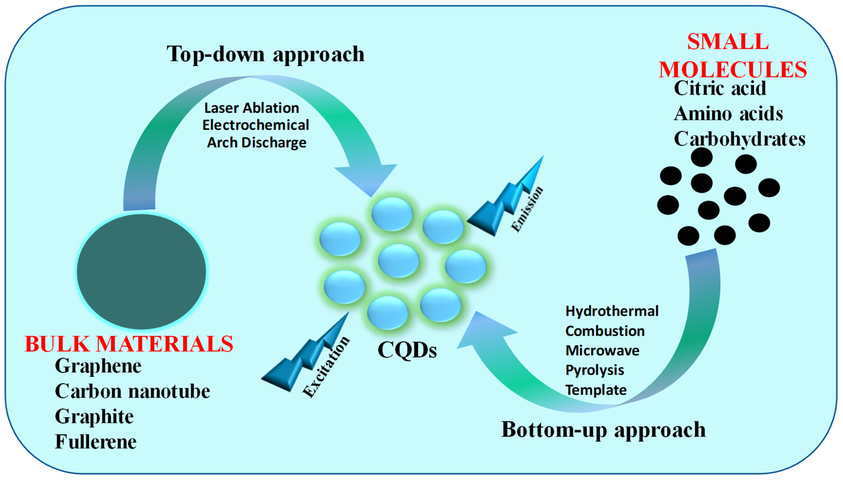
Since the discovery of carbon quantum dots (CQDs), several convenient, cost-effective, size-controlled, and large-scale production approaches have been developed. For the synthesis of CQDs, two general categories, top-down and bottom-up, approaches are utilized (Figure 1). Even though CQDs synthesis is facile, there are definite challenges related to their synthesis, such as an aggregation of nanomaterials, the tuning of surface properties, and controlling the size and uniformity [41]. To adjust the functional groups present on the surface and achieve better CQDs performance, post-treatment can be conducted in both approaches. Quantum yields (QYs) of CQDs can be enhanced after surface passivation, which eliminates the emissive traps from the surface. CQDs doped with heteroatoms (N and P) or metals such as Au or Mg improve solubility and electrical conductivity [42]. Even though for the synthesis of CQDs, both the top-down and bottom-up approaches have been used, the environmentally and cost-effective bottom-up approach is most commonly used [43].Figure 1.
The typical approaches for the synthesis of CQDs.
2.1. Top-Down Approach
In a top-down approach, the larger carbon resources such as carbon nanotubes, fullerene, graphite, graphene, carbon soot, activated carbon, etc., are broken down into smaller constituents with the help of different techniques such as laser ablation and electrochemical and arch discharge [28,44,45,46,47][28][44][45][46][47]. Carbon structures with sp2 hybridization that lack efficient energy gaps or band gaps are commonly used as starting materials for top-down processes. Although the top-down approach is extremely helpful and suitable for microsystem industries, it has some limitations, such as the fact that pure nanomaterials cannot be obtained from the large carbon precursor; their purification is costly and also unable to accurately control the morphology and size distribution of CQDs [48].2.2. Bottom-Up Approach
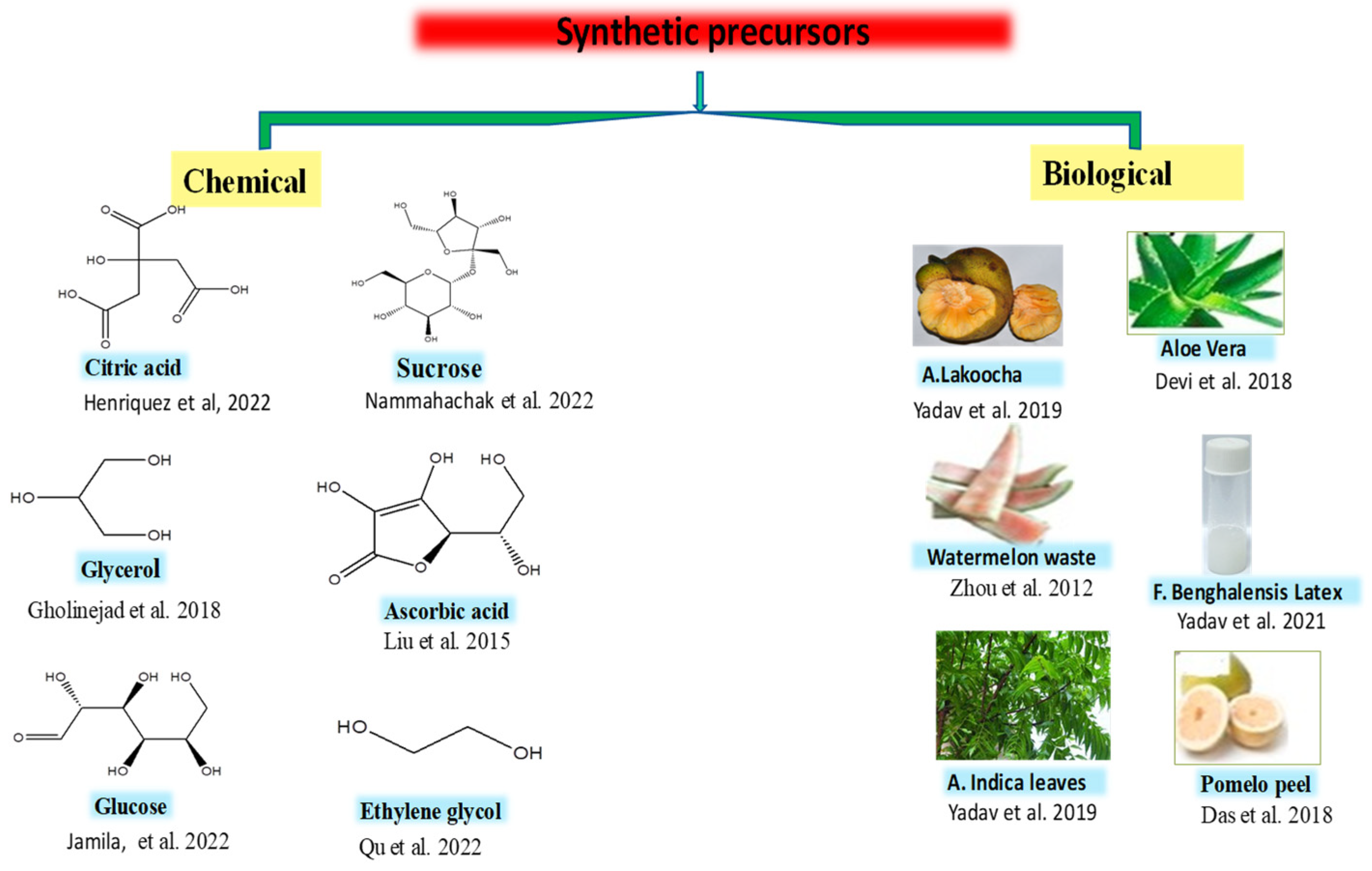
In a bottom-up approach, the smaller carbon resources such as amino acids, polymers, carbohydrates, and waste materials combine to form CQDs by a variety of techniques such as hydrothermal/solvothermal, combustion, pyrolysis, and microwave irradiation. In this method, the size and structure of CQDs depend on a variety of factors such as solvent, precursor molecular structures, and conditions of the reaction (temperature, pressure, reaction time, etc.). The conditions of the reaction are necessary, since they influence the reactants and the extremely casual nucleation and escalation procedure of CQDs. This approach strengthens the material chemistry because of its ease of operation, lower cost, and easier implementation for production in a large scale [61][49]. The precursor used for the synthesis of CQDs may be both chemical and biological, i.e., natural. The chemical precursors include glucose, sucrose, citric acid, lactic acid, ascorbic acid, glycerol, ethylene glycol, etc. [62,63,64,65,66,67,68][50][51][52][53][54][55][56]. The natural sources include Artocarpous lakoocha seeds, rice husks, Azadirachta indica leaves, pomelo peel, the latex of Ficus benghalensis, aloe vera, etc. (Figure 2) [69,70,71,72][57][58][59][60].Figure 2. Chemical and biological precursors utilized for the synthesis of CQDs [61,62,63,64,65,66,67,68,69,70,71,72].
Chemical and biological precursors utilized for the synthesis of CQDs [49][50][51][52][53][54][55][56][57][58][59][60].
3. Structure of CQDs
Tang et al. reported that CQDs have core–shell structures which are either amorphous (mixed sp2/sp3) or graphitic crystalline (sp2), depending upon the extent of the occurrence of sp2 carbon in the core [93][61]. Graphitic crystalline (sp2) cores were reported by several researchers [94,95,96][62][63][64]. The size of cores is very small (2–3 nm), with a characteristic lattice spacing of ~0.2 nm [97][65]. The cores are categorized depending on the technique utilized for the synthesis and the precursors used, as well as other synthetic parameters (such as duration, temperature, pH, etc.) [98][66]. Generally, the graphitization (sp2) structure is obtained at over 300 °C reaction temperatures, while amorphous cores are obtained at lower temperatures, unless sp2/sp3-hybridized C is present in the precursor [99][67]. To determine the core structure of CQDs, various instrumental techniques such as Transmission Electron Microscopy (TEM) or High Resolution (HR) TEM, Scanning Electron Microscopy (SEM), Raman spectroscopy, and X-ray diffraction (XRD) are utilized. To measure the size and morphology of the CQDs, TEM or SEM are carried out [100][68]. The selected area electron diffraction (SAED) patterns reveal the amorphous or crystalline nature of CQDs [101][69]. The XRD pattern also determines the crystal structure of CQDs. The broad peak at 2θ 23° indicates the amorphous nature of CQD, while the occurrence of two broad peaks at 2θ 25° and 44° specifies a low-graphitic carbon structure analogous to (002) and (100) diffraction [102][70]. The general structure and presence of different functional groups on the surface of CQDs are determined using Fourier transform infrared (FT-IR) spectroscopy, X-ray photoelectron spectroscopy (XPS), elemental analysis (EA), and nuclear magnetic resonance (NMR) [103,104][71][72]. Using nitrogen sorption analysis, the surface area of the carbon nanoparticles is calculated [103][71]. To decide the optical properties and qualitative information regarding the presence of C=C and C=O in CQDs, UV-Vis absorption spectroscopy is carried out [105][73]. To determine the positive or negative charge on the surface of CQDs and the extent of the electrostatic interaction between them, zeta potential is conceded [106,107][74][75].
Figure 3 is the typical structure of carbon quantum dots (CQDs), which reveals the presence of different functional groups (such as carbonyl, carboxyl, hydroxyl, amino, etc.) on the surface of CQDs. The presence of these functional groups was confirmed by instrumental techniques such as FTIR and XPS [108][76].
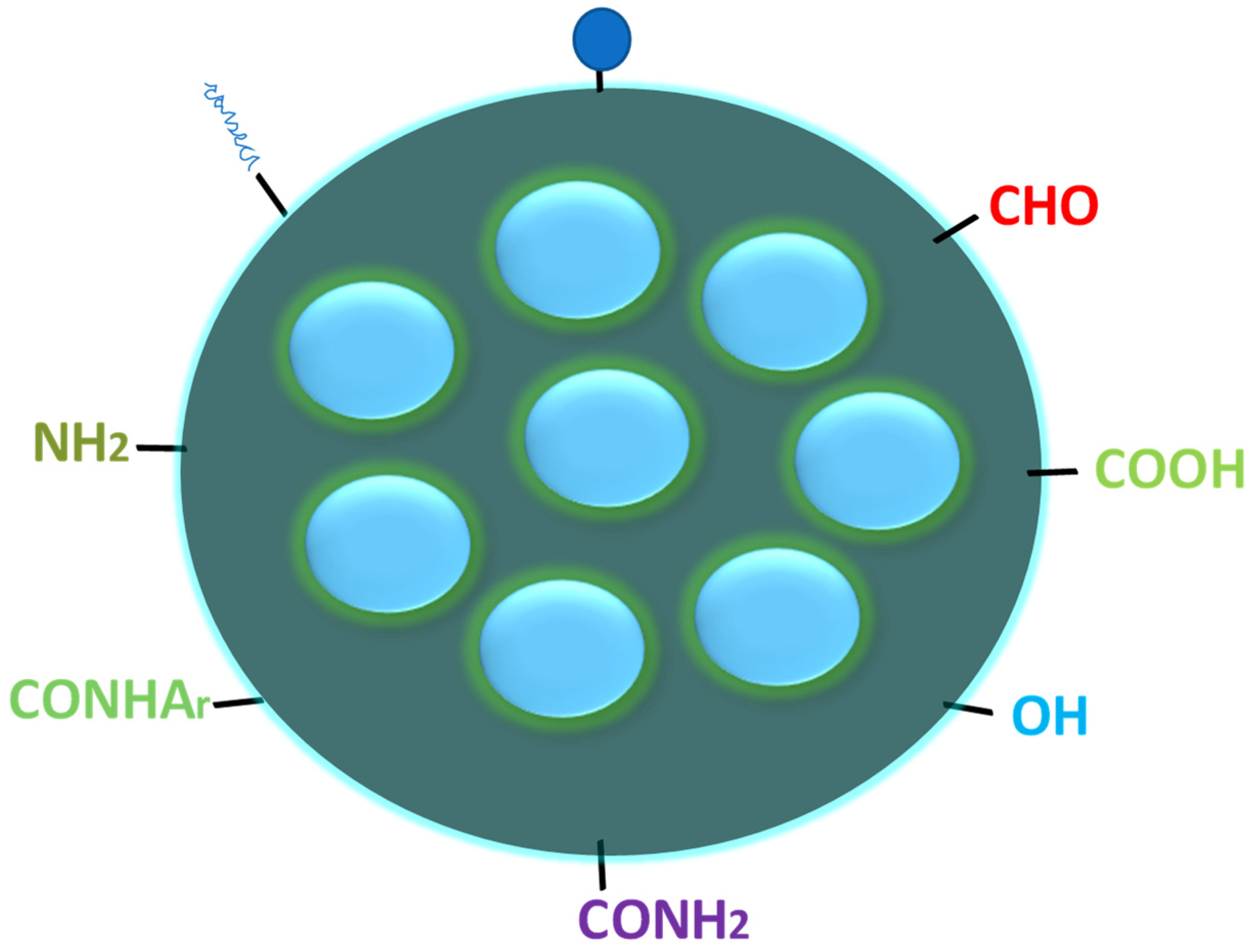
Figure 3.
Typical structure of CQDs with different functional groups on the surface.
4. Optical Properties of Carbon Quantum Dots (CQDs)
4.1. Absorbance
CQDs generally exhibit two absorption bands in the visible region around 280 nm and 350 nm, alongside a tail broadly in the UV region. Hu et al. reported that an absorption band at 280 nm is due to a pi-pi* (π-π*) transition of a C=C bond, and the one at 350 nm is due to an n-π* transition of the C=O bond [109][77]. Figure 4 is the typical UV-visible absorption spectrum of fluorescent CQDs. The absorption properties of CQDs can be influenced by surface modification or surface passivation [110,111,112,113][78][79][80][81]. Depending on the raw precursor and synthesis methodology, the positions of these absorption bands are different to some extent. Doping in CQDs can also alter the absorption wavelength.
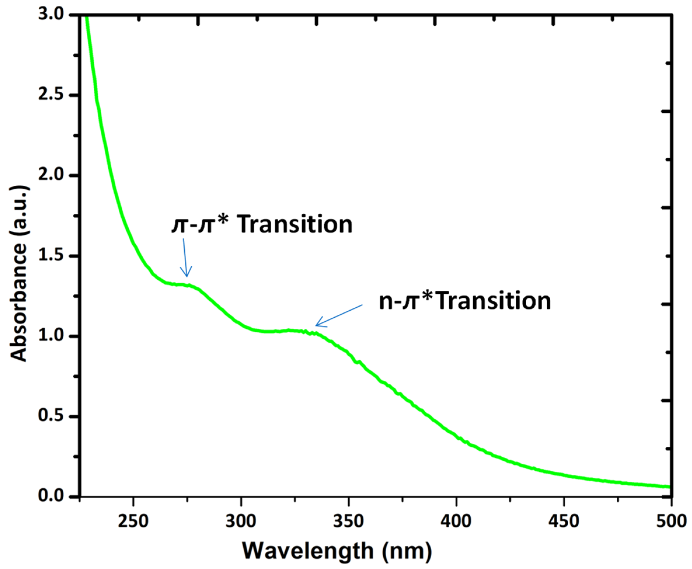
Figure 4.
The UV-visible absorption spectrum of fluorescent CQDs.
The optical properties of CQDs can be customized by doping/co-doping with heteroatoms, functional groups, and surface passivation [114][82]. In the process of surface passivation, a slim insulating (protecting) layer of covering materials such as thiols, thionyl chloride, spiropyrans, and oligomers (polyethylene glycol (PEG), etc.) is formed on the CQDs surface. The important functions of such types of protective layers are to shield CQDs from the adhesion of impurities and to provide stability [115][83]. CQDs with surface-passivating agents become extremely optically active, demonstrating considerable fluorescence from the visible to the near-IR region [116][84]. The quantum yields (QYs) of CQDs can also be enhanced up to 55–60% by surface passivation [114][82]. The absorbance of CQDs improved to longer wavelengths (350–550 nm) after surface passivation with 4,7,10-trioxa-1,13-tridecanediamine (TTDDA) [117][85]. Particle size is associated with the absorption wavelength. As the size of the CQDs increases, absorption wavelength also increases [118,119][86][87]. The CQDs are viable for covalent bonding with functionalizing agents [114][82]. Different functional groups such as amines, carboxyl, hydroxyl, carbonyl, etc., were introduced on the surface of CQDs by surface functionalization. The functionalized CQDs revealed good biocompatibility, high stability, outstanding photoreversibility, and low toxicity compared to undoped CQDs. The efficient technique to modify the CQDs absorption spectrum is doping/co-doping with heteroatoms (such as boron (B), nitrogen (N), fluorine (F), phosphorous (P), and sulfur (S)). The dopant adjusts the bandgap, electronic structure, and, consequently, the optical properties of CQDs by altering the π-π* energy level (related through the core-sp2 carbon system) [120][88]. On increasing N-dopant concentration, a gradual increase in the band gap of the CQDs from 2.2 to 2.7 eV was observed [121][89]. In contrast, it was also found that the doping of N in CQDs results in a reduction in size [122][90]. The CQDs established innovative electronic states, resulting in a reduction in the bandgap of CQDs (about ~48–57%) [123][91]. Zuo et al. synthesized F-doped CQDs using a hydrothermal method which exhibited higher QYs and enhanced the electron transfer and acted as a superior photocatalyst [124][92].
4.2. Photoluminescence
The emission of light from a substance upon the absorption of light (photon) is called photoluminescence (PL). Photoluminescence includes two types, namely fluorescence and phosphorescence. Fluorescent materials emit absorbed light from the lowest singlet excited state (S1) to the singlet ground state (S0). This process is very fast and has a nanosecond lifetime. The transitions that occur among two electronic states in the fluorescence process are allowed because it has the same spin multiplicity. In contrast, in phosphorescence, the transition occurs from the lowest triplet excited state (T1) to singlet ground state (S0), i.e., a forbidden transition occurs according to the spin selection rule.
4.2.1. Fluorescence
The fluorescence properties of CQDs have attracted great attention among researchers because of their several sensing and analytical applications. Numerous mechanisms have been reported to gain deep insight into the cause of fluorescence in CQDs [125,126,127,128,129,130][93][94][95][96][97][98]. Among them, the following two have been found more prominent. The first is that the fluorescence mechanism is due to band gaps’ transitions arising from the π-conjugated domains (sp2-hybridized), which is similar to aromatic molecules employing definite energy band gaps in favor of absorptions and emissions [131][99]. The second cause of fluorescence is related to the surface defects, quantum size effect, carbon core state, surface passivation/functionalization effect, and different emissive traps on the surface of CQDs [132,133,134][100][101][102].
The main reason for the surface defects in CQDs is an unsymmetrical allocation of sp2- and sp3-hybridized carbon atoms, and the existence of heteroatoms such as B, N, P, and S [126,135][94][103]. When this surface defect is independently incorporated into the solid host, it creates surroundings similar to aromatic molecules. These molecules can attract UV light and display various color emissions [131,136][99][104]. CQDs show two types of emission, i.e., excitation-dependent emission (tunable emission) and excitation-independent emission. The tunable emission is due to the presence of various emission sites on the surface of CQDs along with particle size distribution; because of this, most CQDs exhibit tunable emissions [137][105]. The excitation-independent emission is due to the extremely ordered graphitic structure of CQDs [118][86]. CQDs exhibit extensive and unremitting excitation spectra which are highly photostable and have steady fluorescence, in contrast to traditional organic dye [95,138,139][63][106][107].
4.2.2. Phosphorescence
In CQDs, the phosphorescence property is also observed, which was first described by De et al. via dispersing CQDs to polyvinyl alcohol matrix at RT and exciting them with ultraviolet light. The maximum emission obtained was 500 nm, with an average lifetime of 380 ns at a 325 nm excitation [140][108]. Phosphorescence in CQDs arises when the singlet and triplet states of an aromatic carbonyl group in CQDs and polyvinyl alcohol matrix are close in energy to assist spin–orbit coupling, which increases the intersystem crossing (ISC). By using microwave synthesis, Lu et al. synthesized ultra-long phosphorescent carbon quantum dots (P-CQDs). When P-CQDs were excited at 354 nm, they displayed yellow-green phosphorescence (525 nm) for up to 9 s. They concluded that as the pH increases, the phosphorescence intensity of P-CQDs gradually decreases. The reason is that protonation dissociates the hydrogen bonds and distresses the phosphorescent sources. By introducing the tetracyclines (TCs), the phosphorescence of P-CQDs was quenched. They applied P-CQDs as biological and chemical sensing and time-resolved imaging [141][109]. Figure 5 is the typical excitation (black line) and emission (red line) spectrum of fluorescent CQDs.
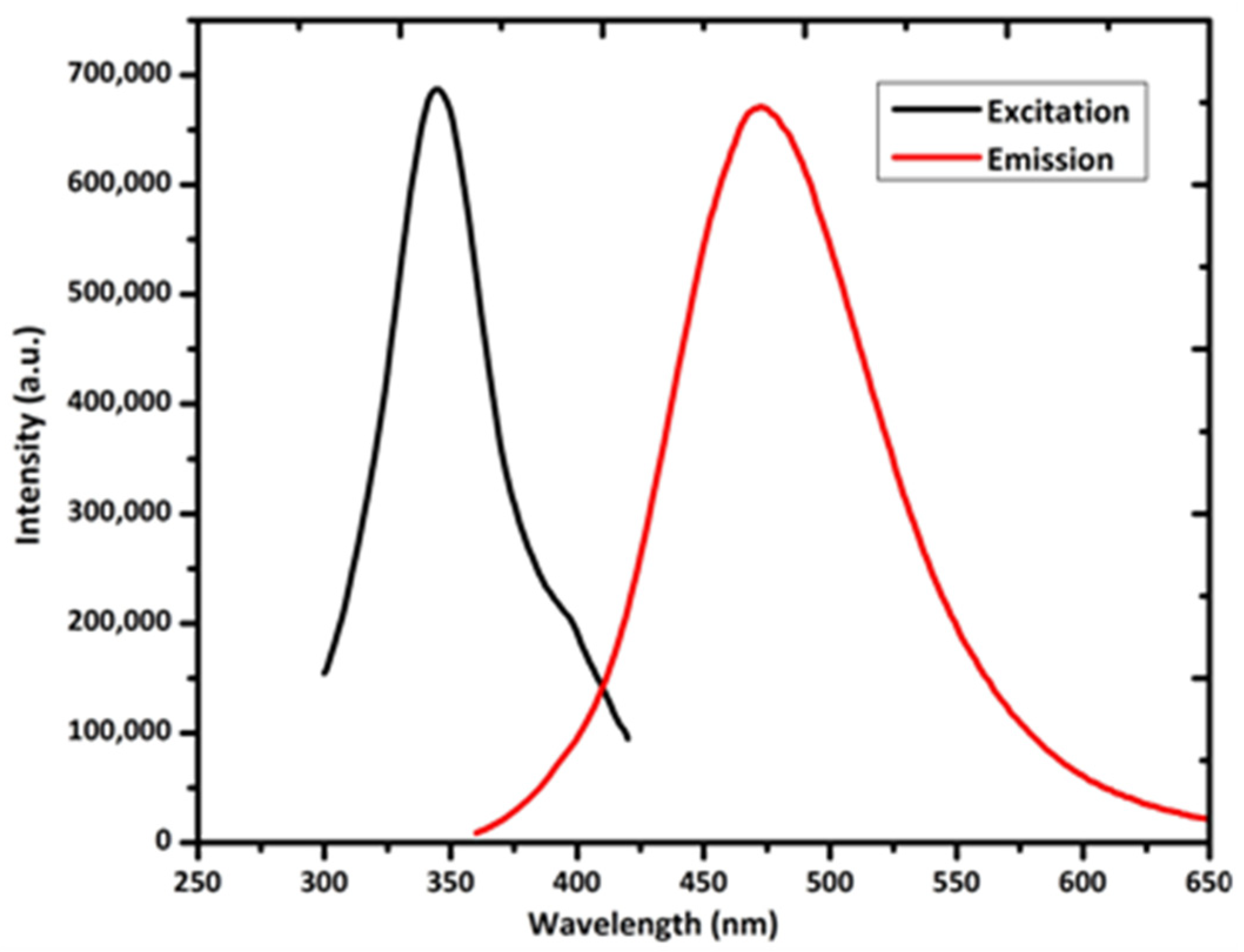
Figure 5.
Excitation and emission spectrum of CQDs.
References
- Clancy, A.; Bayazit, M.K.; Hodge, S.A.; Skipper, N.T.; Howard, C.A.; Shaffer, M.S.P. Charged Carbon Nanomaterials: Redox Chemistries of Fullerenes, Carbon Nanotubes, and Graphenes. Chem. Rev. 2018, 118, 7363–7408.
- Lin, H.-S.; Jeon, I.; Xiang, R.; Seo, S.; Lee, J.-W.; Li, C.; Pal, A.; Manzhos, S.; Goorsky, M.S.; Yang, Y.; et al. Achieving high efficiency in solution-processed perovskite solar cells using C60/C70 mixed fullerenes. ACS Appl. Mater. Interfaces 2018, 46, 39590–39598.
- Georgakilas, V.; Perman, J.A.; Tucek, J.; Zboril, R. Broad Family of Carbon Nanoallotropes: Classification, Chemistry, and Applications of Fullerenes, Carbon Dots, Nanotubes, Graphene, Nanodiamonds, and Combined Superstructures. Chem. Rev. 2015, 115, 4744–4822.
- Rao, R.; Pint, C.L.; Islam, A.E.; Weatherup, R.S.; Hofmann, S.; Meshot, E.R.; Wu, F.; Zhou, C.; Dee, N.; Amama, P.B.; et al. Carbon nanotubes and related nanomaterials: Critical advances and challenges for synthesis toward mainstream commercial applica-tions. ACS Nano 2018, 12, 11756–11784.
- Patel, K.D.; Singh, R.K.; Kim, H.W. Carbon-based nanomaterials as an emerging platform for theranostics. Materials Horizons 2019, 3, 434–469.
- Wang, X.; Feng, Y.; Dong, P.; Huang, J. A Mini Review on Carbon Quantum Dots: Preparation, Properties, and Electrocatalytic Application. Front. Chem. 2019, 7, 671.
- Zuo, J.; Tao, J.; Zhao, X.; Xiong, X.; Xiao, S.; Zhu, Z. Preparation and application of fluorescent carbon dots. J. Nanomater. 2015, 2015, 787862.
- Xu, X.; Ray, R.; Gu, Y.; Ploehn, H.J.; Gearheart, L.; Raker, K.; Scrivens, W.A. Electrophoretic Analysis and Purification of Fluorescent Single-Walled Carbon Nanotube Fragments. J. Am. Chem. Soc. 2004, 126, 12736–12737.
- Sun, Y.-P.; Zhou, B.; Lin, Y.; Wang, W.; Fernando, K.S.; Pathak, P.; Meziani, M.J.; Harruff, B.A.; Wang, X.; Wang, H. Quantum-Sized Carbon Dots for Bright and Colorful Photoluminescence. J. Am. Chem. Soc. 2006, 128, 7756–7757.
- Ahmad, F.; Khan, A.M. Carbon quantum dots: Nanolights. Int. J. Petrochem. Sci. Eng. 2017, 2, 247–250.
- Yang, S.; Sun, J.; Li, X.; Zhou, W.; Wang, Z.; He, P.; Ding, G.; Xie, X.; Kang, Z.; Jiang, M. Large-scale fabrication of heavy doped carbon quantum dots with tunable-photoluminescence and sensitive fluorescence detection. J. Mater. Chem. A 2014, 2, 8660–8667.
- Guo, H.; Liu, Z.; Shen, X.; Wang, L. One-Pot Synthesis of Orange Emissive Carbon Quantum Dots for All-Type High Color Rendering Index White Light-Emitting Diodes. ACS Sustain. Chem. Eng. 2022, 10, 8289–8296.
- Toma, E.E.; Stoian, G.; Cojocaru, B.; Parvulescu, V.I.; Coman, S.M. ZnO/CQDs Nanocomposites for Visible Light Photodegradation of Organic Pollutants. Catalysts 2022, 12, 952.
- Subedi, S.; Rella, A.K.; Trung, L.G.; Kumar, V.; Kang, S.-W. Electrically Switchable Anisometric Carbon Quantum Dots Exhibiting Linearly Polarized Photoluminescence: Syntheses, Anisotropic Properties, and Facile Control of Uniaxial Orientation. ACS Nano 2022, 16, 6480–6492.
- Gu, L.; Zhang, J.; Yang, G.; Tang, Y.; Zhang, X.; Huang, X.; Zhai, W.; Fodjo, E.K.; Kong, C. Green preparation of carbon quantum dots with wolfberry as on-off-on nanosensors for the detection of Fe3+ and l-ascorbic acid. Food Chem. 2022, 376, 131898.
- Chen, Y.; Xue, B. A review on quantum dots modified g-C3N4-based photocatalysts with improved photocatalytic activity. Catalysts 2020, 1, 142.
- Parya, E.; Rhim, J.-W. Pectin/carbon quantum dots fluorescent film with ultraviolet blocking property through light conversion. Colloids Surf. B Biointerfaces 2022, 219, 112804.
- Ajayan, P.M.; Zhou, O.Z. Applications of carbon nanotubes. Carbon Nanotub. 2001, 80, 391–425.
- Baptista, F.R.; Belhout, S.A.; Giordani, S.; Quinn, S.J. Recent developments in carbon nanomaterial sensors. Chem. Soc. Rev. 2015, 44, 4433–4453.
- Chen, D.; Tang, L.; Li, J. Graphene-based materials in electrochemistry. Chem. Soc. Rev. 2010, 39, 3157–3180.
- Cayuela, A.; Benítez-Martínez, S.; Soriano, M.L. Carbon nanotools as sorbents and sensors of nanosized objects: The third way of analytical nanoscience and nanotechnology. TrAC Trends Anal. Chem. 2016, 84, 172–180.
- Pardo, J.; Peng, Z.; Leblanc, R.M. Cancer Targeting and Drug Delivery Using Carbon-Based Quantum Dots and Nanotubes. Molecules 2018, 23, 378.
- Sahar, T.; Abnous, K.; Taghdisi, S.M.; Ramezani, M.; Alibolandi, M. Hybrid car-bon-based materials for gene delivery in cancer therapy. J. Control Release 2020, 318, 158–175.
- Mingjun, C.; Cao, Y.; Zhu, Y.; Peng, W.; Li, Y.; Zhang, F.; Xia, Q.; Fan, X. Oxidation-Modulated CQDs Derived from Covalent Organic Frameworks as Enhanced Fluorescence Sensors for the Detection of Chromium (VI) and Ascorbic Acid. Ind. Eng. Chem. Res. 2022, 31, 11484–11493.
- Murali, G.; Kwon, B.; Kang, H.; Modigunta, J.K.R.; Park, S.; Lee, S.; Lee, H.; Park, Y.H.; Kim, J.; Park, S.Y.; et al. Hematoporphyrin Photosensitizer-Linked Carbon Quantum Dots for Photodynamic Therapy of Cancer Cells. ACS Appl. Nano Mater. 2022, 5, 4376–4385.
- Li, P.; Yu, M.; Ke, X.; Gong, X.; Li, Z.; Xing, X. Cytocompatible Amphipathic Carbon Quantum Dots as Potent Membrane-Active Antibacterial Agents with Low Drug Resistance and Effective Inhibition of Biofilm Formation. ACS Appl. Bio Mater. 2022, 5, 3290–3299.
- Wu, Y.; Qin, D.; Luo, Z.; Meng, S.; Mo, G.; Jiang, X.; Deng, B. High Quantum Yield Boron and Nitrogen Codoped Carbon Quantum Dots with Red/Purple Emissions for Ratiometric Fluorescent IO4– Sensing and Cell Imaging. ACS Sustain. Chem. Eng. 2022, 10, 5195–5202.
- Kaur, A.; Pandey, K.; Kaur, R.; Vashishat, N.; Kaur, M. Nanocomposites of Carbon Quantum Dots and Graphene Quantum Dots: Environmental Applications as Sensors. Chemosensors 2022, 10, 367.
- Sharma, V.; Vishal, V.; Chandan, G.; Bhatia, A.; Chakrabarti, S.; Bera, M. Green, sustainable, and economical synthesis of fluorescent nitrogen-doped carbon quantum dots for applications in optical displays and light-emitting diodes. Mater. Today Sustain. 2022, 19, 100184.
- Wang, Y.; Chen, D.; Zhang, J.; Balogun, M.T.; Wang, P.; Tong, Y.; Huang, Y. Charge Relays via Dual Carbon-Actions on Nanostructured BiVO4 for High Performance Photoelectrochemical Water Splitting. Adv. Funct. Mater. 2022, 32, 2112738.
- Rasooll, M.M.; Zarei, M.; Zolfigol, M.A.; Sepehrmansourie, H.; Omidi, A.; Hasani, M.; Gu, Y. Novel nano-architectured carbon quantum dots (CQDs) with phosphorous acid tags as an efficient catalyst for the synthesis of multisubstituted 4H-pyran with indole moieties under mild conditions. RSC Adv. 2021, 11, 25995–26007.
- Liu, Z.X.; Bin Chen, B.; Liu, M.L.; Zou, H.Y.; Huang, C.Z. Cu(i)-Doped carbon quantum dots with zigzag edge structures for highly efficient catalysis of azide–alkyne cycloadditions. Green Chem. 2017, 19, 1494–1498.
- Fanqing, M.; Wang, Y.; Chen, Z.; Hu, J.; Lu, G.; Ma, W. Synthesis of FeOOH nanoneedles with abundant active edges for efficient electro-catalytic degradation of levofloxacin: Degradation mechanism and toxicity assessment. Appl. Catal. B Environ. 2021, 282, 119597.
- Rezaei, A.; Mohammadi, Y.; Ramazani, A.; Zheng, H. Ultrasound-assisted pseudohomogeneous tungstate catalyst for selective oxidation of alcohols to aldehydes. Sci. Rep. 2022, 12, 3367.
- Preethi, M.; Viswanathan, C.; Ponpandian, N. A metal-free, dual catalyst for the removal of Rhodamine B using novel carbon quantum dots from muskmelon peel under sunlight and ultrasonication: A green way to clean the environment. J. Photochem. Photobiol. A Chem. 2022, 426, 113765.
- Ye, J.; Ni, K.; Liu, J.; Chen, G.; Ikram, M.; Zhu, Y. Oxygen-Rich Carbon Quantum Dots as Catalysts for Selective Oxidation of Amines and Alcohols. Chemcatchem 2017, 10, 259–265.
- Ruihua, L.; Huang, H.; Li, H.; Liu, Y.; Zhong, J.; Li, Y.; Zhang, S.; Kang, Z. Metalnanopar-ticle/carbon quantum dot composite as a photocatalyst for high-efficiency cyclohexane oxidation. ACS Catal. 2014, 1, 328–336.
- Han, Y.; Huang, H.; Zhang, H.; Liu, Y.; Han, X.; Liu, R.; Li, H.; Kang, Z. Carbon Quantum Dots with Photoenhanced Hydrogen-Bond Catalytic Activity in Aldol Condensations. ACS Catal. 2014, 4, 781–787.
- Pradeep Kumar, Y.; Singh, V.K.; Chandra, S.; Bano, D.; Kumar, V.; Talat, M.; Hasan, S.H. Green synthesis of fluorescent carbon quantum dots from azadirachtaindica leaves and their peroxidase-mimetic ac-tivity for the detection of H2O2 and ascorbic acid in common fresh fruits. ACS Biomater. Sci. Eng. 2018, 2, 623–632.
- Li, H.; Sun, C.; Ali, M.; Zhou, F.; Zhang, X.; MacFarlane, D.R. Sulfated Carbon Quantum Dots as Efficient Visible-Light Switchable Acid Catalysts for Room-Temperature Ring-Opening Reactions. Angew. Chem. 2015, 127, 8540–8544.
- Wang, Y.; Hu, A. Carbon quantum dots: Synthesis, properties and applications. J. Mater. Chem. C 2014, 34, 6921–6939.
- Sofia, P.; Palomares, E.; Martinez-Ferrero, E. Graphene and carbon quantum dot-based materials in pho-tovoltaic devices: From synthesis to applications. Nanomaterials 2016, 6, 157.
- Chae, A.Y.; Choi, S.J.; Paoprasert, N.P.; Park, S.Y.; In, I. Microwave-assisted synthesis of fuorescent carbon quantum dots from an A2/B3 monomer set. RSC Adv. 2017, 7, 12663–12669.
- Liu, Y.; Huang, H.; Cao, W.; Mao, B.; Liu, Y.; Kang, Z. Advances in carbon dots: From the perspective of traditional quantum dots. Mater. Chem. Front. 2020, 4, 1586–1613.
- Li, H.; Xu, Y.; Zhao, L.; Ding, J.; Chen, M.; Chen, G.; Li, Y.; Ding, L. Synthesis of tiny carbon dots with high quantum yield using multi-walled carbon nanotubes as support for selective “turn-off-on” detection of rutin and Al3+. Carbon 2018, 143, 391–401.
- Zhao, M.; Zhang, J.; Xiao, H.; Hu, T.; Jia, J.; Wu, H. Facile in situ synthesis of a carbon quantum dot/graphene heterostructure as an efficient metal-free electrocatalyst for overall water splitting. Chem. Commun. 2019, 55, 1635–1638.
- Yuan, F.; Su, W.; Gao, F. Monolayer 2D polymeric fullerene: A new member of the carbon material family. Chem 2022, 8, 2079–2081.
- Zhu, H.; Wang, X.; Li, Y.; Wang, Z.; Yang, F.; Yang, X. Microwave synthesis of fluorescent carbon nanoparticles with electrochemiluminescence properties. Chem. Commun. 2009, 34, 5118–5120.
- Wang, L.; Ruan, F.; Lv, T.; Liu, Y.; Deng, D.; Zhao, S.; Wang, H.; Xu, S. One step synthesis of Al/N co-doped carbon nanoparticles with enhanced photoluminescence. J. Lumin. 2015, 158, 1–5.
- Inderbir, S.; Arora, R.; Dhiman, H.; Pahwa, R. Carbon quantum dots: Synthesis, characterization and biomedical applications. Turk. J. Pharm. Sci. 2018, 2, 219–230.
- Qu, Y.; Li, X.; Zhang, H.; Huang, R.; Qi, W.; Su, R.; He, Z. Controllable synthesis of a sponge-like Z-scheme N, S-CQDs/ TiO2 film with enhanced photocatalytic and antimicrobial activity under visi-ble/NIR light irradiation. J. Hazard. Mater. 2022, 429, 128310.
- Kaixin, C.; Zhu, Q.; Qi, L.; Guo, M.; Gao, W.; Gao, Q. Synthesis and Properties of Nitro-gen-Doped Carbon Quantum Dots Using Lactic Acid as Carbon Source. Materials 2022, 15, 466.
- Henriquez, G.; Ahlawat, J.; Fairman, R.; Narayan, M. Citric Acid-Derived Carbon Quantum Dots Attenuate Paraquat-Induced Neuronal Compromise In Vitro and In Vivo. ACS Chem. Neurosci. 2022, 13, 2399–2409.
- Nammahachak, N.; Aup-Ngoen, K.K.; Asanithi, P.; Horpratum, M.; Chuangchote, S.; Ratanaphan, S.; Surareungchai, W. Hydrothermal synthesis of carbon quantum dots with size tunability via heterogeneous nucleation. RSC Adv. 2022, 12, 31729–31733.
- Jamila, G.S.; Sajjad, S.; Leghari, S.A.K.; Kallio, T.; Flox, C. Glucose derived carbon quantum dots on tungstate-titanate nanocomposite for hydrogen energy evolution and solar light catalysis. J. Nanostruct. Chem. 2021, 12, 611–623.
- Qiu, Y.; Li, D.; Li, Y.; Ma, X.; Li, J. Green carbon quantum dots from sustainable lignocellulosic biomass and its application in the detection of Fe3+. Cellulose 2021, 29, 367–378.
- Aayushi, K.; Maity, B.; Basu, S. Rice Husk-Derived Carbon Quantum Dots-Based Dual-Mode Nano-probe for Selective and Sensitive Detection of Fe3+ and Fluoroquinolones. ACS Biomater. Sci. Eng. 2022, 11, 4764–4776.
- El-Brolsy, H.M.E.M.; Hanafy, N.A.N.; El-Kemary, M.A. Fighting Non-Small Lung Cancer Cells Using Optimal Functionalization of Targeted Carbon Quantum Dots Derived from Natural Sources Might Provide Potential Therapeutic and Cancer Bio Image Strategies. Int. J. Mol. Sci. 2022, 23, 13283.
- Kumari, M.; Chaudhary, G.R.; Chaudhary, S.; Umar, A.; Akbar, S.; Baskoutas, S. Bio-Derived Fluorescent Carbon Dots: Synthesis, Properties and Applications. Molecules 2022, 27, 5329.
- Yao, L.; Zhao, M.-M.; Luo, Q.-W.; Zhang, Y.-C.; Liu, T.-T.; Yang, Z.; Liao, M.; Tu, P.; Zeng, K.-W. Carbon Quantum Dots-Based Nanozyme from Coffee Induces Cancer Cell Ferroptosis to Activate Antitumor Immunity. ACS Nano 2022, 16, 9228–9239.
- Tang, L.; Ji, R.; Cao, X.; Lin, J.; Jiang, H.; Li, X.; Teng, K.S.; Luk, C.M.; Zeng, S.; Hao, J.; et al. Deep Ultraviolet Photoluminescence of Water-Soluble Self-Passivated Graphene Quantum Dots. ACS Nano 2012, 6, 5102–5110.
- Hola, K.; Bourlinos, A.B.; Kozak, O.; Berka, K.; Siskova, K.M.; Havrdova, M.; Tucek, J.; Safarova, K.; Otyepka, M.; Giannelis, E.P.; et al. Photoluminescence effects of graphitic core size and surface functional groups in carbon dots: COO− induced red-shift emission. Carbon 2014, 70, 279–286.
- Sciortino, A.; Marino, E.; van Dam, B.; Schall, P.; Cannas, M.; Messina, F. Solvatochromism un-ravels the emission mechanism of carbon nanodots. J. Phys. Chem. Lett. 2016, 17, 3419–3423.
- Dager, A.; Uchida, T.; Maekawa, T.; Tachibana, M. Synthesis and characterization of Mono-disperse Carbon Quantum Dots from Fennel Seeds: Photoluminescence analysis using Machine Learning. Sci. Rep. 2019, 9, 14004.
- Zhang, W.; Yu, S.F.; Fei, L.; Jin, L.; Pan, S.; Lin, P. Large-area color controllable remote carbon white-light light-emitting diodes. Carbon 2015, 85, 344–350.
- Martindale, B.C.M.; Hutton, G.A.M.; Caputo, C.A.; Prantl, S.; Godin, R.; Durrant, J.R.; Reisner, E. Enhancing Light Absorption and Charge Transfer Efficiency in Carbon Dots through Graphitization and Core Nitrogen Doping. Angew. Chem. 2017, 129, 6559–6563.
- Tingting, Y.; Wang, H.; Guo, C.; Zhai, Y.; Yang, J.; Yuan, J. A rapid microwave synthesis of green-emissive carbon dots with solid-state fluorescence and pH-sensitive properties. R. Soc. Open Sci. 2018, 7, 180245.
- Haitao, L.; Kang, Z.; Liu, Y.; Lee, S.-T. Carbon nanodots: Synthesis, properties and applications. J. Mater. Chem. 2012, 46, 24230–24253.
- Zheng, Y.; Yang, D.; Wu, X.; Yan, H.; Zhao, Y.; Feng, B.; Duan, K.; Weng, J.; Wang, J. A facile approach for the synthesis of highly luminescent carbon dots using vitamin-based small organic molecules with benzene ring structure as precursors. RSC Adv. 2015, 5, 90245–90254.
- Hou, H.; Banks, C.E.; Jing, M.; Zhang, Y.; Ji, X. Carbon Quantum Dots and Their Derivative 3D Porous Carbon Frameworks for Sodium-Ion Batteries with Ultralong Cycle Life. Adv. Mater. 2015, 27, 7861–7866.
- Semeniuk, M.; Yi, Z.; Poursorkhabi, V.; Tjong, J.; Jaffer, S.; Lu, Z.-H.; Sain, M. Future per-spectives and review on organic carbon dots in electronic applications. ACS Nano 2019, 6, 6224–6255.
- Bomben, K.D.; Moulder, J.F.; Stickle, W.F.; Sobol, P.E. Handbook of X-Ray Photoelectron Spectroscopy: A Reference Book of Standard Spectra for Identication and Interpretation of XPS, Physical Electronics, Eden Prairie; Perkin-Elmer Corporation: Waltham, MA, USA, 1995.
- Zhou, Y.; Sharma, S.K.; Peng, Z.; Leblanc, R.M. Polymers in Carbon Dots: A Review. Polymers 2017, 9, 67.
- Kolanowska, A.; Dzido, G.; Krzywiecki, M.; Tomczyk, M.M.; Łukowiec, D.; Ruczka, S.; Boncel, S. Carbon Quantum Dots from Amino Acids Revisited: Survey of Renewable Precursors toward High Quan-tum-Yield Blue and Green Fluorescence. ACS Omega 2022, 45, 41165–41176.
- Qiang, S.; Zhang, L.; Li, Z.; Liang, J.; Li, P.; Song, J.; Guo, K.; Wang, Z.; Fan, Q. New Insights into the Cellular Toxicity of Carbon Quantum Dots to Escherichia coli. Antioxidants 2022, 11, 2475.
- Gayen, B.; Palchoudhury, S.; Chowdhury, J. Carbon Dots: A Mystic Star in the World of Nanoscience. J. Nanomater. 2019, 1–19.
- Hu, C.; Yu, C.; Li, M.; Wang, X.; Yang, J.; Zhao, Z.; Eychmüller, A.; Sun, Y.-P.; Qiu, J. Chemically Tailoring Coal to Fluorescent Carbon Dots with Tuned Size and Their Capacity for Cu(II) Detection. Small 2014, 10, 4926–4933.
- Wang, S.; Kirillova, K.; Lehto, X. Travelers’ food experience sharing on social network sites. J. Travel Tour. Mark. 2016, 34, 680–693.
- Jiang, K.; Zhang, L.; Lu, J.; Xu, C.; Cai, C.; Lin, H. Triple-Mode Emission of Carbon Dots: Applications for Advanced Anti-Counterfeiting. Angew. Chem. 2016, 128, 7347–7351.
- Li, F.; Li, Y.; Yang, X.; Han, X.; Jiao, Y.; Wei, T.; Yang, D.; Xu, H.; Nie, G. Highly Fluorescent Chiral N-S-Doped Carbon Dots from Cysteine: Affecting Cellular Energy Metabolism. Angew. Chem. 2018, 130, 2401–2406.
- Anwar, S.; Ding, H.; Xu, M.; Hu, X.; Li, Z.; Wang, J.; Bi, H. Recent advances in synthesis, optical properties, and bio-medical applications of carbon dots. ACS Appl. Bio Mater. 2019, 6, 2317–2338.
- Jhonsi, M.A. Carbon Quantum Dots for Bioimaging. In State of the Art in Nano-Bioimaging; IntechOpen: London, UK, 2018; pp. 35–55.
- Konstantinos, D. Carbon quantum dots: Surface passivation and functionalization. Curr. Org. Chem. 2016, 6, 682–695.
- Li, L.; Dong, T. Photoluminescence tuning in carbon dots: Surface passivation or/and functionalization, heteroatom doping. J. Mater. Chem. C 2018, 30, 7944–7970.
- Peng, H.; Travas-Sejdic, J. Simple Aqueous Solution Route to Luminescent Carbogenic Dots from Carbohydrates. Chem. Mater. 2009, 21, 5563–5565.
- Wang, L.; Li, W.; Yin, L.; Liu, Y.; Guo, H.; Lai, J.; Han, Y.; Li, G.; Li, M.; Zhang, J.; et al. Full-color fluorescent carbon quantum dots. Sci. Adv. 2020, 6, eabb6772.
- Nisha, P.; Amrita, D. Theoretical study of Dependence of Wavelength on Size of Quantum Dot. Int. J. Sci. Res. Dev. 2016, 4, 126–130.
- Kandasamy, G. Recent Advancements in Doped/Co-Doped Carbon Quantum Dots for Multi-Potential Applications. C 2019, 5, 24.
- Darragh, C.; Rocks, C.; Padmanaban, D.B.; Maguire, P.; Svrcek, V.; Mariotti, D. Environmentally friendly nitrogen-doped carbon quantum dots for next generation solar cells. Sustain. Energy Fuels 2017, 7, 1611–1619.
- Gao, R.; Wu, Z.; Wang, L.; Liu, J.; Deng, Y.; Xiao, Z.; Fang, J.; Liang, Y. Green Preparation of Fluorescent Nitrogen-Doped Carbon Quantum Dots for Sensitive Detection of Oxytetracycline in Environmental Samples. Nanomaterials 2020, 10, 1561.
- Yu, J.; Liu, C.; Yuan, K.; Lu, Z.; Cheng, Y.; Li, L.; Zhang, X.; Jin, P.; Meng, F.; Liu, H. Luminescence Mechanism of Carbon Dots by Tailoring Functional Groups for Sensing Fe3+ Ions. Nanomaterials 2018, 8, 233.
- Zuo, G.; Xie, A.; Li, J.; Su, T.; Pan, X.; Dong, W. Large Emission Red-Shift of Carbon Dots by Fluorine Doping and Their Applications for Red Cell Imaging and Sensitive Intracellular Ag+ Detection. J. Phys. Chem. C 2017, 121, 26558–26565.
- Baker, S.N.; Baker, G.A. Luminescent carbon nanodots: Emergent nanolights. Angew. Chem. Int. Ed. 2010, 49, 6726–6744.
- Gokus, T.; Nair, R.R.; Bonetti, A.; Böhmler, M.; Lombardo, A.; Novoselov, K.; Geim, A.K.; Ferrari, A.C.; Hartschuh, A. Making Graphene Luminescent by Oxygen Plasma Treatment. ACS Nano 2009, 3, 3963–3968.
- Demchenko, A.P.; Dekaliuk, M.O. Novel fluorescent carbonic nanomaterials for sensing and imaging. Methods Appl. Fluoresc. 2013, 1, 042001.
- Zhang, Q.; Wang, R.; Feng, B.; Zhong, X.; Ostrikov, K. Photoluminescence mechanism of carbon dots: Triggering high-color-purity red fluorescence emission through edge amino protonation. Nat. Commun. 2021, 12, 6856.
- Nguyen, H.A.; Srivastava, I.; Pan, D.; Gruebele, M. Unraveling the Fluorescence Mechanism of Carbon Dots with Sub-Single-Particle Resolution. ACS Nano 2020, 14, 6127–6137.
- An, Y.; Liu, C.; Li, Y.; Chen, M.; Zheng, Y.; Tian, H.; Shi, R.; He, X.; Lin, X. Preparation of Multicolour Solid Fluorescent Carbon Dots for Light-Emitting Diodes Using Phenylethylamine as a Co-Carbonization Agent. Int. J. Mol. Sci. 2022, 23, 11071.
- Cao, L.; Meziani, M.J.; Sahu, S.; Sun, Y.-P. Photoluminescence Properties of Graphene versus Other Carbon Nanomaterials. Acc. Chem. Res. 2012, 46, 171–180.
- Fang, Y.; Guo, S.; Li, D.; Zhu, C.; Ren, W.; Dong, S.; Wang, E. Easy Synthesis and Imaging Applications of Cross-Linked Green Fluorescent Hollow Carbon Nanoparticles. ACS Nano 2011, 6, 400–409.
- Shen, J.; Zhu, Y.; Chen, C.; Yang, X.; Li, C. Facile preparation and upconversionlumi-nescence of graphene quantum dots. Chem. Commun. 2011, 9, 2580–2582.
- Li, X.; Wang, H.; Shimizu, Y.; Pyatenko, A.; Kawaguchi, K.; Koshizaki, N. Preparation of carbon quantum dots with tunable photoluminescence by rapid laser passivation in ordinary organic solvents. Chem. Commun. 2010, 47, 932–934.
- Nourbakhsh, A.; Cantoro, M.; Vosch, T.; Pourtois, G.; Clemente, F.; van der Veen, M.; Hofkens, J.; Heyns, M.M.; De Gendt, S.; Sels, B.F. Bandgap opening in oxygen plasma-treated graphene. Nanotechnology 2010, 21, 435203.
- Dekaliuk, M.O.; Viagin, O.; Malyukin, Y.V.; Demchenko, A.P. Fluorescent carbon nanomateri-als:“Quantum dots” or nanoclusters? Phys. Chem. Chem. Phys. 2014, 30, 16075–16084.
- Bibekananda, D.; Karak, N. A green and facile approach for the synthesis of water soluble fluorescent carbon dots from banana juice. RSC Adv. 2013, 22, 8286–8290.
- Dong, Y.; Shao, J.; Chen, C.; Li, H.; Wang, R.; Chi, Y.; Lin, X.; Chen, G. Blue luminescent graphene quantum dots and graphene oxide prepared by tuning the carbonization degree of citric acid. Carbon 2012, 50, 4738–4743.
- Zhi, B.; Yao, X.; Cui, Y.; Orr, G.; Haynes, C.L. Synthesis, applications and potential photoluminescence mechanism of spectrally tunable carbon dots. Nanoscale 2019, 11, 20411–20428.
- De Caluwé, E.; Halamouá, K.; Van Damme, P.; Adansoniadigitata, L. A review of traditional uses, phytochemistry and pharmacology. Afr. Focus 2010, 1, 11–51.
- Lu, C.; Su, Q.; Yang, X. Ultra-long room-temperature phosphorescent carbon dots: pH sensing and dual-channel detection of tetracyclines. Nanoscale 2019, 11, 16036–16042.
More