Your browser does not fully support modern features. Please upgrade for a smoother experience.
Please note this is a comparison between Version 1 by Nabil Grimi and Version 2 by Rita Xu.
The freshwater microalga
Haematococcus pluvialis
is well known as the cell factory for natural astaxanthin, which composes up to 4–7% of its total dry weight. The bioaccumulation of astaxanthin in
H. pluvialis
cysts seems to be a very complex process that depends on different stress conditions during its cultivation. The red cysts of
H. pluvialis
develop thick and rigid cell walls under stress growing conditions.
- microalga
- Haematococcus pluvialis
- biomolecular composition
- electrical treatment
1. Introduction
In recent years, the up and downstream processing problem of H. pluvialis microalgae has attracted great attention [1][2][3][4][5][1,2,3,4,5]. The low-cost bio-production of natural astaxanthin using this microalga is still one of the market’s most concerning issues. It remains a challenge to biosynthesize natural astaxanthin using microalgae such as H. pluvialis on a large scale. The main reason for the high cost of natural astaxanthin is that the recovery of the final product necessitates additional manufacturing steps.
The cultivation of H. pluvialis for natural astaxanthin production (known as a king of antioxidants) involves two phases, green and red. During the second one, the red cysts or aplanospores accumulate a high amount of astaxanthin, which accounts for ≈80% of the total carotenoid content. Accumulation of astaxanthin is a rather complex process that typically includes specific abiotic stress applications such as nutrient starvation [6], pH [7], illumination stresses [8][9][10][8,9,10], temperature [11], irradiation or pressure stresses [12] and a mixture of different stresses [13]. Particularly, the synergetic combination of different factors allowed improving of astaxanthin accumulation [13][14][13,14].
Application of electrotreatments during the growth stages allows genetic transformation, inactivation of culture contaminants, improvement of growth kinetics, accumulation of bioactives and increasing of permeability of cell walls [15][16][17][15,16,17]. The cells of H. pluvialis are covered by extraordinarily thick, rigid and indigestible cell walls, and the availability of internal biomolecules is very limited [18][19][18,19]. Therefore, the selective recovery of these biomolecules, and particularly astaxanthin, from H. pluvialis requires application of rather complex cell disruption and purification techniques. The topic is a very hot issue in contemporary studies of extraction from various microalgae [19]. The processing of H. pluvialis using pulsed electrotechnologies (pulsed electric fields and (or) high-voltage electric discharges) is very attractive because with proper adaptation these techniques allow effective extraction and obtaining of high purity extracts [20].
2. Main Steps Haematococcus pluvialis Biorefinery
At both laboratory and industrial scales, many important steps are included in the processing biorefinery of H. pluvialis microalgae. The general overviews of these steps are presented in Figure 1.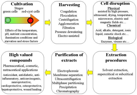
Figure 1. Overview of different steps in H. pluvialis microalgae processing biorefinery.
3. Cultivation and Growth Stages of Haematococcus pluvialis, Effects of Electrotreatment
The freshwater microalga H. pluvialis was described for the first time in 1841 [21][22]. It has been reported that this microscopic object can occur in green and red forms. The green forms were observed in juvenile states. In cold seasons (winter, spring, and autumn), the green forms were mainly observed [22][23][23,24]. A detailed discussion of the early studies of H. pluvialis was presented in 1899 [24][25]. H. pluvialis occurs in different regions of Europe, Africa, and North America [25][26].3.1. Cultivation and Growth Stages
It is still acknowledged that the green microalga H. pluvialis is recognized for its capacity to produce significant quantities of natural astaxanthin via the application of different stress conditions and its high specific growth rate 1.3 d−1 [26][27]. H. pluvialis astaxanthin production for the market is relatively new. The induction of astaxanthin bioaccumulation in H. pluvialis cells is carried out by subjecting the cells to stress growth conditions. In the last two decades, the two-stage strategy for cultivation has been widely adopted for both laboratory and large-scale cultures. The first stage involves growing H. pluvialis under favorable conditions for green biomass production (green stage); this is followed by a second stage (red stage) in which the astaxanthin biosynthesis is induced by changing the favorable growth conditions to unfavorable ones. The natural response to the harsh environmental conditions involves the inhibition of normal growth and the accumulation of astaxanthin for the purpose of protecting the photosynthetic apparatus from photo-oxidative damage by absorbing the excess light. The role of astaxanthin is important to protect cells by scavenging free radicals and reacting with reactive oxygen species. This role is to ensure the maintenance of photosynthesis during red-stage stress [27][28]. Obviously, the life cycle of H. pluvialis microalgae is complicated. The two-stage culturing implies different cell forms. This life cycle can be roughly divided into motile (bi-flagellated cells) and non-motile stages (aplanospores, astaxanthin-accumulating aplanospores, red cysts cells, sporangium and zoospores released from the sporangium) [19][28][29][30][19,29,30,31]. Each transition from the motile to the non-motile cell form involves remarkable changes in the cell wall structure. Photosynthetic activity is highest in the initial vegetative green growth phase (days 1 to 12) [7]. In this green vegetative phase, the cells do not accumulate astaxanthin and their carotenoid pattern is mainly composed of lutein (75–80%) and β-carotene (10–20%) [31][32]. However, the astaxanthin accumulation is triggered in the second phase by specific stress application such as nitrogen starvation, oxidative stress, salinity stress, high temperature and high irradiation. These cited factors significantly affect the astaxanthin accumulation. The recent literature on cultivation and growth conditions of H. pluvialis is rather vast. It includes discussions of the effects of temperature [11], pH [7], nutrient concentration (nitrogen, phosphorus and carbon starvation) [6][32][6,33] and illumination conditions [8][9][10][33][34][35][8,9,10,34,35,36]. The effects of light illumination on the growth of biomass and accumulation of bioastaxanthin were analyzed, and it was demonstrated that red light is more effective for increasing the biomass, whereas blue light supports the accumulation of bioastaxanthin [10]. A comprehensive examination of the effects of different photo-, mixo- and heterotrophic cultivation conditions on H. pluvialis cell germination has been recently presented [36][37]. The effects of temperature (20–30.5 °C) on the cell growth and accumulation of astaxanthin has been discussed [11]. It was demonstrated that increased temperature allows enhanced astaxanthin accumulation combined with nitrogen starvation stress. It was demonstrated that the photosynthetic properties are differentially modulated in response to nitrogen starvation/high-light-illumination stress and a correct balance between these stresses is required for efficient astaxanthin production [37][38]. Effects of nuclear irradiation and a high concentration of carbon dioxide CO2 on enhancing the growth rate and astaxanthin yield were demonstrated [12]. The economical two-stage strategy with low light illumination in the initial phase (growth for biomass production) and a combination of high light illumination and elevated carbon dioxide levels (5 or 15%) in the second phase (astaxanthin accumulation) has been tested [8]. The applied procedures showed a significant increase (2–3 times) in the accumulation of astaxanthin. A two-step process was proposed to maximize algal growth and astaxanthin yield [13]. During the first step (biomass production), the different nitrogen sources were tested. During the second step (carotenogenesis induction), a mix of moderate stressors (mild light, nitrogen limitation, and the addition of sodium acetate) was used. The synergetic combination of different factors allowed the promotion of astaxanthin accumulation. The new sequential heterotrophy–dilution–photoinduction cultivation strategy was shown to be rather effective for production of astaxanthin from H. pluvialis [14]. The effects of continuous and interrupted (light/dark cycles) illumination conditions on the production of vegetative green cells and astaxanthin accumulation were investigated [38][40]. It was demonstrated that light/dark cycles are optimal to produce green cells, whereas continuous illumination is better for astaxanthin accumulation. The effects of fulvic acid on biomass growth and astaxanthin accumulation were investigated [39][41]. It was demonstrated that the astaxanthin content increased by 86.89% in 5 mg/L-treated cells. Recently, the details of laboratory and commercial scale cultivation procedures in the production of natural astaxanthin derived from H. pluvialis have been discussed [40][41][42][42,43,44]. The effects of different cultivation conditions and strategies, technological innovations and types of cultivation systems and an economic assessment for astaxanthin production were also reviewed [43][44][45][46][47][48][49][50][45,46,47,48,49,50,51,52].3.2. Electrotreatments during the Growth Stages
The effects of electrotreatments applied during the growth of H. pluvialis on cell viability, cell number density and accumulation of astaxanthin attract great attention. A general review of the application of different electrotechnologies for genetic transformation, inactivation of culture contaminants and improvement of growth kinetics of different species of microalgae is given in [15]. Application of electrotreatments of H. pluvialis during the different growing stages may reduce the strength and impermeability of cell walls and positively affect the accumulation of bioactives. The effects of pulsed electric field (PEF) treatments with square wave and exponential decay wave pulses on cell viability during the growth stages were discussed in [51][53]. Nanosecond PEF treatment (with very short pulses of 25–50 nm and high electric field strength 40 kV/cm) has been applied to H. pluvialis cells during the growth stage [16]. It was demonstrated that such treatment can modulate the expression of the astaxanthin biosynthesis genes. PEF treatment increased both the cell mortality (by up to 20%) and astaxanthin accumulation (by 20–30%) from the initial values. Such results were explained by the expression of carotenoid-pathway-related genes. Electrical treatment of H. pluvialis cells was studied in the bio-electrochemical chambers [52][53][54,55]. Such treatments enhanced the nitrogen consumption and chlorophyll biosynthesis and resulted in a considerable increase in electro-treated cell number density (by 20%) after cultivation for seven days [52][54]. However, such treatments did not significantly affect the intracellular astaxanthin content (10% increase). The special adaptation of electrotreatment parameters (electric strength and duration) allowed a significant increase in the astaxanthin content in electrostimulated H. pluvialis (by 36.9%) [53][55]. Application of electrical treatment by low-temperature plasma in H. pluvialis biotechnology has been also discussed. It was demonstrated that low-temperature plasma promotes the growth of H. pluvialis [54][56]. The plasma treatment is widely used as a safe and environmentally friendly sterilization technology [55][57] and may be applied during the growth stages as a strong oxidative stress and mutagenesis tool without damaging the microalgae cells [17]. Atmospheric pressure argon dielectric barrier discharge plasma was used for mutation induction in H. pluvialis [56][58]. The presence of enhanced astaxanthin accumulation in the mutants was observed that was attributed to genetic modification by the mechanism of carotenogenesis. It was indicated that plasma mutation is suitable for effective H. pluvialis breeding with promising industrial applications [57][59]. The plasma treatment effectively improved the growth of H. pluvialis and its astaxanthin accumulation; under optimal conditions the astaxanthin content was 51.96% higher than in the starting strain. The possible mechanisms of astaxanthin accumulation induced by plasma treatment were also discussed [58][59][60,61].4. Structure of Cell and Biomolecular Composition of Haematococcus pluvialis
4.1. Cell Structure
Figure 2 gives a schematic presentation of the cell structure of H. pluvialis at the resistant cyst stage. On the final aplanospore stage of cyst formation, the rigid and resistant multilayered cell walls are formed [60][61][62][63][64][65][62,63,64,65,66,67]. The total size of the cells at this stage is approximately 20–30 µm [28][29]. The cell internal compartment contains cytoplasm starch granules, sub-cellular micro-compartments of pyrenoids and astaxanthin deposited in extra-plastidial oil globules (lipid droplets). This internal space is covered by lipid membrane with thickness of ≈5 nm.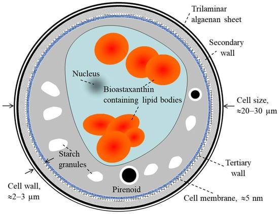
Figure 2. Description of H. pluvialis cell structure at the resistant cyst stage.
4.2. Biomolecular Composition
The chemical constituents of extracellular extracts of H. pluvialis were recently analyzed [66][68]. H. pluvialis cell composition includes ash, protein, carbohydrates, dietary fiber, carotenoids and lipids. Figure 3 presents the relative percentage proportions of ash, protein, carbohydrates total dietary fiber, carotenoids and lipids in the initial biomass and extracts obtained using supercritical CO2 extraction at optimized conditions (pressure of 350 bar, temperature of 50 °C, and CO2 flow rate of 0.47 Kg/min) for the H. pluvialis red phase [67][69]. The total dietary fiber and protein were observed as the main intracellular constituents in the initial biomass. Carotenoids include astaxanthin (66–70%), β-carotene (3.5–6.5%) and lutein (27–28%). The content of proteins, lipids and carotenoids in extracts obtained by supercritical CO2 was higher in comparison to that of the biomass. An inverse situation was observed for the content of total dietary fibers (58% in the biomass compared with 26% in the extract).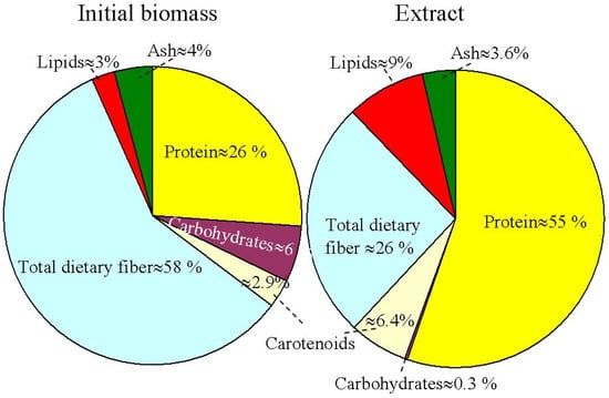
Figure 3. Initial biomass and supercritical CO2 extract relative percentage proportions for different biomolecular component compositions in the H. pluvialis red phase.