Phenolic Compounds as Modulators of Nrf2 in Neuroprotection: Comparison
Please note this is a comparison between Version 3 by Jessie Wu and Version 2 by Jessie Wu.

Neurodegenerative diseases (NDs) are a diverse group of pathologies characterized by a gradual loss in neuron number and function. These pathologies are primarily caused by the accumulation of misfolded proteins, as seen in Alzheimer’s disease (AD), Parkinson’s disease (PD), and Huntington’s disease, and are associated with a decline in cognitive abilities and movement disorders. There has been an increase in the study of the nuclear factor erythroid 2-related factor 2 (Nrf2) pathway and the natural products that positively regulate it to reduce oxidative damage to the nervous system, both in in vitro models with neurons and microglia subjected to stress factors and in vivo models using mainly murine models. Quercetin, curcumin, anthocyanins, tea polyphenols, and other less studied phenolic compounds such as kaempferol, hesperetin, and icariin can also modulate Nrf2 by regulating several Nrf2 upstream activators.

  • Nrf2
  • oxidative stress
  • neurodegeneration
  • phenolic compounds

1. Introduction

The predominant risk factor for neurodegeneration is advanced age, although other factors such as high blood pressure, depression, low educational level, sedentary lifestyle, and oxidative stress (OS) also contribute [1][2][3]. The nervous system is highly sensitive to reactive oxygen species (ROS) which primarily originate in the mitochondria but can also come from external sources. When ROS exceed the neutralization systems of the cell, OS is generated [4]. The organism’s antioxidant capacity is dependent on the presence of enzymes that break down ROS such as superoxide dismutase-1 (SOD-1), heme oxygenase-1 (HO-1), NADPH (nicotinamide adenine dinucleotide phosphate) quinone reductase-1 (NQO-1), catalase (CAT), and peroxidase, as well as internal free radical scavengers such as NADPH, glutathione (GSH), and coenzyme Q, and external such as vitamins A, C, and E [5][6][7]. The ability to modulate antioxidant mechanisms could be a useful tool to mitigate nervous damage associated with OS.
Phytochemicals are associated with an improvement in human health and lifespan due to their antioxidant properties, which decrease OS and reduce the toxicity of diseases such as cancer, cardiovascular diseases, and neurodegenerative diseases (NDs) [8][9]. While several studies support the benefits of natural products such as curcumin, resveratrol, epigallocatechin gallate, and quercetin on NDs, further research is needed to use them in the treatment of conditions such as Alzheimer’s disease (AD or PD) or Parkinson’s disease (PD) [10]. In recent years, there has been growing interest in studying the Nrf2 (nuclear factor erythroid 2-related factor 2) pathway, both in basic research and in clinical applications. The focus is especially on natural compounds that could modulate the activity of the pathway and thus alleviate OS [11].

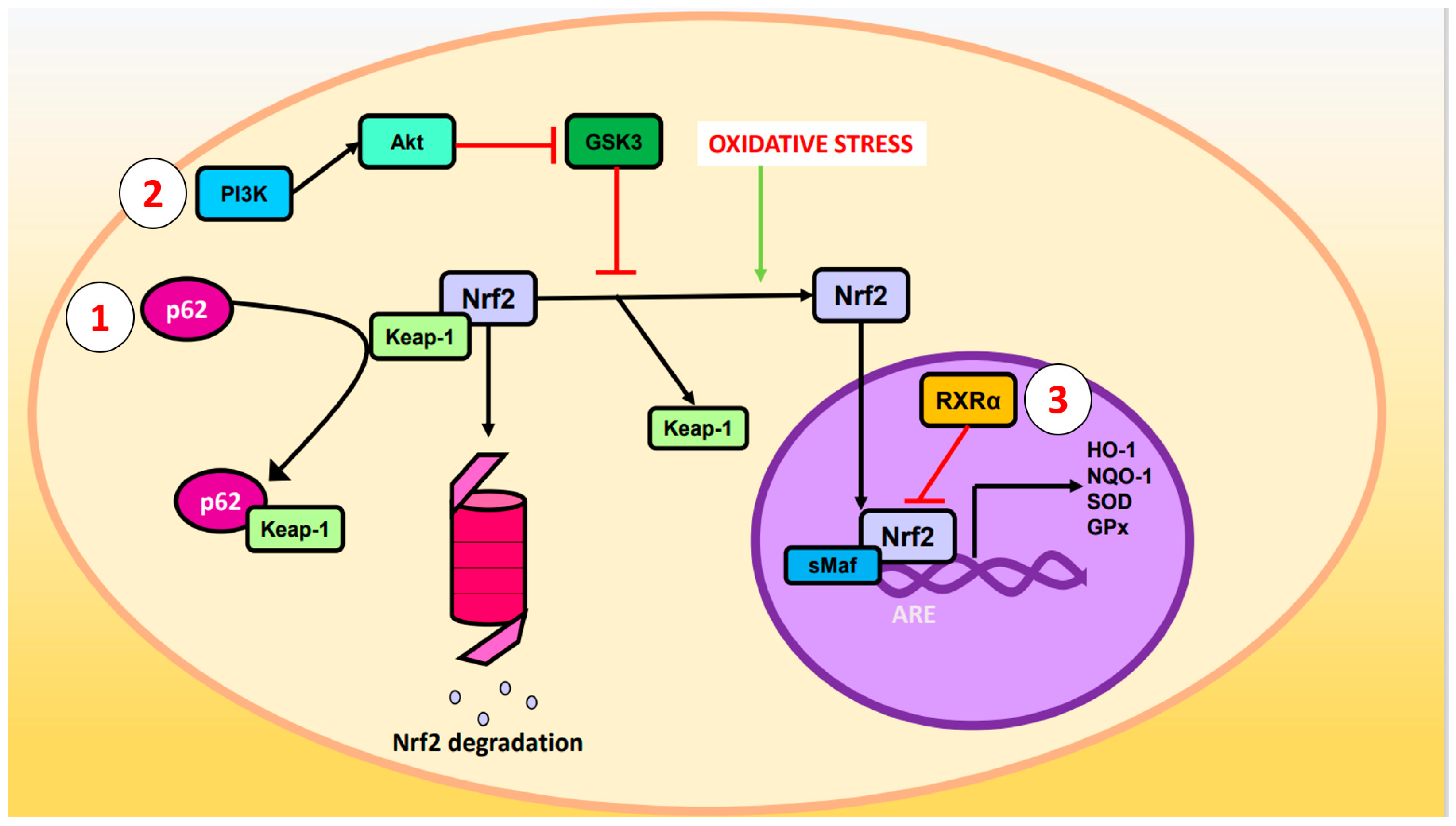
2. Nuclear Factor Erythroid 2-Related Factor 2 Signaling Pathway against OS

The transcription factor nuclear factor erythroid 2-related factor 2 (Nrf2) is a critical cellular protein involved in combating OS and eliminating xenobiotics from the organism [12]. This transcription factor belongs to the “cap’n’collar” subfamily (CNC) with basic leucine zipper (bZIP) that allows it to bind to DNA (deoxyribonucleic acid) [13]. It is a modular protein composed of seven domains known as Neh 1-7 (Nrf-ECH homology domain) [14]. Nrf2 has been linked to resistance to infections, tumor resistance to chemotherapeutics, and, as this review explores, protection against NDs [15][16][17].
Nrf2 is negatively regulated by Keap-1 (Kelch-like ECH-associated protein 1) under normal conditions. Keap-1 binds to the DLG (Asp-Leu-Gly) and ETGE (Glu-Thr-Gly-Glu) motifs of the Neh2 domain and recruits the Cul3 (cullin3)/Rbx1 (RINGBox1) E3 ubiquitin ligase complex, which leads to the ubiquitination of lysine residues of Nrf2 and subsequent degradation in the proteosome [18][19]. However, when cells are subjected to OS, the cysteine residues of Keap-1 become oxidized and release Nrf2, increasing its intracellular levels. Free Nrf2 then penetrates the nucleus, binds to sMaf (small muscle aponeurosis fibromatosis), and couples to cis-regulatory elements of certain genes called ARE specifically to the sequence 5′-TGACXXXGC-3′ [14][20]. The expression of enzymes responsible for ROS scavenging is regulated by the Nrf2-sMaf heterodimer, such as HO-1, SOD, NQO-1, and glutathione peroxidase (GPx), among others [12][21][22].
Other ways by which the pathway is regulated are illustrated in Figure 1:
  • p62 phosphorylated at Ser-351: p62, also known as sequestosome 1, is a multifunctional protein involved in various cellular processes, including autophagy and OS response. Upon phosphorylation, p62 acquires high affinity for Keap-1, preventing Nrf2 ubiquitination and degradation [23].
  • Glycogen synthase kinase-3 (GSK-3): GSK-3 is a Ser/Thr kinase that negatively regulates Nrf2 by phosphorylating Ser residues of the Neh6 domain. The phosphorylated residues are recognized by the E3 adaptor ligase β-TrCP (β-transducin repeat-containing protein), which recruits the Cul3/Rbx complex, ubiquitinates the Nrf2, and leads to its degradation. The PI3K (phosphatidylinositol-3 kinase)/Akt (protein kinase B) pathway can inhibit GSK-3 and prevents Nrf2 phosphorylation. Likewise, PI3K/Akt can be activated by ion channels, growth factors, and G coupled-protein receptor ligands [24].
  • RXRα (retinoid X receptor α): this transcription factor associates with the Neh7 domain of Nrf2 to block the expression of genes related to decreased OS [25].
Figure 1. Regulation of Nrf2. Oxidative stress causes cleavage of Nrf2 and Keap-1 such that Nrf2 ceases to be degraded in the proteasome and enters the nucleus where it forms a heterodimer with sMaf and induces the expression of ARE genes such as HO-1, NQO-1, SOD, and GPx. The Nrf2 pathway can be modulated by the (1) p62, (2) PI3K/Akt/GSK-3, or (3) RXRα pathways.

3. Phenolic Compounds

Phenolic compounds are a group of plant phytochemicals with an aromatic ring linked to hydroxyl groups. These natural compounds are attributed with several benefits on human health such as the prevention of diabetes, cardiovascular diseases, and NDs. They work through antioxidant and anti-inflammatory properties, sometimes by modulating Nrf2 or NF-κB (nuclear factor kappa-light-chain-enhancer of activated B cells) signaling pathways [26]. The findings are succinctly presented in Table 1 for the in vitro models and Table 2 for the corresponding in vivo models, arranged in the order of their occurrence in the manuscript.
Table 1. Modulation of phenolic compounds on Nrf2 pathway in in vitro models. The increase in protein or RNAm levels are represented with (↑) and decrease is represented with (↓).
Treatment Experimental Model Effects on Nrf2 Pathway Ref.
Metanolic extract of Dendropanax morbifera leaves, quercetin, or isoquercetin Glutamate-induced oxidative stress (OS) in HT22 cells. Three treatments ↑ Nrf2 and HO-1 protein levels. [27
Quercetin 42 supplied intracranially in SD rats. ↑ Nrf2, HO-1, SOD, CAT, and GSH protein levels.]
[53] Quercetin 7-ketocholecterol-induced oxiapoptophagy in N2a cells. ↑ Nrf2 and SOD-1 mRNA levels.

↑ Nrf2, SOD-1, SOD-2, CAT, and GPx protein levels.

↑ SOD and GPx activities.
[28]
Streptozotocin supplied intracerebroventricularly in Wistar rats. ↑ HO-1 protein levels (maybe mediated by α7nAChR/Nrf2 pathway). Corticosterone-induced cytotoxicity in primary cortical neurons. Protective effect of quercetin is not mediated by Nrf2. [29]
[54]
Energy-drink-induced neurotoxicity in Wistar rats. ↑ Nrf2 and HO-1 mRNA and protein levels. [55] ΔK280 TauRD-DsRed SH-SY5Y cells.
Traumatic brain injury in SD rats.↑ Nrf2 protein levels (maybe mediated by TRKB/Akt pathway). ↑ Nrf2 nuclear and cytoplasmatic protein levels.

↑ HO-1 protein levels.

↑ SOD, CAT, and GPx activities.[30]
[56] High glucose concentration in SH-SY5Y cells. ↑ Nrf2 and p-NRF2 protein levels (maybe mediated by PKC activation and/or GSK-3β inhibition).
Chronic unpredictable mild stress in Kunming mice.

↑ Nrf2 nuclear levels.
↑ Nrf2, HO-1, p-PI3K, and p-Akt protein levels.[31]


↑ SOD and glutathione-S-transferase (GST) activities. [57] 25-35-induced cytotoxicity in PC12 cells. ↑ HO-1 mRNA and protein levels.

↓ Nrf2 and Sirtuin-1 (SIRT1) mRNA and protein levels.

↑ SOD, CAT, and GPx activities.
[32]
Acrylamide-induced cytotoxicity in C6 cells.
t-MCAO in Wistar rats. ↑ Nrf2, HO-1, and SIRT1 protein levels. [58] ↑ Nrf2 nuclear levels. [33]
Curcumin Traumatic brain injury in ICR mice. ↑ HO-1 and NQO-1 protein levels.

↑ SOD and GPx activities.
[59] Curcumin Immortalized mouse cortical neuronal cells. ↑ Nrf2, HO-1, NQO-1, and GST mRNA and protein levels (maybe mediated by PKCδ/p62/Nrf2 pathway)

↑ Nrf2 nuclear levels.

↑ ARE activity.
[34]
Traumatic brain injury in Nrf2-knockout mice and WT. ↑ Nrf2, HO-1, and NQO-1 mRNA and protein levels in WT mice.

↓ HO-1 and NQO-1 mRNA and protein levels in Nrf2-knockout mice
[60] Ethanol-induced OS in HT22 cells. ↑ Nrf2 and HO-1 protein levels. [35]
↑ Nrf2 nuclear levels.

Chronic unpredictable mild stress in SD rats. ↑ Nrf2, HO-1, and NQO-1 mRNA levels.

↑ Nrf2 nuclear levels.
[61] Methylmercury-induced cytotoxicity in primary rat astrocytes. ↑ Nrf2, HO-1, NQO-1 and GSH protein levels (independently of PKCδ).

↑ Nrf2 nuclear levels.

↑ CAT activity.
[36]
Quinolinic-acid-induced neurotoxicity in Wistar rats. ↑ Nrf2 protein levels (maybe mediated by BDNF/TRKB/ERK pathway).

↑ CAT, GSH, SOD, and GPx activities.
[62] Heme-induced OS in HAPI cells and primary rat cortical microglia. ↑ Nrf2, HO-1, NQO-1, and GPx4 mRNA and protein levels.
Arsenic-trioxide-induced neurotoxicity in Sanshui white ducks. ↑ Nrf2, SOD-1, HO-1, CAT, GPx1, and thioredoxin mRNA and protein levels.

↓Keap1 mRNA and protein levels.
[37]
[63] Pam3CSK4-stimulated BV2 cells ↑ Nrf2 and HO-1 protein levels.

↑ Nrf2 nuclear levels.
Olive dry extract enriched in hydroxytyrosol 20% Caenorhabditis elegans wild-type and C. elegans mutants.[38]
↑ SKIN-1 (same function that Nrf2). [64] Hydroxytyrosol 6-OHDA-induced cytotoxicity in SH-SY5Y cells. No changes in Nrf2 protein levels.

No changes in HO-1 mRNA and protein levels.
[39]
Anthocyanins purified from methanolic extract from Korean black bean Double-mutant APP/PS1 mice as AD model. ↑ Nrf2 nuclear levels (maybe mediated by PI3K/Akt/GSK-3β pathway).

↑ HO-1 and GCLM protein levels.
[41] Mixture of oleuropein, p-coumaric acid, and tyrosol. H2O2-induced oxidative damage in SK-N-SH cells. ↓ Nrf2 protein levels.
Proanthocyanidins Cypermethrin-induced OS in mouse cortical neurons. ↓ Nrf2, HO-1, and NQO-1 mRNA and protein levels.

↑ Keap-1 protein levels.
[40]
[43] Anthocyanins purified from methanolic extract from Korean black bean AβO-induced cytotoxicity in HT22 cells. ↑ Nrf2, p-PI3K, p-Akt, p-GSK-3β, HO-1, and GCLM protein levels (reversed by Nrf2 and p-PI3K inhibitors).
Icariin 6-OHDA-induced neuroinflammation in WT and Nrf2 knockout mice. ↑ Nrf2, HO-1, and NQO-1 mRNA and protein levels in WT mice.

↑ Nrf2 nuclear levels in WT mice.

Nrf2 knockout mice did not have these effects.
[41]
[47] Cyanidin-3-glucoside Glutamate-induced cytotoxicity in HT22 cells. ↑ Nrf2 and ERK protein levels.

↑ SOD-1, SOD-2, CAT, and GPx mRNA levels.
[42]
Hesperetin 1-42-induced neurotoxicity in brain mice. ↑ Nrf2 and HO-1 protein level. [65] Proanthocyanidins Cypermethrin-induced OS in mouse cortical neurons.
Ethanolic extract of Abelmoschus esculentus↓ Nrf2, HO-1, and NQO-1 mRNA and protein levels.

↑ Keap-1 protein levels.
flowers TCIRI-induced OS in Kunming mice. ↑ Nrf2, HO-1, and NQO-1 protein levels.[43]
[66] Kaempferol OGD/R-induced ferroptosis in primary mouse cortical neurons. ↑ Nrf2, SLC7A11, and GPx4 protein levels.

↑ GSH and SOD activities.
[
EGCG CCH-induced cognitive impairments in SD rats.44]
↑ Nrf2, HO-1, PI3K, p-Akt, and SOD protein levels.

↑ HO-1 activity. Tiliroside BV2 and HT22 cells. ↑ Nrf2, HO-1, NQO-1, and SIRT1 protein levels in BV2 cells.

↑ Nrf2, HO-1, and NQO-1 protein levels in HT22 cells.
[45]
Engeletin 1-42-induced OS and neuroinflammation in BV2 cells. ↑ Nrf2 protein levels.

↑ Nrf2 nuclear levels.

↓ Keap1 protein levels.

↑ SOD and GPx activities.
[46]
[67] Icariin 6-OHDA-induced neuroinflammation in mouse neuron–microglia co-culture. Inhibition of Nrf2 reversed protective effect of icariin in microglia cells, but not in neurons. [47]
Isoliquiritigenin AβO-induced neuroinflammation in BV2 cells. ↑ Nrf2, HO-1, and NQO-1 mRNA and protein levels. [48]
Pinocembrin-7-methyleter 6-OHDA-induced neurotoxicity in SH-SY5Y cells. ↑ Nrf2 nuclear levels (maybe mediated by ERK and Akt).

↑ SOD and GPx activities.

↑HO-1 and NQO-1 mRNA and protein levels.
[49]
Garlic acid (GA), epigallocatechin (EGC), epicatechin-3-gallate (EGG), epigallocatechin-3-gallate (EGCG), theaflavin (TF), and tannic acid (TA) HEK293T and SH-SY5Y cells. GA, EGG, and TA ↑ ARE activity in HEK293T.

GA, EGG, and TA ↑ HO-1 protein levels in SH-SY5Y cells.

EGC, EGCG, and TF did not have these effects.
[50]
TF from ethanolic extract of black tea OGD/R-induced OS in PC12 cells. ↑ HO-1 protein levels.

↑ Nrf2 nuclear levels.

↑ SOD activity.
[51]
EGCG CoCl2-induced hypoxia in BV2 cells. ↑ HO-1 protein levels.

↑ Nrf2 nuclear levels.
[52]
Table 2. Modulation of phenolic compounds on Nrf2 pathway in in vivo models. The increase in protein or RNAm levels are represented with (↑) and decrease is represented with (↓).
Treatment Experimental Model Effects on Nrf2 Pathway Ref.
Quercetin is a flavonol found in fruits, vegetables, and tea. It has been studied for its effects on the Nrf2 pathway in in vitro and in vivo models. A methanolic extract from the dried leaves of Dendropanax morbifera was used to isolate and characterize quercetin and isoquercetin which showed a protection against glutamate-induced ROS production in HT22 cells. When the cells were treated with 4 mM glutamate, Nrf2 and HO-1 expression increased, and the addition of isoquercetin (50 and 100 μM), quercetin (5 and 10 μM), or the extract of D. morbifera led to a higher induction of expression, indicating a protective effect of these flavonoids against Glu-generated toxicity [27]. Cholesterol oxidation forms 7-ketocholestrol (7KC) which causes oxidative damage in neurons. In vitro, N2a cells were cultured with 7KC (50 μM for 48 h), and adding polyphenols (apigenin, resveratrol, or quercetin) diminished the cytotoxic effects. All three compounds studied (3.125 μM) increased Nrf2 and SOD-1 mRNA (messenger ribonucleic acid) levels compared to those treated with 7KC alone, but quercetin had the greatest increase in the protein levels of Nrf2, SOD-1, SOD-2, GPx, and CAT over resveratrol and apigenin. However, SOD and GPx activities did not differ significantly between the different polyphenols [28]. Corticosterone (200 μM for 96 h) was used as a stressor on primary rat cerebral cortex cells (neurons and astrocytes) causing morphological changes and apoptosis. Pretreatment with quercetin (3 μM for 24 h) increased viability but did not seem to be mediated by Nrf2 action as the genes regulated by this factor such as HO-1, NQO-1, and glutamate–cysteine ligase catalytic subunit (GCLC) did not show a significant increase in expression. Nrf2 inhibition with trigonelline did not affect the neuroprotective action of quercetin. The decrease in corticosterone-induced cell death was proposed to be due to the increased levels of FKBP5 (FK509 binding protein 5, a negative regulator of the glucocorticoid receptor) produced by quercetin [29]. In ΔK280 TauRD SH-SY5Y cells, treatment with quercetin (5 μM for eight hours) increased Nrf2 levels and reduced OS caused by ΔK280 TauRD. Similar results were seen with apigenin and 7,8-dihydroxyflavone (7,8-DHF). Chiang et al. (2021) proposed that the activation of the Nrf2 pathway may be partly mediated by the activation of tropomyosin receptor kinase B (TRKB), a brain-derived neurotrophic factor (BDNF) receptor, which upregulates Akt activation, an activator of Nrf2, and thus decreases the mutant Tau protein aggregation [30]. Another assay in SH-SY5Y cells stressed by high glucose concentration showed that quercetin improved cell viability through an upregulation of glyoxalase 1 activity. The increased expression of this ARE-regulated gene was due to an increase in the amount of Nrf2, p-Nrf2, and their translocation to the nucleus. In addition, inhibitors of Nrf2 regulatory kinases were employed which led to the conclusion that the activation of this factor could be mediated by protein kinase C (PKC) activation and/or inhibition of GSK-3β [31]. An in vitro study was performed using PC12 cells exposed to Aβ25-35 (amyloid β) (20 μmol/L for 24 h) to simulate AD. Prior treatment with quercetin (80 μmol/L) was found to enhance cell survival and increase antioxidant enzyme activity (SOD, CAT, and GPx). Despite these positive effects, pretreatment with quercetin resulted in decreased levels of Nrf2 and its positive regulator Sirtuin-1 (SIRT1) mRNA and protein, which did not occur with HO-1 [32].
Glioma C6 cells exposed to acrylamide were found to exhibit similar behavior as observed in previous studies. Results obtained through immunocytochemical analysis revealed that treatment with acrylamide led to an increase in Nrf2 levels, and pretreatment with quercetin (2 μM) followed by acrylamide (4 μM) resulted in a further increase in Nrf2 levels with grater nuclear staining than cytoplasmatic staining. Thymoquinone also displayed similar effects, although a higher concentration (3.9 μM) was required [33].
An in vivo study was performed using Sprague Dawley (SD) rats intracranially injected with Aβ42 for 15 days to establish a model of AD. For the next 18 days, one of the groups of rats received daily oral administration of quercetin (100 mg/kg) dissolved in olive oil, which exhibited improved memory, reduced accumulation of Aβ aggregates, and lower OS levels. Nrf2 levels were increased by quercetin treatment, in addition to antioxidant proteins (CAT, SOD, HO-1, and GSH). These effects were more pronounced when quercetin was combined with a DPP4 (dipeptidyl-peptidase 4) inhibitor (sitagliptin) [53]. Streptozotocin (3 mg/kg for three days) given intracerebroventricularly in Wistar rats was used as a model of AD. Rats co-treated with quercetin (50 mg/kg for 18 days) had improved memory and cholinergic dysfunction and decreased Aβ aggregates compared to those treated with streptozotocin alone. Additionally, quercetin showed an increment in α7nAChR (α7 nicotinic acetylcholine receptor) and HO-1 levels. The effects presented by quercetin were reversed by the coadministration of trigonelline (Nrf2 inhibitor) and methyllycaconitine (α7nAChR inhibitor), so a possible α7nAChR/Nrf2/HO-1 signaling pathway activated by quercetin was suggested [54]. The action of quercetin (50 mg/kg/day) to palliate the neurotoxic effects generated by an energy drink (7.5 mL/day) was also studied in Wistar rats. In the group receiving the beverage, an increased release of proinflammatory molecules (IL (interleukin)-1β) and increased OS were experienced. In the group receiving the beverage + quercetin, a decrease in IL-1β and OS was observed, possibly due to augmented transcription and expression of Nrf2 and HO-1 [55]. The same deoxidant and anti-inflammatory response was reported in SD rats with traumatic brain injury (TBI) that received quercetin treatment at the same dose as in the previous experiment. An increase in the protein levels of Nrf2 (in both the nucleus and cytoplasm) and HO-1 was also perceived, in addition to increased CAT, SOD, and GPx activities [56]. Kunming mice subjected to chronic unpredictable mild stress experienced inflammatory and oxidative damage in the hippocampus, in addition to decreased amounts of SOD, GST (glutathione-S-transferase), p-PI3K, p-Akt, Nrf2, and HO-1. Quercetin-treated mice (40 mg/kg/day) experienced an increased expression of the proteins above. The proposed pathway through which quercetin works was PI3K/Akt/Nrf2/HO-1 [57]. In another experiment, Wistar rats were subjected to transient middle cerebral artery occlusion (tMCAO), and the group that received quercetin treatment showed a relief in infarct volume, neuronal impairments, and blood–brain barrier permeability. Quercetin elicited the expression of SIRT1, Nrf2, and HO-1, so the neuroprotective activity was suggested to be mediated by the SIRT1/Nrf2/HO-1 pathway. To test this, a SIRT1 inhibitor (EX527) blocked quercetin protection [58].

3.2. Curcumin

Curcumin, a phenolic compound present in Curcuma longa rhizome, has anti-inflammatory and antioxidant properties. The activation of Nrf2 by curcumin is described in in vitro experiments such as the one performed by Park et al. (2021) in mouse cerebral cortex cells. They demonstrated that cells treated with curcumin (10 μM) had higher expression of Nrf2, NQO-1, GST, and HO-1, as well as exhibiting Nrf2 translocation to the nucleus and the transcriptional activity of ARE genes. The proposed mechanism for this regulation involves the PKCδ activation by curcumin, which phosphorylates p62 at Ser-351, and this prevents the inhibition of Nrf2 by Keap-1 [34]. By providing lower concentrations of curcumin (2 μM) to HT22 cells stressed with ethanol (100 μM), an increase in Nrf2 and HO-1 expression was demonstrated with respect to the ethanol-treated cells. The effect was reversed when Nrf2 was silenced by siRNA (small interfering RNA) [35].
In a study of primary cultures of SD rat astrocytes exposed to methyl-mercury (5 μM for 6 h), pretreatment with curcumin showed a decrease in ROS and an increase in CAT activity, GSH content, Nrf2 (both cytoplasmic and nuclear), HO-1, and NQO-1. However, the effect of curcumin was reversed when Nrf2 siRNA was used. The transduction pathway was tested by PKCδ inhibitors, concluding that curcumin protects against methylmercury independently of this kinase [36].
Microglia studies (HAPI cells and primary cortical microglia culture) attempted to recreate the prooxidative conditions of intracranial hemorrhage by adding heme (20 μM) to the medium. When treatment with heme + curcumin (10 μM) was added, the amount of ROS decreased, but this did not occur in the presence of ML385, an Nrf2 inhibitor. Moreover, these results were complemented by others in which an increase in the transcription and protein amount of Nrf2, HO-1, NQO-1, and GPx4 was observed [37]. Experiments performed on Pam3CSK4-stimulated BV2 microglia revealed that curcumin increased HO-1 expression in a dose-dependent manner, with the maximum induction observed at eight hours with 20 μM treatment. Furthermore, curcumin treatment was found to increase translocation of Nrf2 to the nucleus at two hours. Additionally, the role of curcuminoid in inhibiting pathways associated with NF-κB and p38MAPK (mitogen-activated protein kinase) inflammation was determined [38]. The potential of curcumin analogues and derivatives that show greater stability and ability to activate Nrf2 has also been investigated [68][69].
In an in vivo model of diffuse axonal injury in SD rats, pretreatment with curcumin (20 mg/kg) decreased neurodegeneration and incremented Nrf2 translocation to the nucleus. The transduction pathway was further investigated, and it was concluded that curcumin induces phosphorylation of p-ERK (extracellular-signal-regulated kinase), and this promotes the mobilization of Nrf2 to its target genes [70]. The neuroprotective potential of curcumin was analyzed in a TBI model in ICR mice, and it was shown that this compound scavenged ROS, raised SOD and GPx activities, and provoked Nrf2 translocation to the nucleus where it promotes gene expression (HO-1 and NQO-1) [59]. Another TBI model was performed in Nrf2 knockout mice, in which the protective effect of curcumin was lower compared to wild-type (WT) mice [60]. OS produced by chronic unpredictable mild stress in SD rats was attenuated by daily treatment with curcumin (100 mg/kg for 28 days) which upregulated Nrf2, HO-1, and NQO-1 transcription [61]. Curcumin (400 mg/kg/day for six days) was able to mitigate nerve damage caused by quinolinic acid in Wistar rats. This was demonstrated to be related to the Nrf2 pathway as it increased Nrf2 levels and the levels of genes regulated by it (SOD, CAT, GST, GSH, GPx). It was proposed that curcumin elevates the levels of BDNF, a ligand of TRKB, which activates ERK1/2 and this in turn activates Nrf2, resulting in the expression of genes that protect against neuronal damage [62]. Finally, research has been conducted in non-mammalian in vivo models. Sanshui white ducks were subjected to arsenic-trioxide-induced neurotoxicity, resulting in a decrease in the expression of Nrf2 and the genes regulated by it (HO-1, thioredoxin, GPx, SOD-1, CAT), and an increase in Keap-1 levels. Dietary supplementation with curcumin had the opposite effects, demonstrating its neuroprotective action through the Nrf2 pathway [63].

3.3. Other Phenolic Compounds

3.3.1. Phenolic Compounds of Olive Tree

Hydroxytyrosol is a biologically active compound found in the fruits and leaves of the olive tree (Olea europaea) and is known for its potent antioxidant properties, making it a potential candidate for addressing NDs. Isolated hydroxytyrosol (5 and 10 μM) produced non-significant variations in the amount of Nrf2 and did not neutralize the appearance of ROS generated by H2O2 in SH-SY5Y cells [39]. With these results, it can be hypothesized that the neuroprotective effect of olive oil is due to a synergistic action of its phenolic composition. The antioxidant activity in SK-N-SH cells treated with H2O2 of a mixture of olive oil phenols (oleuropein, p-coumaric acid, and tyrosol) was studied. The result showed that the compendium of the three substances improved cell viability and decreased ROS. However, Nrf2 levels decreased when the mixture was applied [40].
An experiment was conducted using an olive dry extract enriched with 20% hydroxytyrosol on Caenorhabditis elegans. The extract was found to decrease the aggregation of Aβ and tau proteins in C. elegans mutants. Furthermore, the study demonstrated an increase in the nuclear translocation of skinhead-1 (a transcription factor homologous to mammalian Nrf2) resulting in elevated expression [64].

3.3.2. Anthocyanins and Proanthocyanidins

In an in vitro AD model with HT22 cells exposed to AβO (amyloid β oligomer), it was found that anthocyanins (100 μg/mL) improved cell viability and decreased cytotoxicity, but when combined with PI3K and Nrf2 inhibitors these results were not observed [41]. Another experiment performed in HT22 cells evidenced that pretreatment with cyanidin-3-glucoside (C3G) reduced ROS and glutamate-induced cytotoxicity. It was proposed that C3G raised Nrf2 levels by ERK activation, as the levels of both increased in a dose-dependent manner. Moreover, there was an upregulation in the transcription of deoxidative enzymes (SOD-1, SOD-2, GPx, and CAT) [42]. On the other hand, the treatment of mouse cortical neurons with proanthocyanidins, a type of flavanol oligomer, reversed the detrimental effects caused by cypermethrin. However, there was a reduction in Nrf2, HO-1, and NQO-1 expression. The authors explained this as a homeostatic response, as an excessive activation of antioxidant systems can also lead to cellular damage [43].
Korean black bean anthocyanins purified from methanolic extract were studied in murine AD models. Anthocyanin treatment was administered at a dose of 12 mg/kg for 30 days in both WT mice and APP (amyloid-β precursor protein)/PS1 (presenilin-1) double mutants. The results obtained evidenced the capacity of these molecules to increase HO-1, glutamate–cysteine ligase modifier subunit (GCLM), and Nrf2 levels, resulting in an alleviation of OS. Likewise, it was determined that this Nrf2 activation was mediated by the PI3K/Akt/GSK-3β pathway [41].

3.3.3. Flavonoids

The flavonoid concept encompasses a set of secondary metabolites of phenolic nature that have a multitude of biological activities that have a positive impact on human health [71]. Apart from quercetin, studies have been conducted on other isolated flavonoids, although not as assiduously. The neuroprotective capacity of kaempferol was tested in a culture of primary mouse cortical neurons subjected to nutritional stress. Neurons treated with kaempferol (10 μM) showed less oxidative damage and higher amounts of antioxidant systems such as SOD and GSH. Furthermore, it was demonstrated that it had the capacity to induce the expression of Nrf2, GPx4, and Solute Carrier Family 7 Member 1 (SLC7A1), a cysteine–glutamate antiporter. However, when an Nrf2 inhibitor was added, the effect was reversed. It was proposed in this study that the kaempferol-activated Nrf2/SLC7A1/GP×4 pathway might be responsible for its protective action [44].
Tiliroside, a kaempferol-containing glycoside, increased the levels of nuclear Nrf2, HO-1, and NQO-1 in both HT22 cells and BV2 microglia at concentrations between 4 and 6 μM, thus allowing a higher antioxidant capacity of the cell types [45]. Another experiment performed on BV2 cells stimulated by Aβ1-42 indicated that engeletin (dihydrokaempferol 3-rhamnoside) at 20 and 40 μM concentrations increased cell viability, improved the antioxidant capacity of the cells, decreased the release of proinflammatory cytokines, and manifested a raise in the amount of Nrf2 and a downregulation of Keap-1 in a dose-dependent manner. The silencing of Nrf2 by siRNA overrode the above results, demonstrating that engeletin protection occurs through the activation of this transcription factor [46]. A glycoside of kaempferol, icariin (40 μM), decreased the toxicity produced by 6-OHDA (6-hydroxydopamine) in co-cultures of mouse neurons and microglia. Furthermore, it became clear that the effect occurred due to the activation of the Nrf2 pathway in microglia and not in neurons. The upregulation of icariin on Nrf2 was also demonstrated in murine models of PD, as the neuroprotection against 6-OHDA observed in WT mice was not present in Nrf2 knockout mice [47].
Isoliquiritigenin is a flavonoid present in licorice root. This compound at 10 and 20 μM concentrations demonstrated anti-inflammatory and antioxidant activity in microglia BV2 cells stimulated by AβO, as it increased the activation of the Nrf2/HO-1 pathway and inhibited NF-κB. Consequently, nitric oxide production and proinflammatory cytokines that generate neuronal damage in AD were reduced [48].
A subgroup within the flavonoids are the flavanones, among which are hesperetin and pinocembrin. In vitro studies demonstrated the neuroprotective effects of pinocembrin-7-methyleter (PME) against 6-OHDA-induced neurotoxicity in SH-SY5Y cells. The results showed that PME improved cell viability, reduced apoptosis, and enhanced antioxidant activity in a dose-dependent manner. The findings indicated that PME decreased cytoplasmic Nrf2 but increased nuclear Nrf2, activating the ARE promoter and increasing the expression of HO-1 and NQO-1. Nrf2 silencing (siRNA) abolished PME-mediated protection, highlighting its role in modulating the Nrf2/HO-1 pathway. Mechanistically, PME-induced positive regulation of Nrf2 was found to occur in part through phosphorylation of Akt and ERK [49]. To recreate a model of AD, Aβ1-42 was injected into the brains of mice. In one of the experiment groups, Aβ1-42 and hesperetin (50 mg/kg/day for six weeks) were simultaneously injected, and this group presented better memory than mice treated only with Aβ1-42. The mechanisms through which hesperetin acted on the cerebral cortex and hippocampus were a decrease in OS by the activation of Nrf2/HO-1, the alleviation of neuroinflammation by the downregulation of TLR4 (Toll-like receptor 4)/NF-κB, and a reduction in the intensity of apoptosis [65]. Another similar experiment showed the same results but using LPS (lipopolysaccharide) as a stressor [72]. Based on this evidence, the protective action of hesperetin through the Nrf2/ARE pathway is evident.
Abelmoschus esculentus is a plant employed in traditional Chinese medicine, and it contains a wide variety of bioactive flavonoids. The beneficial effects of A. esculentus flower ethanolic extract with high flavonoid content (788.56 mg/g) on OS were studied in transient cerebral ischemia–reperfusion injury (TCIRI). For this purpose, in an in vivo model of TCIRI, Kunming mice were treated with different concentrations of the extract. It was observed that the extract was able to decrease oxidative damage by scavenging ROS and modulating the Nrf2/HO-1 pathway [66].

3.3.4. Tea Polyphenols

Tea is a widely consumed beverage globally, offering a diverse range of phenolic compounds that have been reported to have pharmacological activities that enhance human health, including the prevention of cancer, cardiovascular diseases, diabetes, and NDs [73]. Six tea polyphenols (garlic acid (GA), epigallocatechin (EGC), epicatechin-3-gallate (EGG), epigallocatechin-3-gallate (EGCG), theaflavin (TF), and tannic acid (TA)) were analyzed for their capacity to combat dopamine-induced neurotoxicity characteristic of PD. It was found that the higher the amount of hydroxyl groups and aromatic rings, the greater the protection against damage caused by oxidized dopamine in vitro. Additionally, only GA, EGC, and TA showed the capacity to alleviate OS through Nrf2/ARE. To study the induction of this signaling pathway, plasmids with ARE-luciferase in their sequence, some WT ARE-pGL3 and some ARE-pGL3 mutants, were designed and transfected to SH-SY5Y cells. In cells treated for six hours with GA, EGG, and TA, a significant difference in luciferase activity was reported between WT ARE-pGL3 and ARE-pGL3 mutants. Furthermore, they were found by Western blot to increase HO-1 levels. Thus, GA, EGG, and TA activated the Nrf2 pathway, which did not occur with EGCG, TF, and EGC treatments [50]. However, in experiments in PC12 cells subjected to nutritional stress, it was found that TF isolated from the ethanolic extract of black tea promoted Nrf2 translocation to the nucleus, ARE activation, and elicit HO-1 expression, using lower concentrations compared to the previous experiment [51]. However, it is important to acknowledge that the comparison is between two distinct cell lines, and the type of stressor applied also differed; hence, variations in results are expected.
Other studies contradict that EGCG does not participate in Nrf2 modulation. The protective action of EGCG was studied in vitro in BV2 cells subjected to CoCl2-induced hypoxia. The results showed that EGCG increased cell viability and decreased proinflammatory markers (cyclooxigenase-2, IL-6, and nitric oxide synthase). The underlying mechanism consisted of NF-κB inhibition and Nrf2/HO-1 upregulation [52]. Chronic cerebral hypoperfusion (CCH) generates OS that is palliated by EGCG in in vivo models conducted with SD rats. By Western blot, it was found that the amount of PI3K, p-Akt, Nrf2, and HO-1 increased when CCH rats received EGCG. To confirm that this polyphenol mediated its action through the PI3K/Nrf2/HO-1 pathway, inhibitors were used at different levels of the cascade, and with all of them the effects of EGCG were masked [67]. In [52], a microglial (BV2) cell line was utilized instead of a neuronal cell line (SH-SY5Y), which may explain the divergent observations of the effects of EGCG on Nrf2. However, the positive regulation of EGCG on Nrf2 in the in vivo model reported in [67] further strengthens the evidence for the hypothesis that this compound exerts a neuroprotective effect via this pathway.

References

  1. Alzheimer’s Disease International. World Alzheimer Report 2019. The State of the Art of Dementia Research: New Frontiers; Alzheimer’s Disease International: London, UK, 2019; pp. 1–86.
  2. Sumien, N.; Cunningham, J.T.; Davis, D.L.; Engelland, R.; Fadeyibi, O.; Farmer, G.E.; Mabry, S.; Mensah-Kane, P.; Trinh, O.T.P.; Vann, P.H.; et al. Neurodegenerative Disease: Roles for Sex, Hormones, and Oxidative Stress. Endocrinology 2021, 162, bqab185.
  3. Hou, Y.; Dan, X.; Babbar, M.; Wei, Y.; Hasselbalch, S.G.; Croteau, D.L.; Bohr, V.A. Ageing as a risk factor for neurodegenerative disease. Nat. Rev. Neurol. 2019, 15, 565–581.
  4. Jurcau, A. Insights into the pathogenesis of neurodegenerative diseases: Focus on mitochondrial dysfunction and oxidative stress. Int. J. Mol. Sci. 2021, 22, 11847.
  5. Jacob, R.A. The integrated antioxidant system. Nutr. Res. 1995, 15, 755–766.
  6. Consoli, V.; Sorrenti, V.; Grosso, S.; Vanella, L. Heme oxygenase-1 signaling and redox homeostasis in physiopathological conditions. Biomolecules 2021, 11, 589.
  7. Magesh, S.; Chen, Y.; Hu, L. Small molecule modulators of Keap1-Nrf2-ARE pathway as potential preventive and therapeutic agents. Med. Res. Rev. 2012, 32, 687–726.
  8. Martel, J.; Ojcius, D.M.; Ko, Y.F.; Ke, P.Y.; Wu, C.Y.; Peng, H.H.; Young, J.D. Hormetic effects of phytochemicals on health and longevity. Trends Endocrinol. Metab. 2019, 30, 335–346.
  9. Chen, W.; Jia, Z.; Pan, M.H.; Anandh Babu, P.V. Natural products for the prevention of oxidative stress-related diseases: Mechanisms and strategies. Oxid. Med. Cell Longev. 2016, 2016, 4628502.
  10. Velmurugan, B.K.; Rathinasamy, B.; Lohanathan, B.P.; Thiyagarajan, V.; Weng, C.F. Neuroprotective role of phytochemicals. Molecules 2018, 23, 2485.
  11. Paunkov, A.; Chartoumpekis, D.V.; Ziros, P.G.; Sykiotis, G.P. A bibliometric review of the Keap1/Nrf2 pathway and its related antioxidant compounds. Antioxidants 2019, 8, 353.
  12. Motohashi, H.; Yamamoto, M. Nrf2–Keap1 defines a physiologically important stress response mechanism. Trends Mol. Med. 2004, 10, 549–557.
  13. Moi, P.; Chan, K.; Asunis, I.; Cao, A.; Kan, Y.W. Isolation of NF-E2-related factor 2 (Nrf2), a NF-E2-like basic leucine zipper transcriptional activator that binds to the tandem NF-E2/AP1 repeat of the beta-globin locus control region. Proc. Natl. Acad. Sci. USA 1994, 91, 9926–9930.
  14. Tonelli, C.; Chio, I.I.C.; Tuveson, D.A. Transcriptional Regulation by Nrf2. Antioxid. Redox Signal. 2018, 29, 1727–1745.
  15. Soares, M.P.; Ribeiro, A.M. Nrf2 as a master regulator of tissue damage control and disease tolerance to infection. Biochem. Soc. Trans. 2015, 43, 663–668.
  16. Taguchi, K.; Yamamoto, M. The KEAP1-NRF2 System in Cancer. Front. Oncol. 2017, 7, 85.
  17. Boas, S.M.; Joyce, K.L.; Cowell, R.M. The NRF2-Dependent Transcriptional Regulation of Antioxidant Defense Pathways: Relevance for Cell Type-Specific Vulnerability to Neurodegeneration and Therapeutic Intervention. Antioxidants 2021, 11, 8.
  18. Tong, K.I.; Katoh, Y.; Kusunoki, H.; Itoh, K.; Tanaka, T.; Yamamoto, M. Keap1 recruits Neh2 through binding to ETGE and DLG motifs: Characterization of the two-site molecular recognition model. Mol. Cell Biol. 2006, 26, 2887–2900.
  19. Kaspar, J.W.; Niture, S.K.; Jaiswal, A.K. Nrf2: INrf2 (Keap1) signaling in oxidative stress. Free Radical Biol. Med. 2009, 47, 1304–1309.
  20. Baird, L.; Yamamoto, M. The Molecular Mechanisms Regulating the KEAP1-NRF2 Pathway. Mol. Cell Biol. 2020, 40, e00099-20.
  21. Zhang, D.D. Mechanistic studies of the Nrf2-Keap1 signaling pathway. Drug Metab. Rev. 2006, 38, 769–789.
  22. Ma, Q. Role of nrf2 in oxidative stress and toxicity. Annu. Rev. Pharmacol. Toxicol. 2013, 53, 401–426.
  23. Ichimura, Y.; Waguri, S.; Sou, Y.S.; Kageyama, S.; Hasegawa, J.; Ishimura, R.; Saito, T.; Yang, Y.; Kouno, T.; Fukutomi, T.; et al. Phosphorylation of p62 activates the Keap1-Nrf2 pathway during selective autophagy. Mol. Cell 2013, 51, 618–631.
  24. Cuadrado, A. Structural and functional characterization of Nrf2 degradation by glycogen synthase kinase 3/β-TrCP. Free Radical Biol. Med. 2015, 88, 147–157.
  25. Wang, H.; Liu, K.; Geng, M.; Gao, P.; Wu, X.; Hai, Y.; Li, Y.; Li, Y.; Luo, L.; Hayes, J.D.; et al. RXRα inhibits the NRF2-ARE signaling pathway through a direct interaction with the Neh7 domain of NRF2. Cancer Res. 2013, 73, 3097–3108.
  26. Rahman, M.M.; Rahaman, M.S.; Islam, M.R.; Rahman, F.; Mithi, F.M.; Alqahtani, T.; Almikhlafi, M.A.; Alghamdi, S.Q.; Alruwaili, A.S.; Hossain, M.S.; et al. Role of Phenolic Compounds in Human Disease: Current Knowledge and Future Prospects. Molecules 2021, 27, 233.
  27. Park, H.J.; Kim, H.N.; Kim, C.Y.; Seo, M.D.; Baek, S.H. Synergistic Protection by Isoquercitrin and Quercetin against Glutamate-Induced Oxidative Cell Death in HT22 Cells via Activating Nrf2 and HO-1 Signaling Pathway: Neuroprotective Principles and Mechanisms of Dendropanax morbifera Leaves. Antioxidants 2021, 10, 554.
  28. Yammine, A.; Zarrouk, A.; Nury, T.; Vejux, A.; Latruffe, N.; Vervandier-Fasseur, D.; Samadi, M.; Mackrill, J.J.; Greige-Gerges, H.; Auezova, L.; et al. Prevention by Dietary Polyphenols (Resveratrol, Quercetin, Apigenin) Against 7-Ketocholesterol-Induced Oxiapoptophagy in Neuronal N2a Cells: Potential Interest for the Treatment of Neurodegenerative and Age-Related Diseases. Cells 2020, 9, 2346.
  29. Donoso, F.; Ramírez, V.T.; Golubeva, A.V.; Moloney, G.M.; Stanton, C.; Dinan, T.G.; Cryan, J.F. Naturally Derived Polyphenols Protect Against Corticosterone-Induced Changes in Primary Cortical Neurons. Int. J. Neuropsychopharmacol. 2019, 22, 765–777.
  30. Chiang, N.N.; Lin, T.H.; Teng, Y.S.; Sun, Y.C.; Chang, K.H.; Lin, C.Y.; Hsieh-Li, H.M.; Su, M.T.; Chen, C.M.; Lee-Chen, G.J. Flavones 7,8-DHF, Quercetin, and Apigenin Against Tau Toxicity via Activation of TRKB Signaling in ΔK280 TauRD-DsRed SH-SY5Y Cells. Front Aging Neurosci. 2021, 13, 758895.
  31. Liu, Y.W.; Liu, X.L.; Kong, L.; Zhang, M.Y.; Chen, Y.J.; Zhu, X.; Hao, Y.C. Neuroprotection of quercetin on central neurons against chronic high glucose through enhancement of Nrf2/ARE/glyoxalase-1 pathway mediated by phosphorylation regulation. Biomed. Pharmacother. 2019, 109, 2145–2154.
  32. Yu, X.; Li, Y.; Mu, X. Effect of Quercetin on PC12 Alzheimer’s Disease Cell Model Induced by Aβ25-35 and Its Mechanism Based on Sirtuin1/Nrf2/HO-1 Pathway. Biomed. Res. Int. 2020, 2020, 8210578.
  33. Kacar, S.; Sahinturk, V.; Tomsuk, O.; Kutlu, H.M. The effects of thymoquinone and quercetin on the toxicity of acrylamide in rat glioma cells. J. Biochem. Mol. Toxicol. 2022, 36, e22992.
  34. Park, J.Y.; Sohn, H.Y.; Koh, Y.H.; Jo, C. Curcumin activates Nrf2 through PKCδ-mediated p62 phosphorylation at Ser351. Sci. Rep. 2021, 11, 8430.
  35. Ikram, M.; Saeed, K.; Khan, A.; Muhammad, T.; Khan, M.S.; Jo, M.G.; Rehman, S.U.; Kim, M.O. Natural Dietary Supplementation of Curcumin Protects Mice Brains against Ethanol-Induced Oxidative Stress-Mediated Neurodegeneration and Memory Impairment via Nrf2/TLR4/RAGE Signaling. Nutrients 2019, 11, 1082.
  36. Yang, B.; Yin, C.; Zhou, Y.; Wang, Q.; Jiang, Y.; Bai, Y.; Qian, H.; Xing, G.; Wang, S.; Li, F.; et al. Curcumin protects against methylmercury-induced cytotoxicity in primary rat astrocytes by activating the Nrf2/ARE pathway independently of PKCδ. Toxicology 2019, 425, 152248.
  37. Duan, C.; Wang, H.; Jiao, D.; Geng, Y.; Wu, Q.; Yan, H.; Li, C. Curcumin Restrains Oxidative Stress of After Intracerebral Hemorrhage in Rat by Activating the Nrf2/HO-1 Pathway. Front. Pharmacol. 2022, 13, 889226.
  38. Jin, M.; Park, S.Y.; Shen, Q.; Lai, Y.; Ou, X.; Mao, Z.; Lin, D.; Yu, Y.; Zhang, W. Anti-neuroinflammatory effect of curcumin on Pam3CSK4-stimulated microglial cells. Int. J. Mol. Med. 2018, 41, 521–530.
  39. Funakohi-Tago, M.; Sakata, T.; Fujiwara, S.; Sakakura, A.; Sugai, T.; Tago, K.; Tamura, H. Hydroxytyrosol butyrate inhibits 6-OHDA-induced apoptosis through activation of the Nrf2/HO-1 axis in SH-SY5Y cells. Eur. J. Pharmacol. 2018, 834, 246–256.
  40. Lambert de Malezieu, M.; Courtel, P.; Sleno, L.; Abasq, M.L.; Ramassamy, C. Synergistic properties of bioavailable phenolic compounds from olive oil: Electron transfer and neuroprotective properties. Nutr. Neurosci. 2021, 24, 660–673.
  41. Ali, T.; Kim, T.; Rehman, S.U. Natural Dietary Supplementation of Anthocyanins via PI3K/Akt/Nrf2/HO-1 Pathways Mitigate Oxidative Stress, Neurodegeneration, and Memory Impairment in a Mouse Model of Alzheimer’s Disease. Mol. Neurobiol. 2018, 55, 6076–6093.
  42. Sukprasansap, M.; Chanvorachote, P.; Tencomnao, T. Cyanidin-3-glucoside activates Nrf2-antioxidant response element and protects against glutamate-induced oxidative and endoplasmic reticulum stress in HT22 hippocampal neuronal cells. BMC Complement Med. Ther. 2020, 20, 46.
  43. Zhou, L.; Chang, J.; Zhao, W.; Gao, Y. Proanthocyanidins regulate the Nrf2/ARE signaling pathway and protect neurons from cypermethrin-induced oxidative stress and apoptosis. Pestic. Biochem. Physiol. 2021, 177, 104898.
  44. Yuan, Y.; Zhai, Y.; Chen, J.; Xu, X.; Wang, H. Kaempferol Ameliorates Oxygen-Glucose Deprivation/Reoxygenation-Induced Neuronal Ferroptosis by Activating Nrf2/SLC7A11/GPX4 Axis. Biomolecules 2021, 11, 923.
  45. Velagapudi, R.; El-Bakoush, A.; Olajide, O.A. Activation of Nrf2 Pathway Contributes to Neuroprotection by the Dietary Flavonoid Tiliroside. Mol. Neurobiol. 2018, 55, 8103–8123.
  46. Huang, Z.; Ji, H.; Shi, J.; Zhu, X.; Zhi, Z. Engeletin Attenuates Aβ1-42-Induced Oxidative Stress and Neuroinflammation by Keap1/Nrf2 Pathway. Inflammation 2020, 43, 1759–1771.
  47. Zhang, B.; Wang, G.; He, J.; Yang, Q.; Li, D.; Li, J.; Zhang, F. Icariin attenuates neuroinflammation and exerts dopamine neuroprotection via an Nrf2-dependent manner. J. Neuroinflammation 2019, 16, 92.
  48. Fu, Y.; Jia, J. Isoliquiritigenin Confers Neuroprotection and Alleviates Amyloid-β42-Induced Neuroinflammation in Microglia by Regulating the Nrf2/NF-κB Signaling. Front. Neurosci. 2021, 15, 638772.
  49. Zou, Z.C.; Fu, J.J.; Dang, Y.Y.; Zhang, Q.; Wang, X.F.; Chen, H.B.; Jia, X.J.; Lee, S.M.; Li, C.W. Pinocembrin-7-Methylether Protects SH-SY5Y Cells Against 6-Hydroxydopamine-Induced Neurotoxicity via Modulating Nrf2 Induction Through AKT and ERK Pathways. Neurotox. Res. 2021, 39, 1323–1337.
  50. Zhou, Z.D.; Xie, S.P.; Saw, W.T.; Ho, P.G.H.; Wang, H.; Lei, Z.; Yi, Z.; Tan, E.K. The Therapeutic Implications of Tea Polyphenols Against Dopamine (DA) Neuron Degeneration in Parkinson’s Disease (PD). Cells 2019, 8, 911.
  51. Li, Y.; Shi, J.; Sun, X.; Li, Y.; Duan, Y.; Yao, H. Theaflavic acid from black tea protects PC12 cells against ROS-mediated mitochondrial apoptosis induced by OGD/R via activating Nrf2/ARE signaling pathway. J. Nat. Med. 2020, 74, 238–246.
  52. Kim, S.R.; Seong, K.J.; Kim, W.J.; Jung, J.Y. Epigallocatechin Gallate Protects against Hypoxia-Induced Inflammation in Microglia via NF-κB Suppression and Nrf-2/HO-1 Activation. Int. J. Mol. Sci. 2022, 23, 4004.
  53. Li, Y.; Tian, Q.; Li, Z.; Dang, M.; Lin, Y.; Hou, X. Activation of Nrf2 signaling by sitagliptin and quercetin combination against β-amyloid induced Alzheimer’s disease in rats. Drug Dev. Res. 2019, 80, 837–845.
  54. Singh, N.K.; Garabadu, D. Quercetin Exhibits α7nAChR/Nrf2/HO-1-Mediated Neuroprotection Against STZ-Induced Mitochondrial Toxicity and Cognitive Impairments in Experimental Rodents. Neurotox. Res. 2021, 39, 1859–1879.
  55. Sayed, W.M. Quercetin Alleviates Red Bull Energy Drink-Induced Cerebral Cortex Neurotoxicity via Modulation of Nrf2 and HO-1. Oxid. Med. Cell Longev. 2021, 2021, 9482529.
  56. Song, J.; Du, G.; Wu, H.; Gao, X.; Yang, Z.; Liu, B.; Cui, S. Protective effects of quercetin on traumatic brain injury induced inflammation and oxidative stress in cortex through activating Nrf2/HO-1 pathway. Restor. Neurol. Neurosci. 2021, 39, 73–84.
  57. Guan, Y.; Wang, J.; Wu, X.; Song, L.; Wang, Y.; Gong, M.; Li, B. Quercetin reverses chronic unpredictable mild stress-induced depression-like behavior in vivo by involving nuclear factor-E2-related factor 2. Brain Res. 2021, 1772, 147661.
  58. Yang, R.; Shen, Y.J.; Chen, M.; Zhao, J.Y.; Chen, S.H.; Zhang, W.; Song, J.K.; Li, L.; Du, G.H. Quercetin attenuates ischemia reperfusion injury by protecting the blood-brain barrier through Sirt1 in MCAO rats. J. Asian Nat. Prod. Res. 2022, 24, 278–289.
  59. Dai, W.; Wang, H.; Fang, J.; Zhu, Y.; Zhou, J.; Wang, X.; Zhou, Y.; Zhou, M. Curcumin provides neuroprotection in model of traumatic brain injury via the Nrf2-ARE signaling pathway. Brain Res. Bull. 2018, 140, 65–71.
  60. Dong, W.; Yang, B.; Wang, L.; Li, B.; Guo, X.; Zhang, M.; Jiang, Z.; Fu, J.; Pi, J.; Guan, D.; et al. Curcumin plays neuroprotective roles against traumatic brain injury partly via Nrf2 signaling. Toxicol. Appl. Pharmacol. 2018, 346, 28–36.
  61. Liao, D.; Lv, C.; Cao, L.; Yao, D.; Wu, Y.; Long, M.; Liu, N.; Jiang, P. Curcumin Attenuates Chronic Unpredictable Mild Stress-Induced Depressive-Like Behaviors via Restoring Changes in Oxidative Stress and the Activation of Nrf2 Signaling Pathway in Rats. Oxid. Med. Cell Longev. 2020, 2020, 9268083.
  62. Santana-Martínez, R.A.; Silva-Islas, C.A.; Fernández-Orihuela, Y.Y.; Barrera-Oviedo, D.; Pedraza-Chaverri, J.; Hernández-Pando, R.; Maldonado, P.D. The Therapeutic Effect of Curcumin in Quinolinic Acid-Induced Neurotoxicity in Rats is Associated with BDNF, ERK1/2, Nrf2, and Antioxidant Enzymes. Antioxidants 2019, 8, 388.
  63. Wu, S.; Rao, G.; Wang, R.; Pang, Q.; Zhang, X.; Huang, R.; Li, T.; Tang, Z.; Hu, L. The neuroprotective effect of curcumin against ATO triggered neurotoxicity through Nrf2 and NF-κB signaling pathway in the brain of ducks. Ecotoxicol. Environ. Saf. 2021, 228, 112965.
  64. Romero-Márquez, J.M.; Navarro-Hortal, M.D.; Jiménez-Trigo, V.; Muñoz-Ollero, P.; Forbes-Hernández, T.Y.; Esteban-Muñoz, A.; Giampieri, F.; Delgado Noya, I.; Bullón, P.; Vera-Ramírez, L.; et al. An Olive-Derived Extract 20% Rich in Hydroxytyrosol Prevents β-Amyloid Aggregation and Oxidative Stress, Two Features of Alzheimer Disease, via SKN-1/NRF2 and HSP-16.2 in Caenorhabditis elegans. Antioxidants 2022, 11, 629.
  65. Ikram, M.; Muhammad, T.; Rehman, S.U.; Khan, A.; Jo, M.G.; Ali, T.; Kim, M.O. Hesperetin Confers Neuroprotection by Regulating Nrf2/TLR4/NF-κB Signaling in an Aβ Mouse Model. Mol. Neurobiol. 2019, 56, 6293–6309.
  66. Luo, Y.; Cui, H.X.; Jia, A.; Jia, S.S.; Yuan, K. The Protective Effect of the Total Flavonoids of Abelmoschus esculentus L. Flowers on Transient Cerebral Ischemia-Reperfusion Injury Is due to Activation of the Nrf2-ARE Pathway. Oxid. Med. Cell Longev. 2018, 2018, 8987173.
  67. Deng, Y.; Zhang, X.; Chen, F.; Huang, J.; Zhang, D.; Luo, J. HO-1 mediated by PI3K/Akt/Nrf2 signaling pathway is involved in (-)-epigallocatechin-3-gallate-rescueing impaired cognitive function induced by chronic cerebral hypoperfusion in rat model. Exp. Aging. Res. 2022, 48, 428–443.
  68. Xu, J.; Zhou, L.; Weng, Q.; Xiao, L.; Li, Q. Curcumin analogues attenuate Aβ25-35-induced oxidative stress in PC12 cells via Keap1/Nrf2/HO-1 signaling pathways. Chem. Biol. Interact. 2019, 305, 171–179.
  69. Liao, L.; Shi, J.; Jiang, C.; Zhang, L.; Feng, L.; Liu, J.; Zhang, J. Activation of anti-oxidant of curcumin pyrazole derivatives through preservation of mitochondria function and Nrf2 signaling pathway. Neurochem. Int. 2019, 125, 82–90.
  70. Huang, T.; Zhao, J.; Guo, D.; Pang, H.; Zhao, Y.; Song, J. Curcumin mitigates axonal injury and neuronal cell apoptosis through the PERK/Nrf2 signaling pathway following diffuse axonal injury. Neuroreport 2018, 29, 661–677.
  71. Panche, A.N.; Diwan, A.D.; Chandra, S.R. Flavonoids: An overview. J. Nutr. Sci. 2016, 5, e47.
  72. Muhammad, T.; Ikram, M.; Ullah, R.; Rehman, S.U.; Kim, M.O. Hesperetin, a Citrus Flavonoid, Attenuates LPS-Induced Neuroinflammation, Apoptosis and Memory Impairments by Modulating TLR4/NF-κB Signaling. Nutrients 2019, 11, 648.
  73. Khan, N.; Mukhtar, H. Tea Polyphenols in Promotion of Human Health. Nutrients 2018, 11, 39.
More
Video Production Service