1. Application of NanoparticlePs in Bone Regeneration
As a nanostructured material, bone comprises organic and inorganic components with hierarchical structures ranging from the nano- to the macroscopic level. In addition to traditional treatments, nanomaterials offer a novel strategy for bone repair. Nanostructured scaffolds control cellular proliferation and differentiation, which contributes to the regeneration of healthy tissues, and give cells a more supportive structure comparable to native bone structure
[1][63]. The specific properties of
nanoparticles (NPs
), including their physical properties, chemical properties, and different modifications, as well as their quantum physical mechanisms, make them advantageous over conventional materials
[2][64]. There are plenty of approaches using NPs to regulate bone regeneration. For example, in the initial implantation period, NPs can be an effective enhancer on the surface of biomaterials to acquire good mechanical properties and stability, providing structural function in the injury site for bone healing
[3][65]. NPs can also be incorporated into biomaterials to offer them adjustable mechanical strength (stiffness), stimulating stem cells to take on an extended shape to differentiate preferentially into osteoblasts
[4][5][66,67]. Meanwhile, a CaP ceramic–magnetic NP (CaP-MNP) composite can use magnetic fields to promote bone healing
[6][68]. Moreover, some NPs themselves can directly improve osteogenesis. For instance, titanium oxide nanotubes of 70 nm diameter induced osteogenic differentiation by regulating H3K4 trimethylation
[7][69]. In the deficiency of any osteoinductive factor, one kind of synthetic silicate nanoplatelet can promote the stem cells’ osteogenic differentiation
[8][70]. Another common application of nanotechnology in bone regeneration is to use NPs to load biomolecules/drugs facilitating osteogenesis, including osteoinductive factors (e.g., osteopontin,
bone-morphogenetic proteins (BMPs
), vascular endothelial growth factor (VEGF), VEGF)
[9][10][11][71,72,73]; drugs reducing bone resorption; and inducing osteogenesis (e.g., alendronate, simvastatin, dexamethasone)
[12][13][14][74,75,76], microRNAs (e.g., miR-590-5p, miR-2861, miR-210)
[13][15][16][75,77,78] and others
[17][18][19][55,79,80].
Despite delivering one bioactive factor, combining two growth factors can better mimic the natural process of bone healing. For example, stromal cell-derived factor 1 (SDF-1), a significant chemokine for stem cell migration, plays a crucial role in the recruitment of
mesenchymal stem cells (MSCs
). Meanwhile, BMP-2 is an inducer of osteogenesis in MSCs. Wang et al. introduced a chitosan oligosaccharide/heparin NPs for delivery. They sustained the release of BMP-2 and SDF-1, which sequentially induced migration of MSCs and promoted their osteogenic differentiation for bone repair, an efficient strategy to avoid the rapid degradation of SDF-1 and BMP-2
[20][81]. Another research study by Poth et al. also loaded BMP-2 on bio-degradable chitosan-tripolyphosphate NPs to induce bone formation
[11][73].
VEGF is a kind of growth factor that plays a vital role in the process of angiogenesis
[21][82]. VEGF is primarily expressed during the early stages to promote blood vessel formation and re-establish vascularization throughout normal bone repair and healing. Meanwhile, BMPs are uninterruptedly expressed to stimulate bone remodeling and regeneration
[22][23][83,84]. Many researchers have reported that the synergistic effects of BMP-2 and VEGF would better benefit bone regeneration than one growth factor. VEGF expression in bone defects can upregulate the production of BMP-2, which is indispensable in bone healing
[24][25][85,86]. As a result, more and more studies focused on the co-delivery of VEGF and BMP-2 using NPs. Geuze et al. created poly(lactic-co-glycolic acid) (PLGA) microparticles for sustained release of BMP-2 and VEGF, which achieved improved osteogenesis
[23][84]. Young Park et al. developed 3D polycaprolactone (PCL) structures with hydrogel decorated with both VEGF and BMP-2 and showed more capillary and bone regeneration compared with the delivery of BMP-2 alone
[26][87]. To achieve sequential release of VEGF and BMP-2, some researchers used microspheres (e.g., PLGA microspheres, O-Carboxymethyl chitosan microspheres) loaded with BMP-2 integrated into scaffolds (e.g., poly(propylene) scaffold, hydroxyapatite collagen scaffold) loaded with VEGF. The scaffolds exhibited a substantial initial strong release of VEGF and a sustained release of BMP-2 over the rest of the implantation period. These studies indicated that it is beneficial for bone formation and remodeling to have a sequential angiogenic and osteogenic growth factor secretion
[27][28][88,89].
Nanoemulsification is one of the most common and well-known methods for producing NPs. It is characterized by synthesizing nanosized particle dispersions by combining the polar phase with the non-polar phase when a surfactant is available and enables the production of 100 nm, injectable, 3D-printable with a high specific surface area and limited mass transport restrictions NPs. Hydroxyapatite NPs synthesized via nanoemulsion technology are thoroughly explored as inorganic components of composite bone implant materials. The combination of nano-hydroxyapatite with an elastic biodegradable polymer, which mimics the organic materials of bone extracellular matrix, has been demonstrated to enhance viability, adhesion, and proliferation significantly. Osteogenic differentiation of cells seeded onto implants such as human mesenchymal stem cells (hMSCs), which is attributed to osteoinductive properties of hydroxyapatite nanomaterials
[29][90]. Additionally, the NPs synthesized from hydroxyapatite and metal materials have significant bactericidal properties
[30][91]. Therefore, nano-hydroxyapatite has been used to create osteoinductive coating materials for bone implants, a strategy to facilitate their osseointegration with the host tissue
[31][92]. Bone implants modified with silver NPs synthesized by bioreduction techniques have enhanced antibacterial and antioxidant properties
[32][93].
Recently, many endeavors have been devoted to developing NPs that bind specifically to the bone. Such NPs can accumulate at the targeted sites, increasing therapeutic efficiency, limiting the adverse side effects of the drug delivery to other tissues/organs
[33][94] and can be widely used in diagnosis, bone tissue engineering, and treatment of bone disease
[34][95]. Bone-targeting NPs are typically created by modifying them with compounds with high affinity for bone tissue, such as Ca
2+ ions. Examples of these compounds include bisphosphonates (BP), which comprise two Ca
2+-binding phosphonate groups in their molecules
[35][96], and alendronate, an anti-osteoporotic drug that can bind to hydroxyapatite via multiple Ca
2+ ions
[36][97]. When NPs are functionalized with alendronate, they can selectively target bone, restraining bone resorption and acting as “anchors” to strengthen the interaction of the implant with the host tissue
[37][38][98,99]. For this reason, alendronate has been widely utilized for the functionalization of NPs for bone regeneration applications such as inorganic (e.g., Fe
3O
4, hydroxyapatite, clay)
[19][39][40][80,100,101] and polymer (e.g., poly(g-benzyl-L-glutamate), PLGA) NPs
[17][18][38][55,79,99].
NPs have unique properties, such as a high surface area-to-volume ratio, which can make them more efficient delivery vehicles for drugs and other therapeutic agents. However, their unique properties also raise several safety concerns, primarily related to their biocompatibility, immunogenic properties, and toxicity.
NPs are generally considered biocompatible as long as they do not cause obvious inflammation or irritation. Otherwise, the application of NPs can be limited due to their bio-incompatibility. One study showed that 50 nm-sized particles of Fe
2O
3-NP caused severe oxidative stress in HepG2 cells and extreme damage in rat liver
[41][102]. NPs may be immunogenic if they contain foreign proteins or other molecules the body recognizes as threats. Immunogenic NPs can trigger an immune response, leading to inflammation, cell death, and other adverse reactions
[42][103]. The toxicity of NPs depends on their composition and size. Smaller NPs have a larger specific surface area and therefore are more likely to interact with cellular components and are more likely to enter cells and be taken up by organs, which can result in toxicity. For example, in one study, the effects of silver nanoparticles of different sizes (20, 80, 113 nm) on cytotoxicity, inflammation, genotoxicity, and developmental toxicity were compared in in vitro experiments, and 20 nm silver nanoparticles were more toxic than larger nanoparticles
[43][104]. The released Ag
+ endangers cellular functions, causing damage to deoxyribonucleic acid and cell death
[44][105].
NPs have been frequently used in bone regeneration in recent years. Integrating nanotechnology into tissue engineering applications has created a plethora of new potential for researchers and new clinical applications.
2. Applications of NPs in Osteoimmunomodulation
Osteoimmunomodulation refers to the modulation of the immune system to make the local immune environment beneficial for bone regeneration. It aims to use functional materials to regulate the immune cell responses to sequentially modulate the bone remodeling processes, facilitating bone healing
[45][106]. It involves regulating immune cells or cytokines to influence bone remodeling and maintain bone health
[46][107].
Immune suppression benefits certain conditions, such as allergies, autoimmune disorders, and organ transplants. Immunomodulatory or anti-inflammatory characteristics are required for these applications. Several experimental and characterization methods are used to assess the properties of nanomaterials, such as polymers, ceramics, composites, and metals in osteoimmunomodulation (Table 1).
Table 1.
Experimental Approach for Osteoimmunomodulation Characterization.
Engineered NPs serve as vehicles for delivering anti-inflammatory drugs to phagocytes, lowering therapeutic doses and immune-related adverse effects
[47][108]. Immune system activation is inevitable when NPs invade. The innate immune cells interact with newly initiated NPs immediately and produce complex immune reactions as a first defense against impending threats to the host. Depending on their physicochemical characteristics, NPs can engage the interactions between proteins and cells to stimulate or inhibit the innate immune response and complement system activation or avoidance. NP size, structure, hydrophobicity, and surface chemistry are the major factors that affect the interactions between the innate immune system and NPs
[48][109].
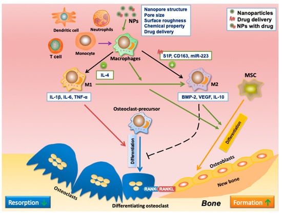
For bone regeneration, immunomodulation is required to generate an ideal environment for the subsequent osteogenesis, which can be achieved by NPs. As explained in
Section 3, macrophage populations are critical regulators of bone regeneration. The pro-inflammatory M1 phenotype of macrophages causes a rise in pro-inflammatory cytokines such as IL-1β, IL-6, and TNF-α, resulting in the inhibition of osteogenesis
[49][50][110,111] and promoting osteoclastogenesis
[51][112]. Alternatively, the anti-inflammatory M2 phenotype can reverse inflammation and secrete osteogenic cytokines, including BMP2 and VEGF, to encourage bone regeneration
[52][53][54][113,114,115]. Hence, targeting macrophages to induce their M2 polarization has been regarded as an efficient way to enhance bone regeneration, and nanomaterials are shown as effective agents for macrophage polarization (
Table 2). Some NPs can efficiently promote M2 polarization, such as gold, TiO
2, and cerium oxide (CeO
2) NPs
[55][56][57][116,117,118]. Moreover, the nanopore structure and pore size were discovered to affect the inflammatory response and release of pro-osteogenic factors of macrophages by influencing their spreading, cell shape, and adhesion
[58][59][119,120]. For instance, Chen et al. ascertained that macrophages grown on larger pore size NPs (100 and 200 nm) were highly anti-inflammatory, demonstrating a decrease in pro-inflammatory cytokine and expression of M1 phenotype surface-marker
[58][119]. One study found that silver NPs with different sizes and shapes showed different effects on bone metabolism and immunity, indicating that controlling the size and shape of nanomaterials can affect their osteoimmunomodulatory effects
[60][121]. NPs with rough surfaces also alter macrophage activation and cytokine release. Research indicated that titanium (Ti) with a smooth surface could induce M1 activation and inflammatory cytokines expression, including IL-1β, IL-6, and TNF-α. Meanwhile, Ti with a rough and hydrophilic surface enhances anti-inflammatory macrophage polarization and the secretion of cytokines such as IL-4 and IL-10
[61][122]. Another way to promote M2 polarization is to modify the composition of NPs surfaces by doping anti-inflammatory elements or decorating bioactive molecules
[62][63][64][123,124,125]. For example, hexapeptides Cys-Leu-Pro-Phe-Phe-Asp
[51][112], peptide arginine-glycine-aspartic acid (RGD)
[65][126], and IL-4
[66][127] have been successfully conjugated on gold NP surfaces to achieve successful anti-inflammation. Besides, CeO
2 NPs have been coated with hydroxyapatite to promote M2 polarization
[67][128]. A previous study indicated that surface modification of hydroxyapatite nanorods with chitosan reduced macrophage activation and enhanced osteoblast proliferation
[68][129]. Moreover, strontium (Sr) or copper (Cu)-decorated bioactive glass particles have been found to enhance M2 polarization and promote osteogenesis
[63][64][124,125]. Zhang et al. synthesized strontium-substituted sub-micron bioactive glasses (Sr-SBG), which have been found to advance the proliferation and osteogenic differentiation of mMSCs
[69][130].
As potential drug delivery systems, NPs have been widely used for bioactive molecule delivery, such as cytokines, growth factors, gene-modulators, and signaling pathway regulators, to stimulate the M1-to-M2 polarization. For instance, IL-4, a widely used anti-inflammatory cytokine, has been frequently adopted as cargo delivered by various nanocarriers to induce M2 polarization
[70][71][72][131,132,133]. One research study introduced an IL-4-incorporated nanofibrous heparin-modified gelatin microsphere, which can alleviate chronic inflammation due to diabetes and improve osteogenesis
[71][132]. Sphingosine-1-phosphate (S1P), as a sphingolipid growth factor, can also stimulate macrophages to polarize to the M2 phenotype
[73][134]. Das et al. synthesized nanofibers composed of polycaprolactone (PCL) and poly (D, L-lactide-co-glycolide) (PLGA) for an S1P synthetic analog delivery, which was found to induce macrophage differentiation to M2 phenotypes, facilitating osseous repair in an animal model of the mandibular bone defect
[81][135]. CD163 is an M2 phenotype marker affiliated with the scavenger receptor cysteine-rich (SRCR) family
[82][136]. One study encapsulated CD163 gene plasmid into polyethyleneimine NPs assembled with a mannose ligand for selectively targeting macrophages and inducing CD163 expression, and further transferring macrophages into their anti-inflammatory phenotype
[74][137]. Upregulation of miR-223 can drive the macrophage polarization toward the anti-inflammatory (M2) phenotype, whereas local-targeted delivery of miRNAs is still challenging due to the low stability of miRNA. To solve this problem, Saleh et al. developed an adhesive hydrogel with NPs loaded with miR-223 5p mimic to regulate macrophage polarization to M2 to promote tissue remodeling
[75][138]. Yin et al. loaded an anti-inflammatory drug, resolvin D1, into the gold nanocages (AuNC) coated with cell membranes from LPS-stimulated M1-like macrophages to facilitate M2 polarization. The overexpressed inflammatory cytokine receptors on the cell membrane can competitively bind to the pro-inflammatory cytokines with cell surface receptors, thereby impeding inflammatory responses
[76][139]. The results indicate that this nanosystem could efficiently inhibit inflammatory responses, stimulate an M2-like phenotype polarization, and promote bone regeneration in the femoral defect.
Despite the crucial role of M2 macrophages in promoting bone tissue regeneration, more and more studies have focused on the importance of M1 macrophages in osteoimmunomodulation. As mentioned, M1 macrophages dominate in the early stage of inflammation, enhancing the early commitment and recruitment of angiogenic and osteogenic precursors. In contrast, M2 macrophages function in the later stage of bone regeneration by facilitating osteocyte maturation and determining the microstructure of the newly formed bone tissue
[83][140]. Therefore, a highly orchestrated immune response comprising sequential activation of M1 and M2 macrophages is essential for subsequent bone healing
[84][141]. Thus, a sequential release of therapeutics from NPs to instruct the timely phenotypic switching of macrophages is deemed necessary. For example, as IFN-γ and IL-4 can induce M1 and M2 polarization, Spillar et al. designed a scaffold with a quick release of IFN-γ to increase the M1 phenotype, subsequently with a release of IL-4 to enhance the M2 phenotype. The sequential release feature was achieved by physically adsorbing IFN-γ onto the scaffolds, while loading IL-4 on the material via biotin-streptavidin binding
[77][142]. In another example, miRNA-155 is highly expressed in M1 and less in M2, while the delivery of miRNA-21 can promote macrophage polarization toward M2 phenotypes
[85][86][87][143,144,145]. Li et al. synthesized NPs through free radical polymerization carrying both miRNA-155 and miRNA-21 to induce macrophages first toward M1 sequentially and then M2 polarization, a new strategy for bone regeneration
[78][146]. Zinc (Zn) is an essential trace element in various immune responses. Zn’s scarcity and low concentration caused inflammation, while a proper concentration of Zn exhibited an anti-inflammatory effect
[88][89][147,148]. Therefore, one study fabricated microcrystalline bioactive glass scaffolds with different doses of ZnO to orchestrate the sequential M1-to-M2 macrophage polarization, taking advantage of varying amounts of Zn
2+ released from the material
[79][149]. Yang et al. incorporated IFN-γ and Sr-substituted nanohydroxyapatite (nano-SrHA) coatings to the surface of native small intestinal submucosa (SIS) membrane, which is widely applied in GBR to direct a sequential M1-M2 macrophage polarization. The nano-SrHA coatings were loaded on the SIS membrane using the sol-gel method, while the IFN-γ was physically deposited. As a result, the physically absorbed IFN-γ released in a burst manner to induce temporary M1 macrophage polarization, then a more sequential release of Sr irons to promote M2 polarization, which intensely improved the vascularization and bone regeneration
[80][150]. Bone marrow macrophages have various receptors on their surface that enable them to recognize molecules such as cytokines, chemokines, lipids, and glycans. NPs to ensure a drug delivery to target bone marrow macrophages can be achieved using strategies such as surface modification of NPs with components interacting with bone marrow macrophage receptors. However, NPs in circulation are removed by the mononuclear phagocyte system (MPS), including the spleen, liver, and Kupffer cells, affecting the NP-based targeted delivery on bone marrow macrophages. Therefore, combining the NPs with bone implants (via approaches such as surface coating, 3D printing, etc.) is suggested instead of systemic administration, which can facilitate the NPs to modulate the local bone healing immune environment and avoid particle clearance due to blood circulation and MPS.
Taken together (
Figure 1), osteoimmunology is a fascinating field focusing on the interconnected molecular pathways between the immune and skeletal systems. Among all the immune cells, macrophages play the most crucial role, secreting cytokines that determine the immune response and modulate the subsequent bone regeneration. Nanomaterials can assist in regulating immune responses by targeting macrophages and managing their polarization, bringing a new strategy for managing bone-related diseases
[90][151].
Figure 1. NPs as drug delivery systems to introduce functional osteoimmunomodulation to promote bone regeneration. Ideally, NPs should modulate the immune system to enable the formation of an ideal immune microenvironment for subsequent osteogenesis and bone regeneration. Macrophage polarization is essential in osteoimmunomodulation. The pro-inflammatory M1 phenotype of macrophages could secrete pro-inflammatory cytokines such as IL-1β, IL-6, and TNF-α to promote osteoclastogenesis. The anti-inflammatory M2 phenotype of macrophages could secrete osteogenic cytokines, including BMP2 and VEGF, to enhance bone regeneration. The timely M1-to-M2 phenotype switch is critical in bone regeneration, which can be induced by NP-based drug delivery. NPs can regulate macrophage polarization through different strategies, such as nanopore structure and size, surface roughness, chemical properties, and delivered drugs. NPs can inhibit M1 polarization, promote macrophage polarization to M2, or enhance M1 to M2 polarization, further promoting bone healing.

