Your browser does not fully support modern features. Please upgrade for a smoother experience.
Please note this is a comparison between Version 1 by Li Lin and Version 2 by Conner Chen.
DNA cytosine methylation is a principal epigenetic mechanism underlying transcription during development and aging. Growing evidence suggests that DNA methylation plays a critical role in brain function, including neurogenesis, neuronal differentiation, synaptogenesis, learning, and memory.
- DNA methylation
- transcriptional regulation
- neurogenesis
1. Introduction
The brain is the most important organ that serves as the center of the nervous system in mammals. The function of the brain is to control and coordinate a wide variety of actions and reactions, especially thought, memory, and emotion in primates. The development and aging of the brain are complex processes that depend on multiple layers of precise regulation. During aging, the brain shrinks and changes structurally and is predisposed to neurodegenerative diseases. Alzheimer’s disease (AD), Parkinson’s disease (PD), and Huntington’s disease (HD) are common neurodegenerative diseases that cause progressive loss of neuronal function and lead to cognitive impairment. Epigenetic mechanisms play essential roles in brain function by regulating gene expression [1][2][3][1,2,3]. As a critical epigenetic modification, DNA methylation affects transcriptional activity by recruiting or inhibiting the binding of transcription factors to DNA. Many brain disorders display altered gene expression, and accumulating evidence implicates dynamic DNA methylation in altered gene expression and pathological processes in the brain [4][5][4,5]. Dysregulation of the epigenome has been reported in neurodevelopmental and neurodegenerative diseases [6].
DNA methylation is a heritable epigenetic mechanism linked to gene expression and the regulation of biological processes [7]. In mammals, 5-methylcytosine (5mC) is formed by DNA methyltransferases (DNMTs) transferring a methyl group from S-adenosylmethionine (SAM) to cytosine at 5-position (Figure 1A). 5mC occurs mainly in the CpG dinucleotides of the mammalian genome. A total of 70–80% of CpGs are modified by 5mC in the human genome [8]. During development and cellular differentiation, the initial establishment of 5mC patterns, termed de novo methylation, relies predominantly on the activity of both DNMT3a and DNMT3b [9], whereas DNMT1 performs a maintenance function over the course of DNA replication to ensure the propagation of 5mC patterns [10].
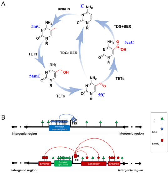
Figure 1. DNA methylation formation and regulation in gene expression. (A) The cycle of DNA methylation. 5mC is formed from C by the catalyzation of DNMTs; then, it can be hydroxylated to 5hmC and further oxidized to 5fC and 5CaC by TET enzymes. 5fC and 5caC are recognized by thymine DNA glycosylase (TDG), which triggers the base excision repair (BER) process, leading to the conversion to unmethylated cytosine. (B) Possible role of 5mC and 5hmC in gene expression. Hypermethylated 5mC at CpG islands of gene promoters generally inhibits gene transcription (blue line). 5hmC of gene enhancers and gene bodies up-regulates gene expression (red line).
5mC has been termed the fifth base of the human genome because it plays a key role in inhibiting transcriptional activities (Figure 1B) [11]. A typical situation is the X chromosome inactivation in placental mammals, where 5mC shuts down the transcription of most genes on one of the female’s X chromosomes to compensate for the different dosages of the X chromosome in males and females [12]. However, some studies have reported that several methylation sites are related to gene transcriptional activation [13][14][13,14], which may be due to the dynamic binding of transcription factors to certain methylated DNA. 5mC patterns are subject to orderly changes during mammalian development and aging. Neurogenesis proceeds from embryonic stages to the adult brain [15][16][15,16]. Dynamic DNA methylation indeed contributes to embryonic and adult neurogenesis, including stem cell maintenance and proliferation, neuronal differentiation and maturation, fate interpretation, and synaptogenesis. Gain or loss of 5mC impacts neuronal development and brain function [17][18][17,18]. Growing evidence supports Horvath’s epigenetic clock theory, which is based on 5mC patterns of specific sites across multiple tissues and provides an estimator for predicting chronological age [19]. During aging, an overall genome-wide decreased 5mC (global hypomethylation) has been reported, and regional hypermethylation within the CpG islands of specific gene promoters has been observed [20][21][20,21]. Abnormal patterns of 5mC commonly disrupt transcriptional regulation and lead to various diseases, such as cancer and neurodegenerative diseases.
Another family, named Ten-eleven translocation (TET) dioxygenases, is responsible for catalyzing the iterative oxidation of 5mC to 5-hydroxymethylcytosine (5hmC), 5-formylcytosine (5fC), and 5-carboxylcytosine (5caC) on DNA. The TET family in mammals contains Tet1, Tet2, and Tet3, which share a homology catalytic domain at their C terminal. 5fC and 5caC are then specifically recognized by thymine DNA glycosylase to trigger the base excision repair (BER) process, leading to their final conversion to unmethylated cytosine (Figure 1A) [22][23][22,23]. Originally, this process was thought to be just an active DNA demethylation with 5hmC, 5fC, and 5caC as transient intermediates. Proteome-wide profiling showed that these modifications may recruit different transcriptional regulators (as readers) to regulate gene expression (Figure 1B) [24][25][24,25]. This suggests that novel forms of DNA methylation provide potential regulatory mechanisms for genome functions. Due to the limitations of detection methods for 5fC and 5caC, most studies have focused on the biological functions of 5hmC.
Large differences in 5hmC levels in different tissues have been reported, ranging from 0.03% to 0.69% [26]. 5hmC is typically enriched in active genes, promoters, transcription factor-binding sites, and brain-specific enhancers but is absent in intergenic regions [27][28][27,28]. At present, a great number of studies have confirmed that 5hmC generally promotes gene expression. Therefore, 5hmC is considered to be a stable epigenetic marker and is thus called the sixth base of DNA [29][30][29,30]. In mammals, 5hmC is the most abundant in the central nervous system (CNS), where mature neurons appear to be the major contributors to 5hmC. 5hmC levels in CNS are approximately 10-fold higher than in embryonic stem cells (ESCs) and 3-fold higher than in peripheral tissues, especially those enriched in Purkinje neurons [26][27][31][26,27,31]. This suggests that high levels of 5hmC are essential for proper neurodevelopment and contribute to the neuropathology of neurodegenerative diseases [5][32][5,32].
In the pathogenesis of various neurodegenerative diseases, the silencing or activation of multiple genes has been characterized by hypomethylation or hypermethylation and abnormal regulation of enzymes related to DNA methylation formation or removal. As epigenetic modifications are invertible processes, this provides insights into therapeutic intervention by correcting the aberrant epigenetic status to achieve epigenetic balance. DNA methylation emerges as a promising target for the selection of therapeutic approaches. To date, DNMT inhibitors (DNMTi) are widely used in preclinical and clinical research, among which 5-Azacytidine (5-Aza-CR) and 5-aza-2′-deoxycitidine (5-Aza-CdR) have been approved by the Food and Drug Administration (FDA) in the United States [33][34][33,34]. Depending on the interaction of epigenetic modifications, the new therapy has also been tried in clinical applications by combining DNMTi and HDACi (histone deacetylases inhibitors) [35][36][37][35,36,37]. Further understanding of the fundamental mechanisms of epigenetic regulation in neuronal development and diseases could promote therapeutic applications.