Your browser does not fully support modern features. Please upgrade for a smoother experience.
Please note this is a comparison between Version 2 by Jason Zhu and Version 3 by Jason Zhu.
Carbon nanotubes have been extensively used as electrochemical sensing composites due to their interesting chemical, electronic, and mechanical properties giving rise to increased performance. Due to these materials' unknown long-term ecological fate, care must be given to make their use tractable.
- carbon nanotubes
- carbon dots
- graphene
- biosensors
- electrochemistry
1. Carbon Nanotube Composites
Carbon nanotubes (CNTs) are divided into two fundamental types: single-walled carbon nanotubes (SWCNTs) and multi-walled carbon nanotubes (MWCNTs) [1].
2. Functionalizing CNTs
Commercialized CNTs with heavily entangled bundles exhibit inherent difficulties in dispersion [2]. To avoid such limitations, various methods have been developed to alter the surface morphology of CNTs, which are important for enhancing the deposition of metal nanoparticles in the fabrication of composite by providing tethering points for their attachment. Such functionalization matters in the context of chemical sensing because it helps to attach metal nanoparticles. The agglomeration of tethered nanoparticles can be minimized and thereby increase the surface area of the composite for greater sensitivity. Furthermore, functionalizing the CNT sidewalls is essential to prevent the agglomeration of nanoparticles (NPs) as O-containing functional groups provide tethering points to improve their dispersion.
Covalent bonding between the layers and small molecules can be carried out using covalent functionalization. General modification of CNTs occurs with small molecules as a pre-modification step. Covalent modification of CNTs with small molecules is required to increase dispersibility to functionalize them with the aim of fabricating biosensing electrocatalysts. The modification affects the compatibility of the matrix and fillers are affected [3][4][5][6][7][8][9][10]. Carbon paste (CPE) and carbon fiber (CFE) electrodes are made in this fashion. To synthesize various O-containing groups on the surface of CNTs, carboxyl, hydroxyl, and epoxy groups are created on the CNTs using oxidation reactions with aqueous H2O2 and concentrated sulfuric acid [11][12][13]. The degree of oxidation on CNTs is controllable using the ratio of sp3 to sp2 hybridized carbon atoms with the increase in the ratio of sp3 to sp2 carbon denoting greater oxidation on the surface of CNTs. Methods include condensation via amidation [14], esterification [15], acylation [16], and silanization reactions with small molecules [17]. The function of such moieties is to serve as tethering points for electrocatalytically active NPs. Oxidized CNTs can also undergo condensation with oxygen-containing functional groups using acid chlorides as intermediates [18] or through carbodiimide-activated coupling [19]. For electrochemical sensing applications, the formation of these functional groups is important for enhancing the surface area of nanocomposites, resulting in enhancing the sensitivity of biological analytes.
One example of attaching NPs to CNT sidewalls is by reducing silver ions to silver NPs; the NP-decorated organic functionalized MWCNTs are used to detect glucose electrochemically [20]. In this covalent functionalization method, MWCNTs were well dispersed in tetrahydrofuran (THF) and SOCl2 and treated with diethylenetriamine via an acylation reaction. MWCNT-CO-NH-cyanuric-NH2 and MWCNT-CO-NH-cyanuric as CNT composites were generated by acylation between CNT-CO-NH2 with cyanuric chloride and diethylenetriamine, respectively.
2.1. Strategies to Modify the Working Electrode Surface
To increase the density of O-containing functional groups to the CNT sidewalls, sonicating the CNTs in the presence of nitric acid and sulfuric acid is commonly used. MWCNTs are functionalized to attach metal nanoparticles, thereby increasing the electroactive surface area, leading to greater electrochemical sensitivity [21]. The cavitation process of sonication is used in this manner to increase the density of -COOH and –CO- moieties on the MWCNT surface. As a result, finely dispersed NPs are attached to the MWCNT sidewalls. Sonication time can be varied to control the surface density of moieties and thereby achieve greater electrocatalytic activity [22]. Pt nanoparticles have successfully been attached to functionalized MWCNTs using this sonication procedure [23]. This sonication approach has been used to attach zinc oxide (ZnO) NPs to the COOH-functionalized MWCNT sidewalls, useful for uric-acid sensing [24]. Sonication was also used to combine CoO NPs with MWCNTs for assaying dopamine using CV [25].
Electrostatic attraction is another important strategy for attaching two different components based on differences in surface charge. The point of zero charge (PZC) is the pH at which the solid surface is electrically neutral under aqueous solution conditions depending on the ability of hydroxylated surfaces to become protonated or deprotonated on the solid-aqueous solution surface, according to electrical double layer theory [26][27]. The PZC of CNTs can be controlled by attaching electron-donating or withdrawing moieties to the sidewalls to deposit well-dispersed metal nanoparticles. Having components with different PZC values requires attachment at an intermediate pH value. Under these conditions, entities with a PZC below the solution pH will adopt a negative charge. Similarly, entities with a PZC above the solution pH will adopt a positive charge. These opposite charge adoptions of the two components serve as the driving force for attachment, forming the composite.
CNT surfaces can adopt a wide range of PZCs existing through chemical functionalization [28]. The PZC of ZnO/MWCNTs and Prussian Blue (PB) were experimentally determined to be 7.3 and 6.0, as shown in Scheme 1, applying a procedure developed by Park and Regalbuto [27]. When such materials [ZnO/MWCNTs (PZC = 7.3) and PB (PZC = 6.0)] were stirred in PBS with pH 6.6, an intermediate pH condition that is between the PZCs of these materials, PB adopted an overall negative charge while ZnO/MWCNTs adopted a positive surface charge as shown in the scheme in Scheme 1 (right-hand drawing) [29]. In this process, the attachment of PB on the surface of ZnO/MWCNTs via Coulombic attraction was successfully accomplished. This action resulted in enhanced electro-composite sensitivity for the detection of H2O2, a biomarker for oxidative stress. Gatabi et al. also applied differing PZC values to attach maghemite NPs onto the surface of ox-MWCNTs (oxidized MWCNTs). The PZC of maghemite is 8 while that of ox-MWCNTs is 2.66. Decorating the MWCNTs with maghemite NPs was achieved in a slightly acidic medium [30]. Maghemite-MWCNT electrochemical composites are useful in assaying resorcinol, a widespread industrial waste product that is also an endocrine disruptor [31]. Selectivity depends not only on the specific potential obtained from CV, but also on the Coulombic interaction, i.e., either attractive or repulsive forces between the pKa value of the analytes and the PZC of the composite. Therefore, the principle of selectivity via altering PZC is extended to the more complex composites. Deb et al. used differences in the PZC of a Pt/MWCNT composite with those of ascorbic acid, acetaminophen, and H2O2 to selectively detect them using CA[32].
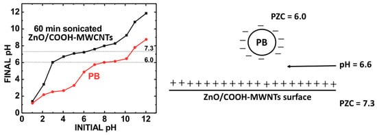
Scheme 1. The determination of PZC of ZnO/MWCNTs and PB. Right: attaching PB with the composite, ZnO/MWCNTs using PZC (left). Reproduced with permission from ref. [29]. Copyright 2019, American Chemical Society.
A final design aspect of the CNT electrode composites is the use of capping agents, which are needed for the working electrode surface to prevent the composites from dislodging from the working electrode. Nafion and Chitosan are commonly used binders to make the composite stable on the surface of glassy carbon electrodes [29][33]. Nafion is a common name for sulfonated tetrafluoroethylene-based fluoropolymer-copolymer. As a result of incorporating trifluorovinyl ether groups terminated with sulfonate onto a tetrafluoroethylene backbone, Nafion displays unique ionic properties. Nafion is a highly conductive material, and it prevents chemical alterations on the GCE surface. In fact, the sulfonate groups of Nafion prevent chemical attack via electrostatic repulsion and hydration effect [34][35]. This additive is required to avoid electro-composites from dislodging the GCE surface.
Nafion, with a 1–5 wt% solution concentration, is used to encapsulate the composite onto the working electrode surface. Similarly, chitosan is a natural polysaccharide synthesized from shrimp shells and characterized by abundant -OH and -NH2 groups. Chitosan and its composites have been widely used in drug and gene delivery, tissue engineering, and biosensing because of their high biocompatibility, biodegradability, hydrophilicity, mechanical strengths, antibacterial properties, and good processability in films. Chitosan enhances both the stability and lifetime of the sensor as it causes dispersion of the biological recognition element in a biomimetic environment [36].
2.2. Biosensing Applications of CNT Composites
Noble Metal-Based Composites
Docetaxel (Dox) is commonly used as an anticancer drug for treating lung, breast, prostate, ovarian, stomach, neck, and head cancer and is registered as the most effective and safe medicine in the world according to the World Health Organization [37][38]. However, monitoring exact levels of Dox in real samples (human urine and human serum) is required due to its various side effects. Najari et al. reported a selective and sensitive method for quantifying Dox at the Au-MWCNTs/GCE surface using CV and differential pulse anodic stripping voltammetry (DPASV) [38]. The introduction of MWCNTs on the GCE surface enhances electric conductivity. In addition, decorating MWCNTs with Au nanoparticles helps to improve the conductivity of MWCNTs-modified electrodes and signal response. This sensor can detect Dox in the range of 0.3–3.3 µmol∙L−1 with the LOD of 90 nmol∙L−1. Such a sensor is selective against Na+, K+, Cl−, Mg2+, Fe3+, CO2−, NO3− ions along with glucose, sucrose, fructose, lactose, ascorbic acid, dopamine, thiourea, and alanine on the DPASV measurements. Only oxidation peaks were observed under the analysis of Dox at Au-MWCNTs/GCE surfaces. The number of electrons shifted during the electrooxidation of Dox on the sensor’s surface becomes equal to the number of protons. The discharge of two electrons and protons was seen under this mechanism. Oxidation current response is proportional to the concentration of the analyte.
A nanocomposite of Pt/Ni(OH)2/MWCNT was synthesized using a simple two-step nitrite sensing method. This sensor showed enhanced electrocatalytic activity and excellent reliability. The nanocomposite was formed from synergistic effects between Pt NPs and Ni(OH)2/MWCNTs composites. In this composite, Ni(OH)2/MWCNTs offer a large surface area which helps to load the Pt NPs and improve the electronic conductivity. This noble nanomaterial-based nitrite sensor exhibits a low detection limit, good reproducibility, and excellent selectivity to quantify nitrite. This analyte is generally found in foods. Monitoring the concentration of nitrite is important to prevent food poisoning. An excess of it leads to irreversible hemoglobin oxidation in the human body, decreasing the blood oxygen-transporting capacity. In addition, nitrite reacts with amines to become carcinogenic nitrosamines, leading to conditions such as hypertension and gastrointestinal cancer [39].
Delerue-Matos et al. studied screen-printed carbon electrodes (SPCEs)-MWCNTs/Au NPs and SPCE-Au NPs to improve the electrochemical analysis of the extracellular domain of the human epidermal growth factor receptor 2 (HER2-ECD) using linear sweep voltammetry [40]. In this analysis, the combination of MWCNT with Au NPs had the highest sensitivity. The antibody concentrations were optimized and applied to develop biosensing strategies on SPCE-AuNP and SPCE-MWCNT/Au NP with LODs of 8.5 ng/mL and 0.16 ng/mL, respectively, well below the cut off value of 15 ng/mL for the breast cancer biomarker. Also, the linear dynamic range of this sensor was reported between 7.5 ng/mL to 50 ng/mL.
Cheng et al. described that MWCNTs/poly(3,4-ethylenedioxythiophene(PEDT))-gold nanocomposites were readily fabricated using a one-pot method [41]. In this process, Au-nanoparticles were synthesized by reducing hydrogen tetrachloroaurate with a 3,4-ethylenedioxythiophene (EDT) monomer, which was simultaneously oxidized and polymerized. The modified sensor prepared from this composite exhibits superior electrochemical sensing performance for luteolin detection using EIS, CV, and SWV. This fabricated luteolin sensor shows good selectivity, stability, and reproducibility. The outcomes indicate that the excellent electrochemical performance of the electrodes with MWCNTs/PEDT-Au nanocomposites are assigned to the synergistic catalytic effect of the high specific area of MWCNTs and catalytically active sites of PEDT-Au. This sensor is used to quantify luteolin (3,4,5,7-tetrahydroxy-flavone), a natural flavonoid compound. Luteolin is abundant in fruits, seed shells, and vegetables, and it has been widely used in clinical applications for the treatments of some serious coronary heart diseases. Some toxic side effects are observed at high doses greater than 20 µM concentrations.
Carboxylic acid-functionalized carbon-allotropic nanomaterials (GO-COOH/MWCNTs-COOH) tethered to gold nanoparticles (Au NPs) have been fabricated for biosensing of 8-hydroxy-2′-deoxyguanine (8-OHdG). The composite sensing materials consist of GO-COOH, MWCNTs-COOH, and polyethyleneimine (PEI). In this composite, Au NPs were synthesized by self-assembly of negatively charged Au NPs onto positively charged PEI-wrapped GO-COOH and MWCNTs-COOH via electrostatic interactions. This composite was generated by simple electrostatic adsorption under sonication and centrifugation. This sensor shows high electrochemical performance for the oxidation of 8-OHdG, a biomarker of oxidative DNA damage. The analysis of this molecule is important for the diagnosis and monitoring of various diseases related to oxidative stress, using differential pulse voltammetry (DPV) [42]. This composite is amenable to urinalysis as it avoids signal interference from uric acid with the assistance of uricase. UA is oxidized to electrochemically inactive allantoin by uricase. The addition of uricase eliminates the interference of uric acid for the quantification of 8-OHdG. The UA peak diminished with the addition of 50 µg∙mL uricase, enhancing the sensor’s selectivity for 8-OHdG. The results of analytical detection of 8-OHdG in human urine samples, trace content, and complex matrices were promising, with a very high recovery percentage (at least 99.43%).
The selectivity of analytes depends on the specific voltage in which the voltages for reduction and oxidation for various analytes are well separated from one another. The measured PZC of the Pt-DEN-PANI-CNT composite was observed to be 6.8 experimentally [32]. Under pH 7.0 conditions, the surface charge of the Pt-DEN-PANI-CNT electrode adopts a negative charge due to a higher degree of surface hydroxylation than protonation. The composite is Coulombically attracted to H2O2 and APAP since their isoelectric points (denoted by their pKa values) are at 11.75 and 9.38, respectively. Such analytes exist in their protonated forms at pH 7.0. On the other hand, ascorbic acid (AA) has an isoelectric point of 4.2 and therefore exists in a deprotonated state at pH 7.0. The deprotonated AA is negatively charged owing to 4 hydroxyl groups within its molecular makeup. AA would be repelled from the Pt-DEN-PANI-CNT surface. Coulombic attraction between composite and APAP and H2O2 served as the driving force for the electrocatalyst’s selectivity to these analytes not present with AA.
The strategy behind this electro-composite fabrication differs in that for SWCNTs. The diameter is too small to incorporate tethered moieties for the much larger electroactive NPs; therefore, another approach is used. Instead, N atoms are used as tethering points to attach dendrimers, which encapsulate the electroactive NPs. The synthesis of nanocomposite is summarized in Scheme 2. In step 1, Pt nanoparticles were encapsulated by adding K2PtCl4 and PAMAM dendrimers in water in the presence of NaBH4 and stirred for 48 h. In step 2, polyaniline was attached to the carbon nanotube sidewalls in the presence of HCl and (NH4)2S2O8 to increase its conductivity. In the final step, poly(sodium-4-styrene sulfonate) was dissolved in a NaCl solution. PANI-CNTs were added to this solution under constant stirring. Then, a Pt-encapsulated dendrimer solution was added. Further stirring was continued. Then, it was centrifuged and washed with deionized water to obtain Pt-DEN-PANI-CNT nanocomposite.
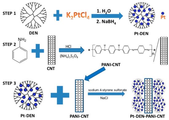
Scheme 2. Fabrication steps to produce the Pt-DEN-PANI-CNT composite. Reproduced with permission from ref. [32]. Copyright 2016, Springer Nature.
Transition Metal Nanoparticle CNT Composites
Dopamine (DA) is a critical neurotransmitter associated with many neurological diseases. Its sensitive and selective detection is important for the early diagnosis of diseases related to abnormal levels of DA [43]. Molybdenum nanoparticles self-supported functionalized multi-walled carbon nanotubes (MoNPs/f-MWCNTs) based core-shell hybrid nanomaterial with an average diameter of 40–45 nm was fabricated for electrochemical DA detection. In this case, O-containing groups from the acid treatment of the MWCNTs served to tether Mo NPs. This composite has a large surface area and numerous electroactive sites. This composite detects DA as low as 0.01 µM quantitatively with its detection limit of 1.20 nM under CV.
Simultaneous detection using electrochemical sensors is emerging as a powerful assay for biomolecular analysis. The rapid and sensitive quantification of six biomolecules offers benefits to pathological research, clinical diagnosis, and pharmaceutical quality control. UA, Xanthine (XA), theophylline (TP), and theobromine (TB) are products emanating from purine metabolism. A lack of AA enhances the risk of scurvy. DA is related to Parkinson’s disease and attention-deficit hyperactivity disorder (ADHD). UA is reported to be a sign of hyperuricemia known to progress into gout. TB, TP, and XA are produced from a Xanthine-based nucleoside from various metabolic pathways. UA is the final oxidized product of the purine catabolism pathway. Since these biomolecules generally exist in urine samples, simultaneous selective detection of these molecules is necessary. Patel et al. reported on the synthesis of a novel nanocomposite of titanium dioxide nanorods with multi-walled carbon nanotubes (TiO2 NRs-MWCNTs) using a solvothermal method [44]. The TiO2 NRs-MWCNTs/GCE as a working electrode detected UA, XA, TP, and TB selectively in the presence of AA and DA. Simultaneous quantification of these six molecules was studied using DPV in a wide potential window ranging from −0.3 to 1.6 V vs. Ag/AgCl at pH 4.0. This observed simultaneous well-separated anodic peaks at 0.13, 0.35, 0.50, 0.85, 1.10, and 1.28 V for AA, DA, UA, XA, TP, and TB, respectively, using TiO2 NRs-MWCNTs/GCE. All six calibration curves of these biomolecules have two segments of a linear relationship, showing a slope for low concentrations and a slope for higher concentrations. A possible reason for a large redox-active area was available on the electrode surface at lower concentrations of the molecules. On the other hand, accessible redox-active areas rapidly saturated at greater concentrations of the biomolecules, resulting in decreased sensitivity in the slope of the second linear segments in their calibration curves. Therefore, concentration breakpoints were seen for all of these analytes. This sensor showed a linear range of AA from 1.5 to 51.0 µM and 51.0 to 191.0 µM with an LOD of 0.51 µM; DA from 0.45 to 30.0 µM and 30.0 to 147.0 µM with an LOD of 0.06 µM; UA from 0.40 to 61.0 µM and 61.0 to 537.0 µM with an LOD of 0.05 µM; XA from 0.5 to 97.0 µM and 97.0 to 586.0 µM with an LOD of 0.09 µM; TP from 1.0 to 203.0 µM and 203.0 to 891.0 µM with an LOD of 0.56 µM; TB from 1.5 to 368.0 µM and 368.0 to 1653.0 µM with an LOD of 0.75 µM. Selective quantification of these molecules at oxidation peak currents does not show any interference with other biomolecules.
A CuCo2O4 nanoparticle modified with nitrogen-doped carbon nanotubes (CuCo2O4/N-CNTs) exhibits a high specific surface area (72.80 m2/g) and good electrical conductivity. N atoms served as the tethering points for the CuCo2O4 NPs. The CuCo2O4/N-CNTs loaded molecularly imprinted polymer (MIP) modified GCE was fabricated for fast and ultrasensitive detection of metronidazole (MNZ) using CV and DPV. Metronidazole is mainly used to treat and prevent amebic disease and echinococcosis caused by anaerobic bacteria. It belongs to the class of 5-nitroimidazole antibiotics. This sensor can selectively detect MNZ in presence of p-nitrophenol, 1-methylimidazole, L-phenylalanine, glycine, carbamide, dimetridazole, and 2-methyl-5-nitroimidazole in biological samples. These interfering analytes were chosen because they have the same chemical structures and sizes as the analyte [45]. In addition, various inorganic ions such as Na+, K+, Ca2+, Cl−, and SO42− were tested with this sensor to prove no significant interferences for the detection of MNZ. The experimental method is summarized in Scheme 3. Oxidation of MWCNTs takes place, and a one-step hydrothermal method is used to synthesize CuCo2O2/N-CNTs. This composite is used to modify the electrode to increase electrochemical signals. Then, the template molecule MNZ with the electrochemically active functional monomer aniline is electropolymerized on the surface of the CuCo2O4/N-CNT electrode.
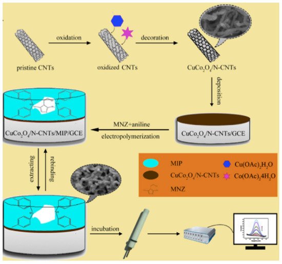
Scheme 3. Schematic diagram for preparation of CuCo2O4/N-CNTs/MIP/GCE and electrochemical detection of metronidazole. Reproduced with permission from ref. [45]. Copyright 2019, Elsevier.
Here, the specific recognition sites for MNZ were designed. With the removal of template molecules, cavities matching the MNZ spatial structure and binding site are achieved. As a result, MNZ is re-adsorbed by stacking and hydrogen bonding interaction. Electrochemical quantification of MNZ was determined by CV and DPV using this sensor.
GCEs modified by synthesized Fe3O4-MWCNTs-Ni NPs have been used to detect glucose with its low detection limit of 6.7 µM. Iron (II, III) oxide (Fe3O4) nanoparticles were in situ loaded on the surface of carboxylated multi-walled carbon nanotubes using chemical co-precipitation procedure. Prior to decorating Fe3O4-MWCNT through ultrasonication, the nickel nanoparticles were synthesized by reducing nickel chloride. This fabricated sensor was used to quantify glucose in honey and energy drinks [46].
Glycans that exist on glycoproteins play important roles in many biological processes such as cell growth and differentiation, cell-cell interactions, and protein folding. A change in glycan expression levels has often been associated with cancer development, progression, and metastasis. Glycans on cell surfaces are considered to be therapeutic targets or clinical biomarkers for the diagnosis of many harmful diseases. A thionine (Th)-bridged multi-walled carbon nanotube/gold nanoparticle (MWCNT/Th/Au NP) composite was fabricated by binding Au NPs to the surface of Th-coated MWCNTs, in which thionine acted as a linker to enable the negatively charged Au NPs to bind to the anionic MWCNT surface. Synthesized MWCNTs/Th/Au NP as a mediator/nanomaterial composite was used as an electrochemical biosensor for glycan assays on living cancer cells. Cell surface glycans, especially mannose, are closely associated with biological processes such as tumor growth and metastasis. Therefore, the study of mannose expression was significant for analyzing its role in cancer development. This sensor can detect mannose in the range of (0.015–0.045) µM. The biosensor was used to quantify 0.03328 µM mannose expression in QGY-7701 cells (at a concentration of (6 × 10² cells per mL) [47]. In this case, the peak current obtained from mannose containing living cancer cells was concurrent with that obtained from standard mannose quantification. The sensor is selective towards mannose in the presence of other biological molecules such as amino acids and proteins. Table 1 shows a representative sampling of bioanalytes detectable using carbon nanotube-based composites. An additional array of CNT-based sensing composites are listed [48][49][50][51][52][53][54][55][56][57][58][59][60].
Table 1. Detectable bioanalytes using carbon nanotube composites.
| Sensor | Technique | Analyte | LOD | Reference |
|---|---|---|---|---|
| Pt/Ni(OH)2/MWCNT/GCE | CV | Nitrite | 0.13 mM | [39] |
| SPCE-MWCNT/AuNPs | LSV | HER2-ECD | 0.16 ng∙mL–1 | [40] |
| MWCNTs/PEDT-Au | EIS, CV, SWV | Luteolin | 0.2 nM | [41] |
| GO-COOH/MWCNT-COOH/PEI/Au | CV | Urinary 8-OHdG |
7.06 nM | [42] |
| Mo NP/f-MWCNTs | CV | Dopamine | 1.26 nM | [43] |
| CuCo2O4/N-MWCNT/GCE | CV | Metronidazole | 0.48 nM | [45] |
| Fe3O4-MWCNT-Ni NPs | CV, CA | Glucose | 6.7 µM | [46] |
| MWCNTs/Th/Au NP | DPV | Mannose | 0.015 µM | [47] |
| GlcDH/PNb-SWNT/GCE | CA | Glucose | 5.0 µM | [48] |
| AlcDH/PNb-SWNT/GCE | CA | Ethanol | 50 µM | [49] |
| DSDH/PMG/MWCNT/GCE | CA | Sorbitol | 100µM | [50] |
| PTBO/MWCNT/GCE | CA | NADH | 0.5 µM | [51] |
| MWCNT/PMB/GCE | CA | H2O2 | 20.7 µM | [52] |
| PmalG/MWCNT/GCE | DPV | AA | 0.23 µM | [53] |
| SWCNT/PTBO/GCE | CA | Nitrite | 0.37 µM | [54] |
| PBG/MWCNT/CFE | DPV | DA | 1.60 µM | [55] |
| PMB/MWCNT/GCE | CV | EP | 69.6 µM | [56] |
| PmalG/MWCNT/GCE | DPV | UA | 0.12 µM | [53] |
| EBNBH modified CNT-CPE | DPV | UA | 15 µM | [57] |
| SWCNT mat grown on Si | CV | DA | 1 µM | [58] |
| CNT paste with 2-PHC | SWV | Epinephrine | 9 nM | [59] |
| SWCNT-inlaying ultrathin CPE | DPV | Xanthine | 0.1 µM | [60] |
References
- Iijima, S. Carbon nanotubes: Past, present and future. Phys. B Condens. Matter 2002, 323, 1–5.
- Deb, A.K.; Chusuei, C.C. Aqueous Solution Surface Chemistry of Carbon Nanotubes. In Physical and Chemical Properties of Carbon Nanotubes; Suzuki, S., Ed.; InTech: Rijeka, Croatia, 2013; pp. 263–283.
- Sahoo, N.G.; Cheng, H.K.F.; Li, L.; Chan, S.H.; Judeh, Z.; Zhao, J. Specific functionalization of carbon nanotubes for advanced polymer nanocomposites. Adv. Funct. Mater. 2009, 19, 3962–3971.
- Moore, E.; Wang, P.Y.; Vogt, A.P.; Gibson, C.T.; Haridas, V.; Voelcker, N.H. Clicking dendritic peptides onto single walled carbon nanotubes. RSC Adv. 2012, 2, 1289–1291.
- Liao, W. Effects of multiwalled carbon nanotubes functionalization on the morphology of mechanical and thermal properties of carbon fiber/vinyl ester composites. ACS Appl. Mater. 2013, 5, 3975–3982.
- Yoonessi, M.; Lebron-Colon, M.; Scheiman, D.; Meador, M.A. Carbon nanotube epoxy nanocomposites: The effects of interfacial modifcations on the dynamic mechanical properties of the nanocomposites. ACS Appl. Mater. Interfaces 2014, 6, 16621–16630.
- Mammeri, F.; Teyssandier, J.; Darche-Dugares, C.; Debacker, S.; Le Bourhis, E.; Chehimi, M.M. Carbon nanotube-poly(methyl methacrylate) hybrid films: Preparation using diazonium salt chemistry and mechanical properties. J. Colloid Interface Sci. 2014, 433, 115–122.
- Zhang, W.; Zhou, Q.; Li, Q.; Chen, G.X. Controlled dielectric properties of polymer composites from coating multiwalled carbon nanotubes with octa-acrylate silesquioxane through Diels-Alder cycloaddition and atom transfer radical polymerization. Ind. Eng. Chem. Res. 2014, 53, 6699–6707.
- Najafi-Shoa, S.; Roghani-Mamaqani, H.; Salami-Kalajahi, M.; Azimi, R. Incorporation of epoxy resin and carbon nanotube into silica/siloxane network for improving thermal properties. J. Mater. Sci. 2016, 51, 9057–9073.
- Abdollahi, A.; Roghani-Mamaqani, H.; Salami-Kalajah, M.; Mousavi, A.; Razavi, B.; Shahi, S. Preparation of organic-inorganic hybrid nanocomposites from chemically modified epoxy and novolac resins and silica-attached carbon nanotubes by sol-gel process: Investigation of thermal degradation and stability. Prog. Org. Coat. 2018, 117, 154–165.
- Stobinski, L. Multiwall carbon nanotubes purification and oxidation by nitric acid studied by the FTIR and electron spectroscopy methods. J. Alloys Compd. 2010, 501, 77–84.
- Shao, Y.; Yin, G.; Zhang, J.; Gao, Y. Comparative investigation of the resistance to electrochemical oxidation of carbon black and carbon nanotubes in aqueous sulfuric acid solution. Electrochim. Acta 2006, 51, 5853–5857.
- Jiang, L.C.; Zhang, W.D. Electrodeposition of TiO2 nanoparticles on multiwalled carbon nanotube arrays for hydrogen peroxide sensing. Electroanalysis 2009, 21, 988–993.
- Hu, X.; Zou, C.; Zou, X. The formation of supramolecular carbon fiber via amidation reaction on the surface of amino single walled carbon nanotubes for selective adsorption organic pollutants. J. Colloid Interf. Sci. 2019, 54, 112–122.
- D’Arlas, B.F.; Goyanes, S.; Rubiolo, G.H.; Mondragon, I.; Corcuera, M.A.; Eceiza, A. Surface modification of multiwalled carbon nanotubes via esterification using a biodegradable polyol. J. Nanosci. Nanotechnol. 2009, 9, 6064–6071.
- Iannazzo, D.; Piperno, A.; Ferlazzo, A.; Pistone, A.; Milone, C.; Lanza, M.; Cimino, F.; Speciale, A.; Trombetta, D.; Saija, A.; et al. Functionalization of multiwalled carbon nanotubes with coumarin derivatives and their biological evaluation. Org. Biomol. Chem. 2012, 10, 1025–1031.
- Zhang, C.; Li, F.; Huang, S.; Li, M.; Guo, T.; Mo, C.; Pang, X.; Chen, L.; Li, X. In-situ facile preparation of highly efficient Copper/Nickel bimetallic nanocatalyst on chemically grafted carbon nanotubes for non-enzymatic sensing of glucose. J. Colloid Interface Sci. 2019, 557, 825–836.
- Peng, H.; Alemany, L.B.; Margrave, J.L.; Khabashesku, V.N. Sidewall carboxylic acid functionalization of single-walled carbon nanotubes. J. Am. Chem. Soc. 2003, 125, 15174–15182.
- Gu, Y.J.; Cheng, J.; Jin, J.; Cheng, S.H.; Wong, W.T. Development and evaluation of pH-responsive single walled carbon nanotube-doxorubicin complexes in cancer cells. Int. J. Nanomed. 2011, 6, 2889–2898.
- Ensafi, A.A.; Zandi-Atashbar, N.; Rezaei, B.; Ghiaci, M.; Chermahini, M.E.; Moshiri, P. Non-enzymatic glucose electrochemical sensor based on silver nanoparticle dcorated organic functionalized multiwall carbon nanotubes. RSC Adv. 2016, 6, 60926–60932.
- Xing, Y.; Li, L.; Chusuei, C.C.; Hull, R.V. Sonochemical oxidation of multiwalled carbon nanotubes. Langmuir 2005, 21, 4185–4190.
- Chusuei, C.C.; Wayu, M. Characterizing Functionalized Carbon Nanotubes for Improved Fabrication in Aqueous Solution Environments. In Electronic Properties of Carbon Nanotubes/Book 5; Marulanda, J.M., Ed.; IntechOpen: Rijeka, Croatia, 2011; pp. 55–66.
- Hull, R.V.; Li, L.; Xing, Y.; Chusuei, C.C. Pt nanoparticle binding on functionalized multiwalled carbon nanotubes. Chem. Mater. 2006, 18, 1780–1788.
- Das, S.C.; Pandey, R.R.; Devkota, T.; Chusuei, C.C. Raman spectroscopy as an assay to disentangle zinc oxide carbon nanotube composites for optimized uric acid detection. Chemosensors 2018, 6, 65.
- Kader, M.S.; Chusuei, C.C. A cobalt (II) oxide carbon nanotube composite to assay dopamine. Chemosensors 2020, 8, 22.
- Zhao, X.; Aoki, K.J.; Chen, J.; Nishiumi, T. Examination of the Gouy-Chapman theory for double layer capacitance in deionized latex suspensions. RSC Adv. 2014, 4, 63171–63181.
- Park, J.; Regalbuto, J.R. A simple and accurate determination of oxide PZC and the strong buffering effect of oxide surfaces at incipient wetness. Colloid Interface Sci. 1995, 175, 239–252.
- McPhail, M.R.; Sells, J.A.; He, Z.; Chusuei, C.C. Charging Nanowalls: Adjusting the Carbon Nanotube Isoelectric Point via Surface Chemical Functionalization. J. Phys. Chem. C 2009, 113, 14102–14109.
- Pandey, R.R.; Guo, Y.; Gao, Y.; Chusuei, C.C. A Prussian Blue ZnO carbon nanotube composite for chronoamperometrically assaying H2O2 in BT20 and 4T1 breast cancer cells. Anal. Chem. 2019, 91, 10573–10581.
- Gatabi, M.P.; Moghaddam, H.M.; Ghorbani, M. Point of zero charge of maghemite decorated multiwalled carbon nanotubes fabricated by chemical precipitation method. J. Mol. Liq. 2016, 216, 117–125.
- Manasa, G.; Bhakta, A.K.; Mekhalif, Z.; Mascarenhas, R.J. Voltammetric study and rapid quantification of rescorcinol in hair dye and biological samples using ultrasensitive maghemite/MWCNT modified cargon paste electrode. Electroanalysis 2019, 31, 1363–1372.
- Deb, A.K.; Das, S.C.; Saha, A.; Wayu, M.B.; Marksberry, M.H.; Baltz, R.J.; Chusuei, C.C. Ascorbic acid, acetaminophen, and hydrogen peroxide detection using a dendrimer-encapsulated Pt nanoparticle carbon nanotube composite. J. Appl. Electrochem. 2016, 46, 289–298.
- Wang, X.; Wu, M.; Tang, W.; Zhu, Y.; Wang, L.; Wang, Q.; He, P. Simulataneous electrochemical determination of ascorbic acid, dopamine, and uric acid using a Palladium nanoparticles/graphene/chitosan modified electrode. J. Electroanal. Chem. 2013, 695, 10–15.
- Heitner-Wirguin, C. Recent advances in perfluorinated ionomer membranes: Structures, properties, and applications. J. Mem. Sci. 1996, 120, 1–33.
- Mauritz, K.A.; Moore, R.B. State of understanding of Nafion. Chem. Rev. 2004, 104, 4535–4586.
- Poletti, F.; Favarette, L.; Kovtun, A.; Treossi, E.; Corticelli, F.; Gazzano, M.; Palermo, V.; Zanardi, C.; Melucci, M. Electrochemical sensing of glucose by chitosan modified graphene oxide. J. Phys. Mater. 2020, 3, 14011.
- Kuppens, I.; Van Maanen, M.; Rosing, H.; Schellens, J.; Beijen, J. Quantitative analysis of docetaxel in human plasma using liquid chromatography coupled with tandem mass spectrometry. Biomed. Chromatogr. 2005, 19, 355–361.
- Najari, S.; Bagheri, H.; Monsef-Khoshhesab, Z.; Hajian, A.; Afkhami, A. Electrochemical sensor based on gold nanoparticle-multiwall carbon nanotube nanocomposite for the sensitive determination of docetaxel as an anticancer drug. Ionics 2018, 24, 3209–3219.
- Sheng, Q.; Liu, D.; Zheng, J. A nonenzymatic electrochemical nitrite sensor based on Pt nanoparticles loaded Ni(OH)2/multiwalled carbon nanotube composites. J. Electroanal. Chem. 2017, 796, 9–16.
- Freitas, M.; Nouws, H.P.A.; Delerue-Matos, C. Electrochemical sensing platforms for HER2-ECD breast cancer biomarker detection. Electroanalysis 2019, 31, 121–128.
- Cheng, W.; Zeng, P.; Ma, C.; Peng, H.; Yang, J.; Huang, J.; Zhang, M.; Cheng, F. Electrochemical sensor for sensitive detection of luteolin based on multi-walled carbon nanotubes/poly(3,4-ethylenedioxythiophene)-gold nanocomposites. New J. Chem. 2020, 44, 1953–1961.
- Yi, J.; Qiao, J.; Wang, Y.; Gao, K.; Zhao, R.; Meng, X. Electrochemical sensor platform for 8-hydroxy-2′-deoxyguanosine detection based on carboxyl-functionalized carbon-allotropic nanomaterials wrapped gold nanoparticles modified electrode. Int. J. Electrochem. Sci. 2019, 14, 9098–9111.
- Keerthi, M.; Boopathy, G.; Chen, S.M.; Chen, T.W.; Lou, B.S. A core-shell molybdenum nanoparticles entrapped f-MWCNTs hybrid nanostructured material based non-enzymatic biosensor for electrochemical detection of dopamine neurotransmitter in biological samples. Sci. Rep. 2019, 9, 13075.
- Patel, B.R.; Imran, S.; Ye, W.; Weng, H.; Noroozifar, M. Simultaneous voltammetric detection of six molecules using a nanocomposite of titanium dioxide nanorods with multi-walled carbon nanotubes. Electrochim. Acta 2020, 362, 137094.
- Wang, Y.; Yao, L.; Liu, X.; Cheng, J.; Liu, W.; Liu, T.; Sun, M.; Zhao, L.; Ding, F.; Lu, Z.; et al. CuCO2O4/N-doped CNTs with molecularly imprinted polymer for electrochemical sensor: Preparation, characterization, and detection of metronidazole. Biosens. Bioelectron. 2019, 142, 111483.
- Nontawong, N.; Amatatongchai, M.; Jarujamrus, P.; Tamuang, S.; Chairam, S. Non-enzymatic glucose sensors for sensitive amperometric detection based on simple method of nickel nanoparticles decorated on magnetite carbon nanotubes modified glassy carbon electrodes. Int. J. Electrochem. Sci. 2017, 12, 1362–1376.
- Zhang, X.; Huang, C.; Jiang, Y.; Shen, J.; Geng, P.; Zhang, W.; Huang, Q. An electrochemical glycan biosensor based on a thionine-bridged multiwalled carbon nanotube/gold nanoparticle composite-modified electrode. RSC Adv. 2016, 6, 112981–112987.
- Du, P.; Wu, P.; Cai, C. A glucose biosensor based on electrocatalytic oxidation of NADPH at single-walled carbon nanotubes functionalized with poly(Nile blue A). Electroanal. Chem. 2008, 624, 21–26.
- Du, P.; Liu, S.; Wu, P.; Cai, C. Single-walled carbon nanotubes functionalized with poly(nile blue A) and their application to dehydrogenase-based biosensors. Electrochim. Acta 2007, 53, 1811–1823.
- Wang, Z.; Etienne, M.; Pöller, S.; Schuhmann, W.; Kohring, G.-W.; Mamane, V.; Walcarius, A. Dehydrogenase-based reagentless biosensors: Electrochemically assisted deposition of sol-gel thin films on functionalized carbon nanotubes. Electroanalysis 2012, 24, 376–385.
- Zeng, J.; Wei, W.; Wu, L.; Liu, X.; Liu, K.; Li, Y. Fabrication of poly(toluidine blue O)/carbon nanotube composite nanowires and its stable low-potential detection of NADH. J. Electroanal. Chem. 2006, 595, 152–160.
- Peña, R.C.; Bertotti, M.; Brett, C.M.A. Methylene blue/multiwall carbon nanotube modified electrode for the amperometric determination of hydrogen peroxide. Electroanalysis 2011, 23, 2290–2296.
- Raoof, J.B.; Ojani, R.; Baghayeri, M. Fabrication of layer-by-layer deposited films containing carbon nanotubes and poly(malachite green) as a sensor for simultaneous determination of ascorbic acid, epinenphrine, and uric acid. Turk. J. Chem. 2013, 37, 36–50.
- Gligor, D.; Walcarius, A. Glassy carbon electrode modified with a film of poly(toluidine blue O) and carbon nanotubes for nitrite detection. J. Solid State Electrochem. 2014, 18, 1571–1580.
- Ghica, M.E.; Winsterstellar, Y.; Brett, C.M.A. Poly(brilliant green)/carbon nanotube-modified carbon film electrodes and application as sensors. J. Solid State Electrochem. 2013, 17, 1571–1580.
- Yogeswaran, U.; Chen, S.-M. Multi-walled carbon nanotubes with poly(methene blue) composite film for the enhancement and separation of electroanalytical responses of catecholamine and ascorbic acid. Sensor Actuat. B. 2008, 130, 730–749.
- Mazloum-Ardakani, M.; Beitollahi, H.; Ganjipour, B.; Naeimi, H.; Nejati, M. Electrochemical and catalytic investigations of dopamine and uric acid by modified carbon nanotube paste electrode. Bioelectrochemistry 2009, 75, 1–8.
- Dumitrescu, I.; Edgeworth, J.P.; Unwin, P.R.; Macpherson, J.V. Ultrathin carbon nanotube mat electrodes for enhanced amperometric detection. Adv. Mat. 2009, 21, 3105–3110.
- Beitollahi, H.; Karimi-Maleh, H.; Khabazzadeh, H. Nanomolar and selective determination of epinephrine in the presence of nonepinephrine using carbon paste electrode modified with carbon nanotubes and novel 2-(4-oxo-3-phenyl-3, 4-dihydro-quinaZolinyl-)N’-phenyl-hydrazinecarbothioamide. Anal. Chem. 2008, 80, 9848–9851.
- Wang, Z.H.; Dong, X.Y.; Li, J. An inlaying ultra-thin carbon paste electrode modified with functional single-wall carbon nanotubes for simultaneous determination of three purine derivatives. Sens. Actuators B-Chem. 2008, 131, 411–416.
More