1. Introduction
The microenvironment, immune system, and signaling pathways are abnormal in cancer and other diseases and have been shown to be closely related to metabolic events
[1][28]. In particular, subpopulations of lesions themselves are particularly dependent on specific nutrients and increase the rate of absorption and generation. Because of this property, many radiotracers that have been developed based on metabolites can be used for visualization, localization, and quantification (
Figure 1 and
Figure 2).
Figure 1. Available metabolism-based radiotracers and their background. The schematic model shows that monosaccharides (glucose and galactose) and glutamine can be taken up by cells, followed by cascade catalysis and synthesis. Glucosamine, pyruvate, glutamate, acetate, and choline may be intermediates or have further consequences. Furthermore, hypoxia is a metabolic reprogramming event with corresponding detectable targets. The blue color represents the derived radiotracer products.
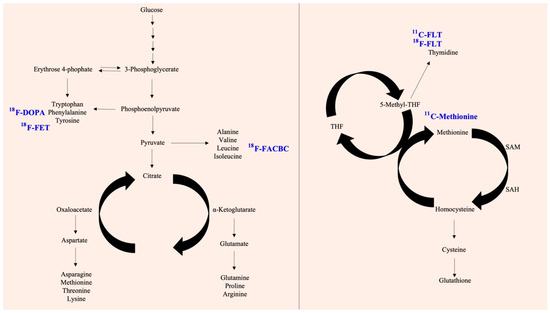
Figure 2. Available metabolism-based radiotracers and their background. The schematic model shows glycolysis and the further TCA cycle, amino acid biosynthesis. In the path, there are some potential targets with radiotracers (left panel). In addition, the coupling of the folate cycle to the methionine cycle is shown (right panel). The blue color represents the derived radiotracer products.
2. Glucose
Glycolysis is a pathway that uses glucose for metabolism and catalysis, and there are many related studies and evidence. Scientists have found that there are many diseases and cancers associated with abnormal glycolysis. The unbalanced proliferation activity of tumor cells results in increased glucose uptake by tumor cells. This includes not only an increase in the glucose uptake rate, but also an increase in the efficiency of the transporter responsible for the membrane
[2][29].
Several clinical studies, such as The Cancer Genome Atlas (TCGA) and Gene Expression Omnibus, have demonstrated that certain cancer types exhibit selective overexpression of glucose transporters (GLUTs). It was found in Kim’s research that the expression of GLUT2 was significantly higher in hepatocellular carcinomas than in normal tissues when compared with other submembers of the GLUT family. Accordingly, this difference is positively correlated with poor prognosis or clinical stage of patients, thus it may serve as a prognosis factor
[3][30]. It has been found that GLUT3 is overexpressed in several carcinomas, including adenocarcinoma and glioblastoma
[4][5][6][31,32,33].
The downstream genes responsible for the 10 steps of the glycolysis response will also follow this pattern by increasing their enzyme activity and expression levels
[7][34], consequently activating several signaling transduction pathways and transcription factors. This process is encapsulated in the Warburg effect theory to accelerate glucose/lactate conversion to respond to oxygen concentrations in tumor cells
[8][35].
In highly proliferating specific cells, there is a specific difference between glucose uptake and glycolysis rates. This has led to the development of molecules that target glucose for use as markers. Fluorodeoxyglucose (FDG) is an analog of a glucose molecule, similar in structure to glucose. FDG is taken up via the GLUT-1 transporter and phosphorylated to FDG-6-phosphate by hexokinase
[9][36]. Because of the lack of C-2 hydroxyl groups, further metabolism is impeded, and the FDG-6-phosphate is trapped within cells, accumulates at high absorption, and is eventually metabolized. Many basic and clinical studies have used FDG as an inhibitor to interfere with the normal glycolysis reaction by selecting specific cells with high sugar uptake. Radiation scientists also use the abovementioned imaging equipment. Radiolabeling technology has been used to design products such as
18F-FDG, which is derived from PET and can track the FDG signal to locate the tumor location. The advantage of FDG-PET is that it can be used as an early assessment of whether the tumor metabolism has changed and to evaluate whether a change in treatment strategy is necessary
[10][37]. As a matter of fact, this depends on the high glycolytic rate of the tumor or disease model, if the glycolytic rate is insufficient, there will be no significant difference in the degree of contrast in imaging
[10][11][37,38].
There are many other modifications and improvements of FDG, including radioisotopes, peptides (linear and cyclic), and some conjugations
[12][39]. Namavari et al. used
18F-FDG as a prosthetic set to design
18F-FDG-RGD and
18F-FDG-cyclo (RGDDYK) by one-step radiosynthesis. This design can increase the binding affinity to the integrin α
vβ
3. The imaging of integrin αvβ3 expression in vivo is a potentially useful method for diagnosing tumors and their metastases to provide a better understanding of tumor angiogenesis and monitor the effects of target-specific antiangiogenic therapy. Senisik et al. also describe a single-step, high-efficiency conjugation of glycylglycine (GlyGly), which is a small peptide, with [
18F] FDG, without the need for purification. Researchers without cyclotron facilities can easily perform efficient radiolabeling and use [
18F] FDG-GlyGly to improve biodistribution studies
[13][40].
Whole body scans are mainly used for tumor examination. It is recommended that the patient fast for a minimum of six hours before the test and maintain a blood sugar level less than 120 mg/dL. After intravenous injection of about 5 to 10 millicuries (mCi) of FDG, the subject must lie down and rest for 45 min, so that the FDG can accumulate in the tumor, be adequately eliminated from normal tissues, and pass through the kidneys and bladder
[14][41]. After 45 min of excretion, after removal of urine, the patient lies down on the scanning table for examination. Generally, it takes about an hour to scan from the head to the upper third of the thigh
[15][42]. To reduce the influence of body tissue on the attenuation effect of gamma rays, a
68Ge radiation source is generally used for another transmission scan to correct the tissue attenuation effect, and an additional 15 to 30 min of examination time is required. After scanning, tomographic image reconstruction and analysis can be performed
[16][43]. If necessary, the absorption rate of FDG by the lesion, such as the standard uptake value (SUV), can be measured. There is the option for a local scan to be added, and the examination time for this is approximately 20 min, to allow for the differential diagnosis of benign and malignant lesions in specific organs (such as lung masses).
3. Pyruvate
In the glycolytic pathway, lactate and pyruvate act as two branches, leading to glycolysis and anaerobic lactic fermentation or aerobic oxidative phosphorylations (OXPHOs)
[17][44]. The conversion of glucose to pyruvate occurs through glycolysis in cancer, whereas lactate is produced by lactate dehydrogenase (LDH)
[18][45]. In this manner, cancer cells are exploited for their unique properties. It has been reported that some studies have attempted to increase detection sensitivity in solution-state nuclear magnetic resonance experiments by using dynamic nuclear polarization of
13C-labeled pyruvate
[19][46]. In vivo imaging of the spatial distribution and metabolism of a labeled molecule following intravenous injection in mouse models is possible by using
13C magnetic resonance spectroscopic imaging (MRSI). In a reaction catalyzed by LDH, the decrease in label flux between hyperpolarized [1-
13C]pyruvate and lactate can be measured to determine whether mouse lymphomas are responding to drug treatment. There may be several reasons for the reduction in flux, such as loss of cell enzymes, reduced tumor cellularity, or reduced concentrations of lactate and NAD(H) in the tumor. Therefore, it is important to compare these results with FDG-PET to improve the value of FDG-PET in its current board-certified clinical application
[20][47].
4. Galactose
A variety of cancers have been successfully treated using the combination of
11C-acetate and FDG PET/CT. However, the detection sensitivity of HCC types for the small primary subtype is low
[21][48]. Several studies have demonstrated that HCC has a greater effect on food intake and galactose production within the body. The expression of galactose metabolism related enzymes such as galactokinase (GALK1) and galactose-1 phosphate uridylyltransferase (GALT) in HCC is higher than that of normal liver tissues and is critical for HCC proliferation
[22][49]. It has been demonstrated that galactose is metabolized specifically in hepatocytes, and thus a proof-of-concept imaging test has been developed for the diagnosis of liver cancer
[23][50]. Compared with other tracers, 2-deoxy-2-[
18F]fluoro-D-galactose ([
18F]FDGal) was found to accumulate in the liver more frequently
[24][25][51,52]. FDGal can be synthesized using commercially available FDG production kits with only minor modifications, enabling this process to be implemented at most clinical PET facilities
[26][53].
18F-FDGal PET/CT was able to detect previously unknown extrahepatic diseases and regions when used in conjunction with contrast enhancement CT (ce-CT) for liver lesions.
[23][50]. Despite this, the
18F-FDGal PET/CT images are still limited by the extremely high uptake of
18F-FDGal in normal liver tissue as well as by the heterogeneity of uptake in cirrhosis patients. A comparison and discussion of three
18F-FDGal PET protocols was conducted by Horsager et al., in order to achieve the highest tumor/background ratio and optimal observations
[27][54]. Furthermore, some studies have noted that coadministration does not improve the interpretation, detection, or statistical values of
18F-FDGal PET/CT images
[28][55].
5. Choline
A nutrient that builds cell membranes and phospholipids, choline, is necessary for brain development and memory, and is a precursor to acetyl-CoA, which is involved in muscle control and neuronal transmission. The activity of choline metabolism-related metabolic enzymes is abnormally increased in cancer and is related to malignancy. Activated phosphatidylcholine-specific phospholipase D (PC-PLD) and phospholipase C (PC-PLC) are associated with abnormal tumor proliferation in ovarian cancer
[29][56]. Increased levels of glycerophosphorylcholine (GPC) and phosphorylcholine (PC) promote epithelial-to-mesenchymal transition (EMT) in ovarian cancer
[30][57]. In some studies, highly saturated phosphatidylcholine species, CDP-choline species, and phosphocholine species are significantly higher in HCC tumors than in adjacent liver and intrahepatic cholangiocarcinoma (ICC) tumors
[31][58].
Fluorine (18F) has been labeled for PET and CT to detect chronic liver diseases and liver cancers
[32][59]. Despite having a similar function to
18F-FDGal,
18F-fluorocholine uptake shows significant associations with liver uptake and neuroinflammatory and fibrotic changes found in chronic liver disease patients’ histology
[32][59].
Choline can also be labeled with carbon-11 (
11C) as a positron emitter for molecular imaging
[33][60]. Among several short-lived positron-emitting radionuclides used in PET imaging,
11C has a unique probability (t1/20.4 min,
Eβ+ = 1.98 MeV) that is easily distinguished from the nonradioactive
12C molecule. In addition,
11C has a short half-life (20.33 min) and most
11C will decay into
11B through β decay, which is less harmful to the human body and has increased applicability
[34][61]. Evaluation of
N-methyl-[
11C]choline in PET in patients with recurrent prostate cancer and localized parathyroid adenomas that exhibit biochemical recurrence has been performed. It has been evidenced that the choline kinase alpha (XKα) overexpression can be found in primary hyperparathyroidism, parathyroid hyperplasia, and neoplastic lesions
[35][62]. Liu et al. evaluated the value of
11C-choline PET in patients with primary hyperparathyroidism and indicated that this tool had potential for the location of parathyroid adenomas when ultrasound and
99mTc-sestamibi imaging yielded negative or discordant results
[36][63]. Although the study by Noltes et al. was a retrospective analysis, the results were still promising, showing high accuracy and sensitivity
[37][64]. Furthermore,
11C-choline PET can also be applied to prostate cancer. Although serum prostate specific antigen (PSA) testing is often used to monitor disease recurrence after definitive therapy for prostate cancer
[38][65], Krause et al. identified a positive correlation between
11C-choline PET detection rate and serum PSA levels
[39][66]. Several clinicopathological factors of prostate cancer can be statistically significant for
11C-choline uptake, even when the PSA value and kinetics are low
[40][67]. Despite some evidence,
18F-FDG is rarely used in the evaluation of prostate cancer, because
18F-FDG PET has a low sensitivity for the detection of prostate cancer, as exemplified by the fact that its sensitivity for detection of bone metastases is lower than that of bone scintigraphy
[41][68]. This also increases the application value of
18F/
13C-choline.
6. Acetate
As with lactate, pyruvate can be converted into acetate through keto acid dehydrogenase. Acetate can serve as an intermediate product of shuttle metabolism in cancer cells that interacts with surrounding cells or is capable of resisting external stress
[42][69]. Studies have shown that cancer cells are capable of adapting to low oxygen environments through the conversion of acetate. Specifically, mitochondria-localized acetyl-CoA synthetases (ACSS 1/2) convert acetate into acetyl-CoA for use in the TCA cycle or fatty acid synthesis
[43][70]. The abnormal expression of ACSS2 has also been observed in several cancers, making it a potential target for cancer therapy
[44][45][46][71,72,73]. Acetate, in addition to serving as a buffer for metabolic intermediates, can also play the function of compensating for acidic PH environments through its role in histone acetylation
[43][70].
Similar to choline, acetate is a precursor for lipid biosynthesis
[47][74]. Studies have shown that it is an important bioenergetic fuel for tumor cells. It has been claimed to promote histone acetylation and other epigenetic modifications. Despite the lower circulating concentrations of acetate, it can still surround the intracellular circulation and tumor microenvironment
[43][70]. The difference in kinetics between normal myocardium, normal renal parenchyma, and renal cancer tissue has been proven. Myocardial and normal renal tissue showed rapid washing, consistent with the predominant oxidation to CO
2 through the tricarboxylic acid cycle. In contrast, the tracer was retained in tumor tissue, probably because of the use of
11C-acetate as an important substrate for the generation of membrane lipids
[48][75]. Through comparison of multiple organs and conditions using imaging tools, acetate has been verified as suitable for the detection of prostate cancer.
For prostate cancer,
11C-acetate can be used as a predictive, prognostic, and intermediated endpoint biomarker.
11C-acetate can be detected under low PSA conditions, but it is hard to differentiate between benign prostatic hyperplasia and prostate cancer. In contrast,
11C-acetate PET can be used to assess the regional lymph node and recurrence status
[49][76]. As mentioned previously, the sensitivity of the diagnostic range of
11C-acetate and choline PET probes is important in the evaluation of prostate cancer. Although
11C-acetate has the advantage of low urinary excretion,
18F-choline is more widely used because of its longer physical half-life.
11C-acetate and
11C-choline can only identify about 50% of recurrence sites, although they both have limitations in patients with PSA levels below 1ng/mL
[50][77].
7. Pivalic Acid
In some clinical studies, glucose is not a suitable tracer, because cells can also produce energy through other metabolic pathways, such as fatty acid oxidation. Some de novo biosynthesis or short-chain fatty acid salvage pathways are important sources of nutrients for cell growth and proliferation
[51][78]. These include acetate and
18F-fluoropivalate (3-
18F-fluoro-2,2-dimethylpropionic acid,
18F-FPIA). Unlike acetate, pivalate cannot be oxidized to carbon dioxide in mammalian cells because it carries a
tert-butyl substituent
[52][79]. Pivalate is esterified in vivo in normal tissue, and the resulting ester enters plasma and is absorbed by cells inhibited by L-carnitine or rapidly eliminated in urine
[53][80]. Dubash et al. evaluated the safety, biodistribution, and internal radiation dosimetry profile of
18F-FPIA in healthy volunteers
[54][81]. A current phase 2 clinical trial (NCT04717674) has enrolled some solid tumor patients who are undergoing
18F-FIPA PET imaging. Their
18F-FIPA tumor uptake will be calculated, and several histological markers of tumor metabolism measured. These biomarkers include Ki-67 and typical enzymes related to fatty acids (SDHA, CPT1/CAT1, CACT, SLC22A2, SLC22A5, and SLC25A20).
8. Cyclobutanecarboxylic Acid
Fluoro-18 (
18F) fluciclovine (anti-1-amino-3–
18F-fluorocyclobutane-1-carboxylic acid [FACBC]) is a radiolabeled amino acid analog that is upregulated by amino acid transport in a variety of cancer cells, and prostate cancer in particular
[55][56][57][82,83,84]. With its low radioactivity background, it is effective for detecting prostate progression, even in cases of lymph node or bone metastasis below 5 mm
[58][85].
It can be taken up through the human L-type amino acid transporter and alanine-serin-cysteine transporter systems
[59][86]. By the same principle, FACBC is analogous to FDG in that it is a metabolite that, when taken up by cells, cannot be metabolized and remains in the tumor area. The sample application principle also includes
18FDG, FACBC, and
11C-choline. They each use glucose transporter (GLUTs), L-type amino acid transporter (LAT/ASCT2), and choline transporter, respectively. The
68Ga PSMA-11 and
111In-Capromab pendetides target the specific antigen PSMA
[60][87].
9. Methionine
Methionine is a natural essential amino acid involved in various types of biosynthesis. It is possible for methionine cycle biosynthesis to convert methionine into S-adenosylmethionine (SAM) in order for methyl donors to be synthesized for genetic processes through methionine adenosyltransferase (MAT)
[61][62][88,89]. The Hoffman effect is a phenomenon that describes proliferating cells relying on methionine for growth
[63][90]. Methionine has been found to be present in high amounts in certain cancer cells
[64][91]. Compared with normal cells, cancer cells deprived of exogenous methionine in the environment grow and move more slowly
[65][92]. Consequently, labeling methionine is considered an effective method for identifying cancer cells. For use as a radiotracer, methionine is labeled using carbon-11 and its distribution is usually detected using PET. MET images of brain tumors often show increased uptake in tumors with the presence of amino acid transporters (sodium-independent L-transporters, LAT1, 2, and 3), MET metabolism, tumor vascular bed-dependent blood flow, microvessel density, and blood–brain barrier distribution. This property has made
11C-Met a popular imaging agent in brain cancer patients
[66][93]. The conditions to which it has been applied range from genetic alterations and relapse status to post-chemotherapy evaluations
[67][68][69][70][71][72][94,95,96,97,98,99].
Park et al. also described the application of
11C-Met in intracranial germinoma, observing that
11C-Met PET parameters had a significant association with tumor location, sex,
KRAS variant, and symptoms
[73][100]. Morales-Lozano et al. also compared
11C-Met and
18F-FDG in multiple myeloma, and they confirmed that
11C-Met PET/CT was more sensitive than
18F-FDG for evaluation of myeloma tumors
[74][101]. Currently, a clinical trial is underway to evaluate the response of a more comprehensive range of cancer types (NCT00840047).
10. Glutamine
Glutamine is the most abundant and versatile amino acid in the body. Many metabolic events are expected to be associated with glutamine use. Even in immune cells, its frequency of use approaches or exceeds that of glucose. In energy metabolism, glutamine is a supplementary pathway other than glucose and can be involved in the TCA cycle and events within the mitochondria. Glutamine is an essential nutrient for lymphocyte proliferation and cytokine production, macrophage phagocytosis and secretory activities, as well as neutrophil bacterial killing
[75][102].
As well as its role as an energy source, glutamine also plays an important role in maintaining a balance between the pressures produced by antioxidants and oxidative stress. Meanwhile, it has been found that Glutamine is reliant on Methionine in some cancer cells
[76][103]. This phenomenon is especially evident in clear cell renal cell carcinomas and breast cancer, in which both tumor microenvironment cells and tumor cells will deplete glutamine levels. This depletion limits the harmful effects of peripheral immune cells on cancer
[77][78][104,105]. As a result, targeting Glutamine selectively is an effective method of diagnosis.
To evaluate the safety and tumor imaging characteristics of fluorine 18-(
2S,4R)-4-gluoroglutamine (FGln), the Dunphy Group performed scans on cancer patients
[79][106]. Cohen et al. reported that
11C-Gln could be applied to metastatic colorectal cancer. The status quo is that some targeted therapies are used in combination with glutamine metabolism inhibitors, but the study lacked appropriate diagnostic tools for assessment of early response. Therefore, calculated glutamine influx and glutamate efflux (
11C-Gln and
18F-FSPG, respectively) have been recruited for further safety and biodistribution evaluation (NCT03263429)
[80][107]. Glioma study indicates that although
18F-FGln is not superior to many current neuroimaging modalities, it may provide complementary biological information specifically about metabolic nutrient uptake relevant to glioma pathology
[81][108].
18F-FGln uptake in gliomas is positively correlated with glioma progression, and can be indicative of gene alterations (
PTEN or
IDH1) events or response to chemotherapy or radiation therapy. More recently, the glutamine-derived development,
18F-labeled glutamate (4
S)-4-(3-
18F-fluoropropyl)-1-glutamate, has been considered a PET tracer in preclinical models and human subjects
[82][109]. However, the disadvantages of
18F-FGln still include neuroinflammation or disruption of the BBB that cannot be clearly observed
[81][108].
11. Fluoropropyl-L-Glutamic Acid (FSPG)
Glutamic acid and glutamine are both interconvertible. Furthermore,
N-acetylglutamate, α-ketoglutarate, 1-Pyrroline-5-carvoxylate,
N-Formimino-L-glutamate, and
N-acetylaspartylglutamic acid (NAAG) can produce glutamic acid through their corresponding enzymes
[83][110]. Glutamic acid and glutamine form the glutamate metabolism that comprises several reversible and irreversible reactions.
FSPG could be applied to measure system XC-transporter activity, because it contributes to the important function of glutathione biosynthesis and the glutaminolytic pathway. These transporter families include SLC1A5, SLC38A1/A2, and SLC7A11
[84][111]. Unlike glycolysis, the glutaminolytic, glutathione biosynthesis, and redox balance pathways are dominant metabolic reprogramming events in cancer
[85][112]. According to previous research, it can be detected and quantified in several types of cancer, including head and neck, colorectal, and non-Hodgkin lymphoma, and can play a complementary role in insignificant and ineffective types of
18FDG. There are many unknowns in the related research, but it has development potential for future systems.
12. L-Tyrosine
L-tyrosine arises from amino acid biosynthesis and is considered one of the necessary amino acids. In addition to being associated with Huntington’s disease and phenylketonuria, abnormal tyrosine metabolism has also been demonstrated to be associated with malignancy in several cancers
[86][113]. It is evident from the method of blood detection that tyrosine levels will increase abnormally in HCC
[87][114]. Based on the analysis of large sets of data such as TCGA and GEO, it appears there is a strong correlation between genes associated with tyrosine metabolism and poor prognosis in HCC
[88][115]. A study conducted by Sun also suggests that acetyl-CoA generated by tyrosine metabolism may promote stemness activity in HCCs, thereby making them resistant to therapeutic treatment
[89][116].
With amino acid PET, one can obtain information on the metabolism of tumor cells, which is complementary to structural imaging using magnetic resonance images
[90][117]. Common amino acid-type radiotracers include
18F-FET and
11C-Met. Because
18F-FET has lower uptake by inflammatory cells than
11C-methionine or 18F-FDG, it is useful for differentiating tumors from treatment-induced necrosis
[91][118]. Labeled with L-tyrosine fluoroine-18 fluorine, the approach with MRI can achieve 93% sensitivity and 94% specificity in glioma tissue
[92][119].
In current glioblastoma treatments, temozolomide (TMZ) is considered the standard chemotherapy regimen for patients, but it leads to drug resistance and survival prolongation is limited. However,
18F-FET may be a valuable tool for predicting the outcome of therapy before the commencement of TMZ maintenance therapy
[93][120]. Merging this evidence,
18F-FET has been identified as having a good diagnostic performance for the initial assessment of patients with new isolated brain lesions
[94][121]. A previous retrospective study also claimed that a combination of static and kinetic
18F-FET parameters achieved a higher diagnostic accuracy than conventional MRI in distinguishing between recurrent or progressive disease and treatment-related changes
[95][122].
13. Thymidine
Thymidine is the DNA nucleoside that pairs with deoxyadenosine in double-stranded DNA
[96][123]. In cell biology, it is used to synchronize cells in the S phase, so its production is closely related to the cell cycle
[97][124]. Cancer cells often exhibit abnormal cell proliferation, and many treatments have been developed to inhibit genetic replication. Inhibitors of thymidylate synthase (TS), such as 5-fluorouracil (5-FU), are available
[98][125]. It has been demonstrated that combining 5-FU and leucovorin or oxaliplatin significantly increases the chance of TS and improves outcomes, but it also has great side effects on patients with advanced CRC
[99][100][126,127]. It is, therefore, beneficial to develop early detection of patients in order to increase the diagnosis rate and formulate further treatment strategies.
In addition to several common thymidine analogs (BrdU and EdU) that have proliferation monitors, the thymidine analog 3′-deoxy-3′[
18F] fluorothymidine (FLT) has been developed as a proliferation marker for cancer research. Interestingly, the rate-limiting enzyme of FLT metabolism, the pyrimidine-metabolizing enzyme thymidine kinase-1 (TK-1), is overexpressed in pancreatic cancer cell lines and pancreatic cancer
[101][128].
18F-FLT is transported through the cell membrane and trapped inside the cell after phosphorylation by thymidine kinase 1 (TK1). Although
18F-FLT is not incorporated into DNA, it is still considered a surrogate marker of cellular proliferation because TK1 activity is closely regulated by the cell cycle
[102][129]. A close relationship could be demonstrated between tumor retention and cell proliferation, suggesting that
18F-FLT is a promising marker for monitoring treatment response. Unlike
18F-FDG,
18F-FLT is not taken up by inflammatory cells, which could reduce the rate of false positive findings in both in pancreatitis and inflammatory tissue after therapy
[101][128]. A comparative study revealed that
18F-FLT was more sensitive than
18F-FDG and
18F
-fluorethylcholine in a human pancreatic xenograft model using SCID mice
[101][128]. Similarly, TK-1 and DTYMK kinase and some related transporters, SLC28A1 and SLC29A3, increased significantly in the PDAC group
[101][128]. The use of
18F-FLT is not limited to pancreatic cancer, but has some additional applications in different types and combinations. The Cieslak group has shown that pharmacological ascorbate (AscH
−) induces cytotoxicity and oxidative stress in pancreatic cancer and then contributes to the radiosensitizer function monitored by
18F-FLT PET
[103][130]. Collet et al. also claimed that
18F-fluoro-
L-thymidine could provide an additional indicator for the staging of gliomas, in particular distinguishing between stage II or stage III (low-grade glioma) and stage IV (glioblastomas)
[104][131]. Furthermore, there is an ongoing study to evaluate 18F-FLT uptake in non-small cell lung cancer following pemetrex treatment. Because of the response and activation of the dexamethasone/pemetrexed thymidine salvage pathway inhibited by dexamethasone/pemetrexed
[105][132], the authors hypothesize that this strategy can be detected as an increase in FLT tumor uptake that subsequently decreases with reduced proliferation. In this study, the FLT response had a good overall survival rate that was twice that of no response. However, the study needs to recruit more case numbers and statistics with clinicopathological factors
[106][133].
14. Dihydroxyphenylalanine (DOPA)
In addition to the well-known carboxylate metabolism, there is fatty acid biosynthesis and amino acid biosynthesis. Neurotransmitters are considered an important aspect, involving signaling pathways in the brain, reflex actions, nerve networks, memory, emotions, etc. Normal and tumoral neuroendocrine cells can uptake and decarboxylate amine precursors (such as L-DOPA and 5-hydroxytryptophan) to produce biogenic amines, such as catecholamines and serotonin
[107][134]. For neuroendocrine tumors (NET), fluorine-18-dihydroxyphenylalanin (
18F-DOPA) has been engineered to capture signals because these tumors can accumulate and decarboxylate biogenic amines. Through activation of large amino acid transporters (LAT, SLC7A5/A8),
18F-DOPA is absorbed into tumors and further decarboxylated by DOPA decarboxylase to produce
18F-DOPAmine, which is then transported by vesicular monoamine transporters (VMAT, SLC18A1/A2) and trapped in cells
[108][135].
18F-DOPA has comprehensive applications in different neuroendocrine tumors, including medullary thyroid cancer (MTC), pancreatic NET, and paragangliomas/phaeochromocytomas
[109][136]. Furthermore,
18F-DOPA can differentiate between Parkinson’s disease and dopaminergic transmission disorder
[110][137].
18F-DOPA enables presynaptic dopaminergic function to be quantified, and specific regions of linear reduction in
18F-DOPA uptake in idiopathic Parkinson’s disease to be observed
[110][137].
In addition to
18F-DOPA, gallium-68-somatostatin analogs (DOTA-NOC, DOTA-TOC or DOTA-TATE) have also shown high diagnostic NET imaging on PET/CT scans targeting the accuracy of the somatostatin receptor (SSR). Several studies have compared the uptake and additional functional activity of SSR PET/CT and
18F-DOPA PET/CT. Although patients with gastroenteropancreatic and thoracic NETs may prefer SSR PET/CT as a priority strategy,
18F-DOPA PET/CT remains another viable option
[111][138].
15. Glucosamine
Glycosylation is a hallmark of various neurological disorders. A current study found that N-linked protein glycosylation in the brain led to glucosamine metabolism through glycogen catabolism
[112][139]. Brain glycogen contains 25% glucosamine, and the mass spectrometry imaging method can reveal the distribution of brain glycogen
[112][139]. Additionally, glycogen is a branched glucose polymer that is synthesized from the activated glucose donor, uridine diphosphoglucose (UDP)
[112][139]. In some cancer cells, researchers can observe that the cell is deficient in energy stores that require glucose uptake. There is also evidence that glycogen is more than a glucose cache, and is a critical storage macromolecule for the brain protein N-glycosylation, which impacts myriad subsequent cellular processes. Therefore, a fluorescent tool labeled fluorine-18 has been developed to detect glycogen
[113][140].
Returning to the metabolic background, the glucosamine analog, N- (methyl-2-fluoroethyl) -1H- [12,3] triazole-4-yl) glucosamine (NFTG) has been applied to some animal models. Witney’s group confirmed that some cancer cells do not have high glucose uptake, but have strong glucose storage and glycogen (glycogenesis). Annotation of glycogenogenesis, glycogen, and glycogen synthase 1 expression in cancer cells with
18F-NFTG is advantageous in understanding the potential ability of
18F-NFTG to specifically image certain quiescent cells (G
0–G
1 phase). Furthermore,
18F-NFTG provides higher-specificity tumor imaging under inflammatory conditions than
18F-FDG
[114][141].