Detecting VOC Biomarkers in the Exhaled Breath: Comparison
Please note this is a comparison between Version 3 by Catherine Yang and Version 2 by Catherine Yang.

In general, volatile organic compounds (VOCs) have a high vapor pressure at room temperature (RT). It has been reported that all humans generate unique VOC profiles in their exhaled breath which can be utilized as biomarkers to diagnose disease conditions. The aforementioned discussions show that the human breath contains a variety of VOCs that serve as biomarkers for various diseases and metabolic problems. Therefore, the real-time monitoring of such VOCs in the exhaled human breath is highly essential to enable non-invasive illness detection. In the following subsections, aA detailed discussion has been carried out to understand various techniques developed to detect VOCs in very low concentrations of part per million volumes (ppmv), part per billion volumes (ppbv), and part per trillion volumes (pptv).

  • breath analysis
  • sensor
  • volatile organic compound
  • biomarker
  • molecularly imprinted polymer

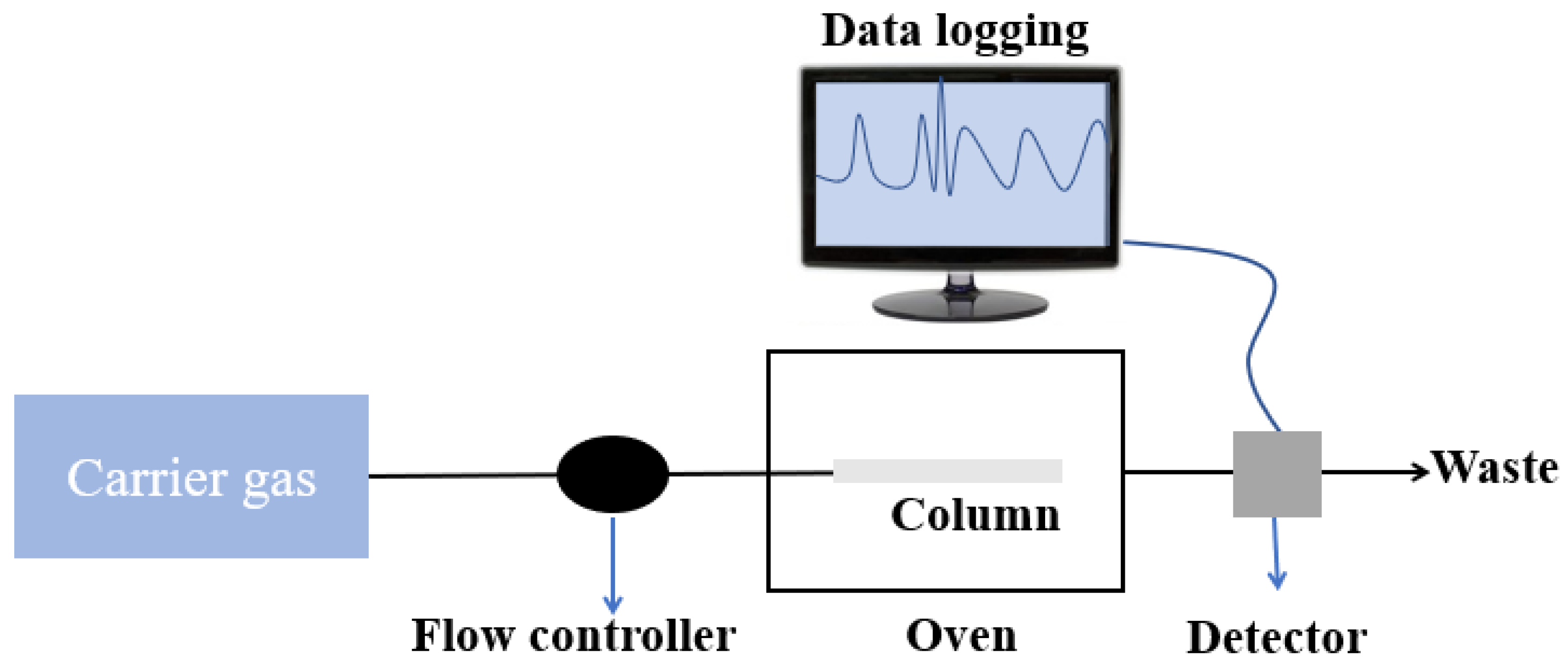
1. Gas Chromatography–Mass Spectrometry (GC–MS) Techniques

GC–MS is a technique in which a mixture of molecules of various compounds travel by a carrier gas (normally helium) via a column that separates molecules and detects them by a detector. In the past decades, this technique has been utilized widely along with various types of detectors to detect VOCs in exhaled breath for the purpose of health monitoring. A block schematic diagram of GC–MS is shown in Figure 1. In 2003, Sanchez et al. detected 25 VOCs in human exhaled breath using a series couple column which includes some of the important biomarkers such as ethanol, methanol, acetone, isoprene, pentane, etc. [1]. The limit of detection (LOD) was observed to be 1–5 ppb in 0.8 L of the exhaled breath. Lord et al. also developed a GC–MS-based analytical detection system which could selectively detect acetone and ethanol in the exhaled breath and reduce the moisture effect to a large extent [2]. Later, Giardina et al. also proposed a low-temperature glassy carbon-based solid-phase mass extraction microfiber which was capable of extracting at least five types of cancer related to VOC biomarkers from the simulated breath. The extracted sample was analyzed using GC–MS with good sensitivity [3]. Lamote et al. utilized e-Nose and GC–MS to distinguish between malignant pleural mesothelioma patients and asymptomatic asbestos-exposed people at a risk of the mentioned disease. Schnabel et al. utilized GC–Time of flight–mass spectrometry (GC–TOF–MS) to demonstrate the non-invasive monitoring of ventilator associated pneumonia in ICU patients by exhaled breath analysis [4]. They identified nearly 12 VOC biomarkers for this purpose. GC–MS was also utilized by Acevedo et al. for gastric cancer [5]. The study proposed the approach to distinguish between healthy people and patients suffering from gastric cancer by the exhaled breath analysis. From all this literature, the researchers can say that GC–MS is considered a potential technique for quantitative analysis for non-invasive detection.
Figure 1. Block schematic diagram of a GC–MS.

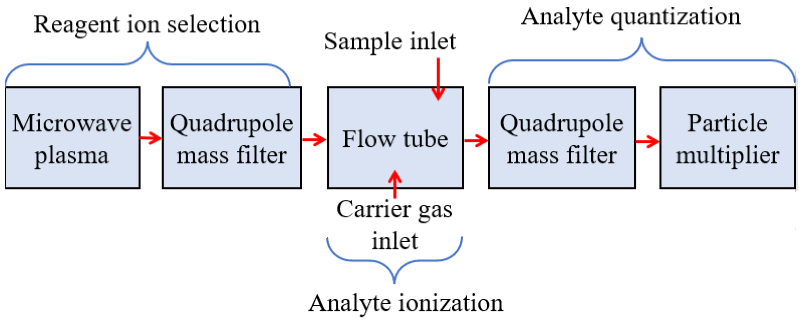
2. Selected-Ion Flow-Tube Mass Spectrometry (SIFT-MS)

SIFT-MS is a tool of analytical chemistry which is similar to gas chromatography for the quantitative monitoring of VOCs [6]. In this technique, the VOC samples are ionized by the reagent ions, such as NO+, H3O+, O2+, etc., which can be later analyzed by a quadrupole mass spectrometer. Figure 2 shows the block diagram of SIFT-MS. SIFT-MS was first reported to (i) determine the VOC trace presented in the exhaled human breath for the prognosis of the disease and (ii) understand pathophysiological and physiological conditions. In 1996, Smith et al. used SIFT-MS to detect ammonia from the exhaled breath of a known Helicobacter pyroli-infected person which was observed to be increased by ∼4 ppm after an oral dose of 2 g nonradioactive urea [7]. In 1999, Spanel et al. utilized SIFT-MS and O2+ as reagent ions in order to quantitatively detect isoprene in the exhaled breath [8]. Later in 2002, Diskin et al. investigated the variation in concentration of common breath VOC biomarkers, such as ammonia, isoprene, ethanol, acetaldehyde, and acetone over a period of 30 days using SIFT-MS with healthy individuals [9]. In the same year, Abbott et al. utilized SIFT-MS to detect acetonitrile in the breath and urinary samples of several smokers and nonsmokers [10]. The result exhibited that the acetonitrile concentration in the exhaled breath was achieved within the range of 17 ppb to 124 ppb, while the urinary acetonitrile concentrations were obtained within the range of 0 μg/L to 150 μg/L that were close to the concentrations previously determined in the blood. Other researchers also utilized SIFT-MS to determine the concentration of various VOCs and diagnosed the stage of related disorders [11][12][13].
Figure 2. Block schematic diagram of a SIFT-MS.

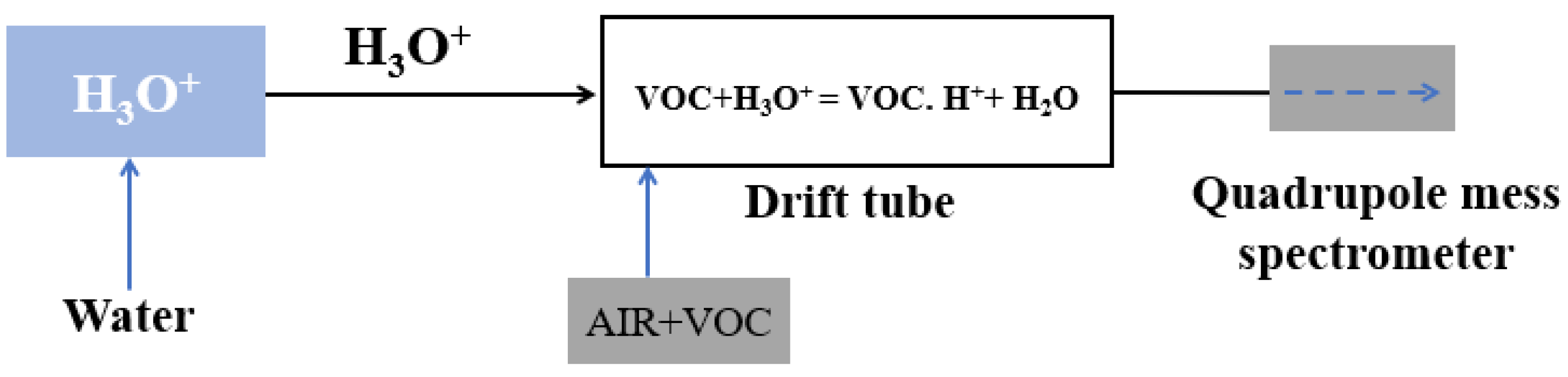
3. Proton-Transfer-Reaction Mass Spectrometry (PTR-MS)

Classical PTR-MS is a tool of analytical chemistry which utilizes gas-phase hydronium ion as n ion with purity >99.5% of source reagent. The PTR-MS is utilized to monitor the absolute concentration of the selected VOCs as low as pptv without any calibration [14]. Figure 3 illustrates the block schematic diagram of PTR-MS. PTR-MS exhibits excellent sensitivity and selectivity which play a crucial role in the exhaled breath analysis to monitor the pathophysiological and physiological state of the human subjects. In 2004, Amann et al. utilized PTR-MS to study the variation in the concentration of different VOCs exhaled in patients during sleep with carbohydrate malabsorption, and inter- and intra-subject variability of a certain mass [15]. Kar et al. utilized PTR-MS to detect cholesterologenesis by investigating the level of isoprene in the exhaled breath [16]. Later, Schmutzhard et al. reported the potential of PTR-MS to diagnose neck and head squamous cell carcinoma by monitoring isoprene in the exhaled breath [17]. Overall, this technique has been exploited widely to detect various VOCs and hence diagnosed the stages of corresponding diseases.
Figure 3. Block schematic diagram of a PTR-MS.

4. Advantages and Limitations of Classical VOC Detection Techniques

These techniques utilize the ion-pair extraction of the analytes and quantification by mass spectrometry to detect the VOC concentrations. However, a limitation of these techniques is that they can be time-consuming and expensive. They also require a skilled technician to operate which can be done only for off-site analysis. A detailed discussion of the advantages and limitations of these techniques is provided in Table 1.
Table 1. Advantage and limitations of classical detection techniques.

References

  1. Sanchez, J.M.; Sacks, R.D. GC Analysis of Human Breath with A Series-Coupled Column Ensemble and a Multibed Sorption Trap. Anal. Chem. 2003, 75, 2231–2236.
  2. Lord, H.; Yu, Y.; Segal, A.; Pawliszyn, J. Breath Analysis and Monitoring by Membrane Extraction with Sorbent Interface. Anal. Chem. 2002, 74, 5650–5657.
  3. Giardina, M.; Olesik, S.V. Application of Low-Temperature Glassy Carbon-Coated Macrofibers for Solid-Phase Microextraction Analysis of Simulated Breath Volatiles. Anal. Chem. 2003, 75, 1604–1614.
  4. Schnabel, R.; Fijten, R.; Smolinska, A.; Dallinga, J.; Boumans, M.-L.; Stobberingh, E.; Boots, A.; Roekaerts, P.; Bergmans, D.; van Schooten, F.J. Analysis of volatile organic compounds in exhaled breath to diagnose ventilator-associated pneumonia. Sci. Rep. 2015, 5, 17179.
  5. Durán-Acevedo, C.M.; Jaimes-Mogollón, A.L.; Gualdrón-Guerrero, O.E.; Welearegay, T.G.; Martinez-Marín, J.D.; Caceres-Tarazona, J.M.; Sánchez-Acevedo, Z.C.; de Jesus Beleño-Saenz, K.; Cindemir, U.; Österlund, L.; et al. Exhaled breath analysis for gastric cancer diagnosis in Colombian patients. Oncotarget 2018, 9, 28805–28817.
  6. Adams, N.G.; Smith, D. The selected ion flow tube (SIFT); A technique for studying ion-neutral reactions. Int. J. Mass Spectrom. Ion Phys. 1976, 21, 349–359.
  7. Smith, D.; Spanel, P. The Novel Selected-ion Flow Tube Approach to Trace Gas Analysis of Air and Breath. Rapid Commun. Mass Spectrom. 1996, 10, 1183–1198.
  8. Španěl, P.; Davies, S.; Smith, D. Quantification of breath isoprene using the selected ion flow tube mass spectrometric analytical method. Rapid Commun. Mass Spectrom. 1999, 13, 1733–1738.
  9. Diskin, A.M.; Španěl, P.; Smith, D. Time variation of ammonia, acetone, isoprene and ethanol in breath: A quantitative SIFT-MS study over 30 days. Physiol. Meas. 2003, 24, 107–119.
  10. Abbott, S.M.; Elder, J.B.; Španěl, P.; Smith, D. Quantification of acetonitrile in exhaled breath and urinary headspace using selected ion flow tube mass spectrometry. Int. J. Mass Spectrom. 2003, 228, 655–665.
  11. Walton, C.; Patel, M.; Pitts, D.; Knight, P.; Hoashi, S.; Evans, M.; Turner, C. The use of a portable breath analysis device in monitoring type 1 diabetes patients in a hypoglycaemic clamp: Validation with SIFT-MS data. J. Breath Res. 2014, 8, 037108.
  12. Alkhouri, N.; Cikach, F.; Eng, K.; Moses, J.; Patel, N.; Yan, C.; Hanouneh, I.; Grove, D.; Lopez, R.; Dweik, R. Analysis of breath volatile organic compounds as a noninvasive tool to diagnose nonalcoholic fatty liver disease in children. Eur. J. Gastroenterol. Hepatol. 2014, 26, 82–87.
  13. Samara, M.A.; Tang, W.H.W.; Cikach, F.; Gul, Z.; Tranchito, L.; Paschke, K.M.; Viterna, J.; Wu, Y.; Laskowski, D.; Dweik, R.A. Single Exhaled Breath Metabolomic Analysis Identifies Unique Breathprint in Patients with Acute Decompensated Heart Failure. J. Am. Coll. Cardiol. 2013, 61, 1463–1464.
  14. Hansel, A.; Jordan, A.; Holzinger, R.; Prazeller, P.; Vogel, W.; Lindinger, W. Proton transfer reaction mass spectrometry: On-line trace gas analysis at the ppb level. Int. J. Mass Spectrom. Ion Process. 1995, 149–150, 609–619.
  15. Amann, A.; Poupart, G.; Telser, S.; Ledochowski, M.; Schmid, A.; Mechtcheriakov, S. Applications of breath gas analysis in medicine. Int. J. Mass Spectrom. 2004, 239, 227–233.
  16. Karl, T.; Prazeller, P.; Mayr, D.; Jordan, A.; Rieder, J.; Fall, R.; Lindinger, W. Human breath isoprene and its relation to blood cholesterol levels: New measurements and modeling. J. Appl. Physiol. 2001, 91, 762–770.
  17. Schmutzhard, J.; Rieder, J.; Deibl, M.; Schwentner, I.M.; Schmid, S.; Lirk, P.; Abraham, I.; Gunkel, A.R. Pilot study: Volatile organic compounds as a diagnostic marker for head and neck tumors. Head Neck 2008, 30, 743–749.
More
ScholarVision Creations