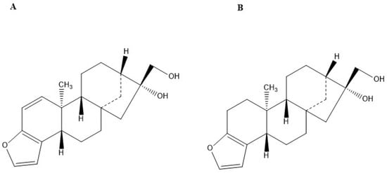
2. Kahweol and Cafestol Effects on Several Cancer Cell Lines
2.1. Lung Cancer
Kahweol and cafestol were tested in vitro against different lung cancer cell lines, and kahweol-induced apoptosis was observed in all cell lines. With regard to mesothelioma, MSTO-211H cells and H28 cells were examined
[7]. The apoptosis of cancerous cells was activated by the upregulation of Bax, alongside the downregulation of Bcl-xL by kahweol and the cleavage of Bid, caspase-3, and PARP by cafestol. Kahweol’s effects were also tested on the lung adenocarcinoma cell line A549, demonstrating DNA fragmentation effects and apoptosis, a decrease in STAT3 expression, as well as an increase in caspase-3 cleavage
[22].
Finally, non-small cell lung cancer was also tested, specifically, the cell lines NCI-H358 and NCI-H1299
[23]. Apoptosis induced by kahweol was achieved, overall, through a very similar molecular targeting that seems to be common between mesotheliomas, lung adenocarcinomas, and non-small cell lung cancers and that involves an increase in the cleavage of both PARP and caspase-3, eventually leading to apoptosis.
2.2. Oral Squamous Cancer
The oral squamous cancer cell lines HN22 and HSC4 were used to help assess the effects of kahweol in an in vitro environment
[24]. Suppression of the transcription factor Sp1, which helps in cell differentiation, cell growth, apoptosis, response to DNA damage, and chromatin remodelling, helped achieve cancer cell apoptosis induced by kahweol.
2.3. Prostate Cancer
The in vitro impact of kahweol acetate and cafestol was assessed in the human prostate cancer cell lines PC-3, DU145, and LNCaP
[25]. Kahweol acetate and cafestol significantly inhibited proliferation and migration in addition to enhancing apoptosis in the cell lines. Cleaved caspase-3 and its downstream target cleaved PARP, both pro-apoptotic proteins, were upregulated in all cell lines, while the anti-apoptotic proteins STAT3, Bcl-2, and Bcl-xL were diminished. Androgen receptor (AR), a strong driver of proliferation in prostate cancer, was downregulated following the treatment with kahweol acetate and cafestol. Moreover, the levels of CCL-2 and CCL-5, in addition to those of their receptors CCR-2 and CCR-5, were decreased.
Kahweol acetate and cafestol also exhibited anti-oncogenic activity in vivo. In a xenograft study where human cell lines DU-145 were injected in SCID mouse models, the oral intake of kahweol and cafestol significantly reduced tumour growth.
2.4. Breast Cancer
Kahweol has been shown to exert an anti-tumour effect on breast cancer cell lines. In vitro treatment of MDA-MB231 with kahweol resulted in the inhibition of cell proliferation along with the induction of apoptosis
[26]. The levels of the pro-apoptotic proteins caspase-3/7 and 9, as well as of the haemeprotein cytochrome c, were increased by kahweol treatment. Kahweol also led to an increase in H
2O
2 cytotoxicity in a dose-dependent manner
[26].
Kahweol’s effects on the MDA-MB231 cell line can be traced back to the increased levels of phosphorylated Akt (p-Akt) and extracellular-signal-regulated kinase (ERK), activating a signalling pathway that regulates multiple cellular processes such as proliferation and apoptosis
[26]. The migratory ability of the cell line was also investigated following kahweol treatment, showing reduced levels of matrix metalloproteinase-9 (MMP-9) and urokinase-type plasminogen activator (uPA).
Kahweol treatment resulted in reduced proliferation and increased apoptosis in the HER2-overexpressing SKBR3 cell line
[27]. It also led to the downregulation of HER2, a growth factor essential to proliferation. To evaluate kahweol’s effect on HER2, two molecular targets were assessed: PEA3, which is a suppressor of HER2 transcription, and AP-2, which upregulates HER2. Kahweol treatment increased the levels of PEA3 while reducing those of AP-2, confirming that the two pathways were targeted by kahweol. In addition, fatty acid synthase (FASN), normally elevated in HER2-overexpressing breast cancers, was decreased in concentration, which was attributed to kahweol’s modulation of sterol regulatory element-binding protein-1c (SREBP-1c) activity. Kahweol also led to a reduction in the levels of p-Akt and of its downstream targets mTOR and cyclin D1, which had an additional impact on the downregulation of FASN
[27].
2.5. Colorectal Cancer
Colorectal cancer cell lines have also been shown to be susceptible to kahweol. Kahweol induced apoptosis in HCT116 cells in vitro through the overexpression of ATF3, which is mediated by CREB1, ERK1/2, and GSK3β
[28]. In addition, kahweol suppressed the proliferation in vitro of HCT116 and SW480 cells, as evidenced by their reduced levels of cyclin D1, a cell cycle protein that promotes progression through the cell cycle
[29]. Kahweol-mediated degradation of cyclin D1 was achieved through the phosphorylation of cyclin D1 at threonine-286 (Thr286). This effect was attenuated upon the inhibition of any of kahweol’s molecular targets ERK1/2, JNK, and GSK3β.
Kahweol’s anti-apoptotic characteristics were also observed in the human colon adenocarcinoma HT-29 cells
[30][31][30,31]. Cell death was induced by kahweol treatment in a dose-dependent manner, which was further proven by the elevation of the pro-apoptotic markers cleaved caspase-3 and PARP, as well as the reduction of the anti-apoptotic markers Bcl-2 and p-AKT. Furthermore, the levels of heat shock proteins (HSP40, HSP70, and HSP90) were diminished following kahweol treatment. HSPs are a family of molecular chaperones that prevent cell death, and their downregulation is associated with increased cytotoxicity of tumour cells.
2.6. Renal Carcinoma
Kahweol’s anti-tumour properties were assessed in several renal carcinoma cell lines in vitro, with a predominant focus on Caki cells. TRAIL, a natural ligand for death receptors found on cancer cells, exerted its apoptotic function on Caki cells more effectively when it was combined with kahweol
[32][33]. Co-treatment with TRAIL and kahweol strongly activated DEVDases and reduced the levels of c-FLIP and Bcl-2, which are anti-apoptotic proteins. The JNK and p38 MAPK pathways were found to mediate these effects. Similarly, the treatment of Caki cells with melatonin and kahweol exhibited comparable findings, which included the induction of apoptosis and the activation of DEVDases
[33][32]. The levels of the pro-apoptotic Bcl-2 protein from the p53 Upregulated Modulator of Apoptosis (PUMA) protein family were also upregulated in response to the treatment. This upregulation was achieved through the C/EBP homologous protein (CHOP), a pro-apoptotic transcription factor. Furthermore, co-treatment with kahweol and sorafenib, a tyrosine kinase inhibitor, had analogous effects on renal carcinoma cells
[34]. Caspase-dependent apoptosis and downregulation of c-FLIP and Mcl-1 were induced by those compounds when tested on Caki cells as well as on other renal carcinoma cell lines, including A498 and ACHN cells.
Cafestol was also shown to exhibit its anti-carcinogenic effect in renal Caki cells. Cafestol-induced apoptosis was found to be mediated by the activation of caspase-2 and 3, the upregulation of the pro-apoptotic proteins Bim and Bax, and the downregulation of anti-apoptotic proteins, including c-FLIP, Bcl-2, Mcl-1, and Bcl-xL
[31]. The apoptotic effect was also achieved by the inhibition of both STAT3 activation and the PI3K/Akt pathway.
The combined anti-tumour activity of kahweol acetate and cafestol was documented in renal Caki and ACHN cell lines
[35]. In these cells, the induction of apoptosis and epithelial–mesenchymal transition upon administration of the diterpenes inhibited proliferation and migration. The inhibition of STAT3 activation and the downregulation of Bcl-2- and Bcl-xL-mediated the apoptosis was exerted by the diterpenes, alongside the upregulation of Bax. Moreover, the diterpenes inhibited Akt and ERK phosphorylation, which are both known to accelerate metastasis and tumour growth.
Finally, cafestol was tested on the Caki cells both in vitro and in vivo using BALB/c-nude mice as the cancer cells recipients
[36]. Anti-cancer effects were induced via the synergistic impacts of cafestol and ABT-737, a Bcl-2 family inhibitor. The inactivation of Bcl-2 proteins (Bcl-2, Bcl-xL, and Bcl-w) helped induce apoptosis. PARP cleavage was also increased by the combined actions of cafestol and ABT-737. Mcl-1 protein was also downregulated by this combination in the in vivo setting, leading overall to a pro-apoptotic effect on Caki cells.
2.7. Leukaemia
The U937 leukaemia cell line was tested on in vitro to evaluate kahweol’s apoptotic effect, which was found to be mediated by the activation of caspase-3 and the downregulation of Bcl-2
[37][38]. The AKT and JNK pathways were also found to be associated with this effect.
Other cell lines such as NB4, K562, HL60, and KG1 were also tested in vitro using cafestol and Ara-c, which is an anti-leukemic agent that was used as a positive control
[38][39]. Increased caspase-3 cleavage was observed in HL60 cells, which helped induce apoptosis via cafestol. Finally, cafestol also helped increase the expression of CD15 and CD11b and decrease the formation of ROS.
Kahweol and cafestol were also shown to enhance the activity of the NK cell line KHYG-1 and its cytolytic effect on the NK-sensitive leukaemia cell line K562. The two diterpenes increased granzyme B expression in NK cells, likely through the phosphorylation of ATF-2, c-Jun, and CREB, transcription factors involved in the regulation of granzyme B. The net result was the enhancement of the cytolytic activity of NK cells in leukaemia cell lines
[39][40].
2.8. Fibrosarcoma
Kahweol acetate was found to attenuate cancer formation, proliferation, and migration by the fibrosarcoma HT-1080 cell line by inhibiting MMP-9, which is usually upregulated by PMA, 12-phorbol 13-myristate acetate, a synthetic compound known for its oncogenic effect
[30]. Kahweol acetate was found to act by inhibiting MMP-9, which is usually upregulated by PMA. This inhibition was achieved by the suppression of PMA-induced NF-κB activity, alongside the suppression of the Akt, p38 MAPK, and JNK1/2 signalling pathways, which were also found to activate MMP-9.
2.9. Hepatocellular Carcinoma
In vitro, kahweol exerted apoptotic effects as well as inhibited cell proliferation in the hepatocellular carcinoma cell lines Hep3B, SNU182, and SNU42
[40][37]. The Src signalling pathway, which is activated upon the phosphorylation of Src, is highly functional in HCC. The Src pathway was blocked by kahweol by inhibiting the expression of p-Akt, p-mTOR, p-p70S6K, and p-4EBP1 in Hep3B and SNU182 cells. This had a direct apoptotic and anti-proliferative effect on the cancer cells. mTOR, which has a proliferative function, was also inhibited by kahweol. Finally, STAT3 was also blocked by kahweol, which induced a pro-apoptotic effect in the HCC cell lines.
2.10. Head and Neck Squamous Cell Carcinoma
Upon the addition of cafestol in vitro, the cell lines of SCC25, CAL27 and FaDu were found to undergo apoptosis in a dose-dependent manner. When combined with cisplatin, cafestol displayed an antagonistic effect in all cell lines; moreover, its addition to radiation therapy showed an additive effect in the cell lines SCC25 and CAL27
[41]. Very limited studies have been performed on head and neck squamous cell carcinoma, which calls for more extensive research on the topic.

