You're using an outdated browser. Please upgrade to a modern browser for the best experience.
Curcumin and Inflammasome: Comparison
Please note this is a comparison between Version 2 by Conner Chen and Version 1 by Chiara Porro.

Curcumin, a traditional Chinese medicine extracted from natural plant rhizomes, has become a candidate drug for the treatment of different diseases due to its anti-inflammatory, anticancer, antioxidant, and antibacterial activities. Curcumin is generally beneficial to human health, with anti-inflammatory and antioxidative properties, as well as antitumor and immune regulation properties. Inflammasomes are NLR family, pyrin domain-containing 3 (NLRP3) proteins that are active in response to a variety of stress signals and promote the proteolytic conversion of pro-interleukin-1β and pro-interleukin-18 to active forms, which are central mediators of the inflammatory response; inflammasomes can also induce pyroptosis, a type of cell death. The NLRP3 protein is involved in a variety of inflammatory pathologies, including neurological and autoimmune disorders, lung diseases, atherosclerosis, myocardial infarction, and many others. Different functional foods may have preventive and therapeutic effects in a wide range of pathologies in which inflammasome protein results activated. In this review, we have focused on curcumin and evidenced its therapeutic potential in inflammatory diseases such as neurodegenerative diseases, respiratory diseases and arthritis by acting on inflammasome.

  • curcumin
  • natural flavonoid
  • inflammasome

1. Introduction

Curcumin, a natural monomer extracted from plants, has gained popularity in recent decades due to its therapeutic benefits in a wide range of human pathological conditions. The medicinal plant Curcuma longa Linn, a perennial herb of the Zingiberaceae family known as “golden spice” for its broad spectrum of pharmacological properties, contains curcumin as one of its most active constituents [1].
Curcumin, with a chemical formula of C21H20O6C21H20O6 and molecular weight of 368.38 g/mole, is also known by its IUPAC name (1E,6E)-1,7-bis (4-hydroxy-3-methoxyphenyl)-1,6-heptadiene-3,5-dione 1. It is a polyphenolic compound derived from the rhizome of the herb Curcuma longa, and it has been shown to have antioxidative, anti-inflammatory, anticancer, chemopreventive, and anti-neurodegenerative properties [2]. Curcumin’s anti-inflammatory properties have been supported by numerous preclinical and clinical studies in a variety of inflammatory diseases.
Due to its anti-inflammatory and antioxidant properties, curcumin is considered a promising drug candidate for the treatment of inflammatory diseases. Inflammation is the body’s response to certain stimuli, such as viral or bacterial infections, mechanical damage, or an excessive immune response. The purpose of the inflammatory response is to remove stimulating factors and accelerate tissue repair, but excessive inflammatory responses can lead to tissue damage, organ dysfunction, and potentially life-threatening disease.
In addition, the chemical structure of curcumin makes it an excellent scavenger of reactive oxygen and nitrogen species (ROS and RNS, respectively) [3]. As a result, curcumin can attenuate or prevent exercise-induced oxidative stress and inflammation [4,5][4][5]. Curcumin activates the Nrf2 pathway, which is important for the activation of antioxidant enzymes such as thioredoxin reductase, Hsp70, and sirtuins [6].
Curcumin’s therapeutic activity is limited by its poor water solubility, fast biological metabolism, and low bioavailability due to insufficient absorption, chemical instability, and rapid metabolism in the body, which is one of the limitations of its use as a potential therapeutic agent [7]. One approach to addressing these issues is through the use of nanocarriers, which are drug delivery systems optimized for improving curcumin bioavailability and thus its therapeutic effects. It activates caspase-1, which results in the processing and secretion of critical inflammatory molecules.
Due to their nanometric size and chemical properties, nanoparticles, liposomes, micelles, phospholipid vesicles, and polymeric nanoparticles can increase the effectiveness of curcumin. Exosomes are extracellular vesicles that are normally released from cells by exocytosis after multivesicular bodies have matured [8]. They are natural nanocarriers for drug delivery [9].
The NLR family, pyrin domain-containing 3 (NLRP3) protein is an intracellular signaling molecule that recognizes a wide range of pathogens, environmental factors, and host-derived factors, including ATP [10]. The NLRP3 inflammasome consists of the NLRP3, the ASC adaptor, and caspase-1, which is activated by various danger signals [11]. When activated, it activates caspase-1, which leads to the processing and secretion of critical inflammatory molecules such as the pro-inflammatory cytokines IL-1β and IL-18 [12].
Normal activation of the NLRP3 inflammasome contributes to host defense, but abnormal activation is pathogenic in inherited disorders such as cryopyrin-associated periodic syndrome (CAPS) and complex diseases such as multiple sclerosis, type 2 diabetes, Alzheimer’s disease, and atherosclerosis [13].
Both inflammasome formation and its activity play a critical role in a variety of pathologies, including cardiovascular, metabolic, renal, digestive, and central nervous system (CNS) diseases.

2. Inflammasome

Inflammasomes are multimeric protein complexes that are widely expressed in the cytoplasm of various cell types, including immune and non-immune cells [14]. Inflammasomes mediate the host’s innate immune responses to microbial infection and cellular damage. Cells are activated through the intervention of germline-encoded pattern-recognition receptors (PRRs) on the surface of the cell membrane. Pathogen-associated molecular patterns (PAMPs) are recognized by PRRs. Examples of conserved microbial factors detected by PRRs include the bacterial secretion system, microbial nucleic acids, and elements of the microbial cell wall. PRRs are also triggered by endogenous danger-associated molecular patterns (DAMPs) generated in the setting of cellular injury or tissue damage [15], such as ATP, uric acid crystals, heat-shock proteins hsp70 and hsp90, and the high-mobility group box 1 (HMGB1) [16]. The binding between PRRs and PAMPs or DAMPs induces NF-κB activation and the expression of the inflammasome [17]. Inflammasome protein complexes are classified into canonical and non-canonical. The classification depends on the activation of cysteinyl and the mode of activation of cysteinyl aspartate specific proteinase (Caspase) during inflammasome formation. Canonical inflammasomes are constructed by the nucleotide-binding oligomerization domain (NOD) leucine-rich repeat (LRR)-containing protein receptors (NLRs). The canonical inflammasomes include Nlrp3, Nlrp1b, Nlrc4, the ALR member absent in melanoma 2 (AIM2), and pyrin. [18]. These assemble canonical inflammasomes that promote activation of the cysteine protease caspase-1 (Figure 1). The non-canonical inflammasome promotes activation of procaspase-11 (caspase-4 and caspase-5 in human). Recently, extracellular lipopolysaccharide (LPS) has been found to trigger the activation of the non-canonical inflammasome. LPS induces the expression of pro-IL-1β and NLRP3 via the TLR4-MyD88-dependent pathway and type I interferon via the TLR4-TRIF-dependent pathway. Type I interferon results in a feedback loop and activates type I interferon receptor (IFNAR) to induce caspase-11 expression [19].
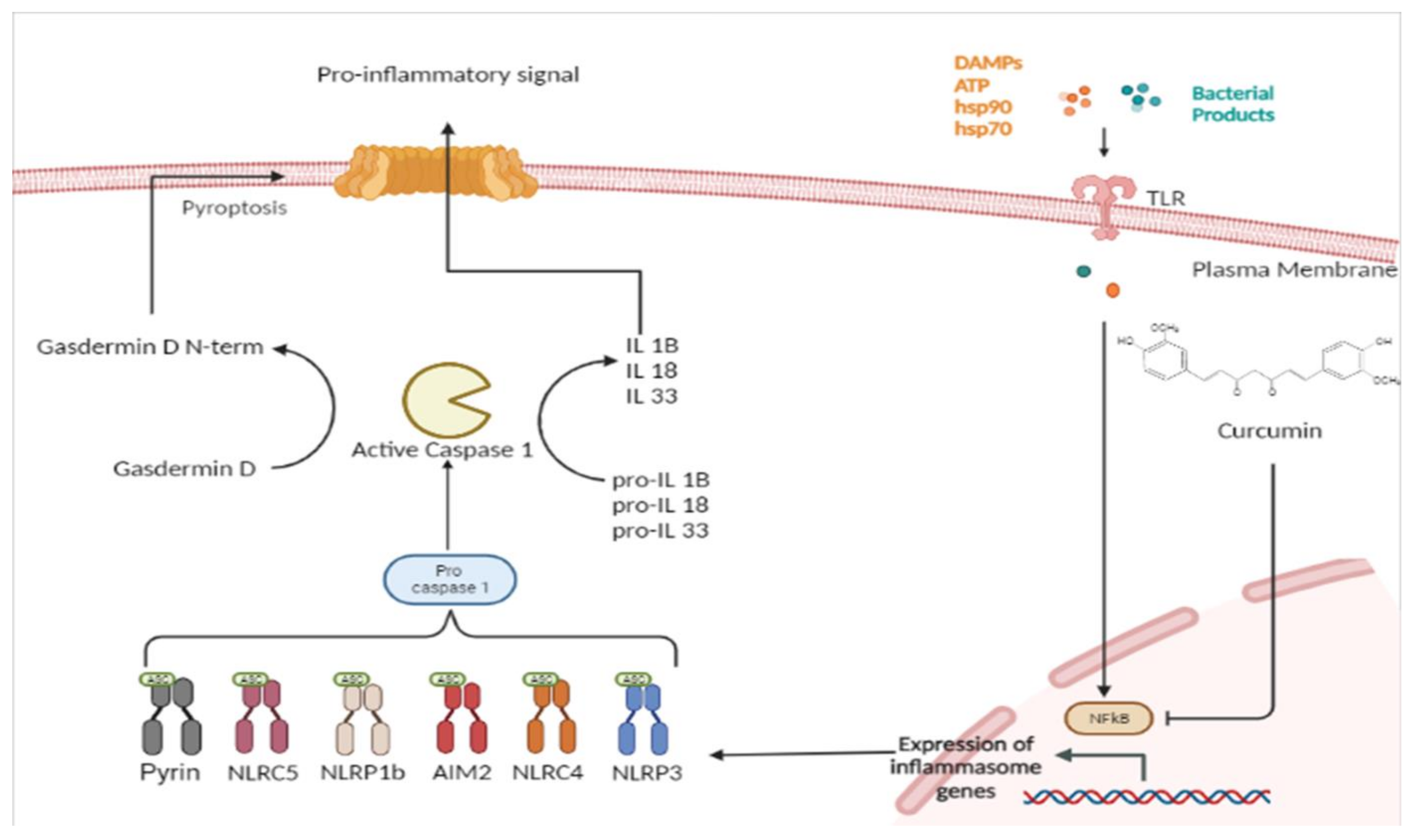
Figure 1.
Curcumin inhibition of canonical inflammasome.
Additionally, AIM2 is a protein that is the founding member of the AIM2-like receptor (ALR) family. ALRs are typically bipartite proteins with one or more HIN domains following the N-terminal pyrin domain (PYD). Caspase-1 is mainly activated by inflammasome assembly. Nucleotide-binding domain and leucine-rich repeat receptors (NLRs) or absent in melanoma 2 (AIM2)-like receptors (ALRs) initiate the assembly of the canonical inflammasome complex [14]. NLRs and ALRs mediate host recognition of PAMPs released during infection or DAMPs released during cell damage. In the case of some of these PRRs, such as NLRP1, NLRP3, and AIM2, the skeleton protein consists mainly of a carboxyl-terminal containing leucine-rich repeats (LRRs) that recognizes the ligand PAMPs. A central domain includes the NACHT cassette, which hydrolyzes adenosine triphosphates (ATP) by activating a deoxyribonucleoside triphosphate (dNTP) enzyme, and an amino acid terminal domain consisting of caspase-recruitment domains (CARD), a pyrin domain (PYD), an acid transactivator, and baculovirus inhibitor repeats (BIRs). Two signals are usually required for the activation of the canonical inflammasome. Ligands such as PAMPs and DAMPs that activate inflammasomes make up the second signal. PAMPs and DAMPs are recognized by the host through the action of NLRs and ALRs. The N-terminal CARD facilitates the recruitment of pro-caspase-1 to the inflammasome complex by attracting apoptosis-associated speck-like protein, which contains a caspase-recruitment domain (ASC) [17]. A subset of these receptors subsequently assembles cytosolic protein complexes known as inflammasomes, which activate proinflammatory caspase-1 and -11. In most cases, activated NLRs and ALRs recruit a bipartite protein ASC to engage caspase-1 activation. In macrophages or dendritic cells, inflammasome-forming NLRs and ALRs induce the reorganization of cytoplasmic ASC into a single ‘speck’ of 0.8–1 μm, which is thought to be a hallmark of inflammasome assembly. ASC is critical for the recruitment of caspase-1 to facilitate the proteolytic processing of pro-IL-1β and pro-IL-18. In fact, upon activation of inflammasomes, only caspase-1 produces mature IL-1β, a pyrogenic cytokine that also promotes adaptive T helper 1 (Th1), Th17, and humoral immunity. IL-1β is an important proinflammatory mediator that is generated at sites of injury or immunological challenges to coordinate programs as diverse as a cellular recruitment to a site of infection or injury. In addition, caspase-1 produces mature IL-18, which is important for IL-17 expression by Th17 cells and, when combined with other cytokines, can polarize T cells towards Th1 or Th2 profiles. Finally, caspase-1-processed IL-33 mainly induces Th2 cells to release IL-13 and IL-5 [20]. In addition, the activation of caspase-1 and caspase-11 triggers the cleavage of the gasdermin D protein (GSDMD). GSDMD splits into two fragments: the N-terminal domain and the C-domain. The N-terminal domain of GSDMD (N-GSDMD) oligomerizes and forms plasma membrane pores on lipid membranes and induces pyroptosis that mediates cell death and the secretion of mature forms of IL-1β and IL-18 [14]. Pyroptosis is a non-homeostatic and lytic mode of cell death that requires caspase-1 or -11 enzymatic activities. Like caspase-independent necroptotic cell death, pyroptotic cell death is characterized by cytoplasmic swelling and plasma membrane rupture [16,21][16][21]. Excessive pyroptosis may promote sepsis by inducing immunosuppression while amplifying the inflammatory response. NLRP1 was the first member of the NLR family identified to form an inflammasome complex [11]. Humans have a single NLRP1 gene, while the mouse genome encodes three paralogs: Nlrp1a, Nlrp1b, and Nlrp1c. The Nlrp1 inflammasome is an important defense mechanism against Bacillus anthraces, as this variant responds to lethal toxins. Nlrp1b is the key locus that determines whether macrophages undergo pyroptosis and IL-1β secretion in response to bacterial toxins. Toxins and muramyl dipeptide (MDP) as PAMPs lead to an efflux of intracellular potassium ions (K+), activate the NLRP1 inflammasome, and induce IL-1β secretion by macrophages. The NLRP3 inflammasome is currently the best-characterized inflammasome. NLRP3 is activated when exposed to a wide range of PAMPs and DAMPs, such as bacterial messenger RNAs, bacterial DNAs, MDP, DNA and RNA viruses, and several host-derived molecules indicative of injury, including extracellular ATP released by injured cells. The activation of the NLRP3 inflammasome requires two separate signals. Signal one (priming process) is triggered by activation of Toll-like receptor (TLR) 4 or tumor necrosis factor (TNF) signaling, which subsequently leads to the transcriptional activation of NLRP3. TLRs, IL-1Rs, and TNFRs induce transcription and translation of NF-κB to produce proforms of IL-1β and IL-18 [22]. As a second step, enzymatic cleavage is required to secrete these pro-inflammatory ILs into the extracellular space. This requires oligomerization of NLRP3 with ASC, facilitating proteolytic cleavage of pro-caspase-1 to its active caspase-1. Caspase-1 then cleaves the pro-cytokines into mature IL-1β and IL-18, which can be secreted. However, recent studies have shown that caspase-11 (caspase-4 and caspase-5 in humans) is required for the non-canonical activation of caspase-1 in macrophages infected with the enteric bacteria Escherichia coli, Citrobacter rodentium, and Vibrio cholerae, whereas caspase-11 is dispensable for caspase-1 activation by canonical NLRP3 activators such as ATP and nigericin. Furthermore, the activation of the NLRP3 inflammasome in LPS-induced endotoxaemia is caspase-11 dependent. The activation signal two (protein complex assembly) is induced by various pathogen-associated molecular patterns (PAMPs) and damage-associated molecular patterns (DAMPs), including extracellular ATP, pore-forming toxins, RNA viruses, and particulate matter. Interleukin 1β (IL-1β)/IL-1R1, LPS/Toll-like receptor 4 (TLR4), tumor necrosis factor (TNF)/TNF receptor (TNFR), sphingosine 1-phosphate (S1P)/S1P receptor 2 (S1PR2), adenosine diphosphate (ADP)/P2Y12, α-synuclein/CD36, and bromodomain-containing protein 4 (BRD4) inhibitor. JQ1 activates NF-κB and then upregulates the transcription of the component required for NLRP3 inflammasome formation. NLRP3 interacts with a wide range of activators. It is unlikely that NLRP3 interacts physically with its activators. NLRP3 may instead detect a common cellular signal induced in response to NLRP3 activators [23]. The identity of this signal is currently under intensive debate. Several molecular and cellular events, including K+ efflux, Ca2+ signaling, reactive oxygen species (ROS), mitochondrial dysfunction, and lysosomal rupture have been proposed as the trigger(s) for NLRP3 inflammasome activation, [22]. Several studies showed that nigericin, a potassium ionophore, stimulates IL-1β maturation in LPS-stimulated murine macrophages. Furthermore, nigericin promotes K+ efflux, and a decrease in cytosolic K+ concentration is sufficient to activate the NLRP3 inflammasome. Furthermore, studies showing that nigericin induces Ca2+ mobilization during the NLRP3 inflammasome activation process [23] support the involvement of Ca2+ signaling in NLRP3 inflammasome activation. NLRP3 has been shown to reside in the endoplasmic reticulum (ER) and cytosol. Following activation by various stimuli, the NLRP3 inflammatory complex is assembled at MAMs, which are highly specialized contact sites between the ER and mitochondria. The localization of NLRP3 to MAMs/mitochondria may contribute to the immediate recognition of mitochondrial damage, mitochondrial DNA (mtDNA) translocation, and cardiolipin, followed by a response. It was recently proposed that mtDNA released into the cytosol is the “ultimate” trigger of the NLRP3 inflammasome under various “priming” and “activation” conditions [24]. The co-localization of NLRP3 and cytoplasmic mtDNA induces the production of IL-1β. In the cytosol, the presence of oxidized mtDNA is a more potent inducer of IL-1β secretion [24]. Importantly, NLRP3 localization to the MTOC leads to its interaction with the centrosome-localized mitotic kinase NEK7, allowing NLRP3 inflammasome assembly [24]. A crucial protein is the mitotic serine/threonine-protein kinase NEK7. NEK7 interacts with NLRP3, which causes oligomerization of NLRP3 [25]. A recent structural modeling study revealed that the molecular mechanism of NEK7–NLRP3 interactions bridges adjacent NLRP3 subunits and promotes NLRP3 inflammasome oligomerization. It is unclear whether NEK7 is absolutely required for NLRP3 oligomerization and further inflammasome assembly [25]. Recent research has highlighted the importance of the Golgi apparatus and its lipid mediators in the aggregation of NLRP3 and the activation of NLRP3 inflammasome assembly [25]. Indeed, when exposed to specific stimuli, NLRP3 causes the trans-Golgi network (TGN) to disassemble into the dispersed TGN (dTGN). Notably, K+ efflux-independent stimuli cause NLRP3-dTGN activation, which leads to aggregation and activation of the NLRP3 inflammasome [25]. In addition, NLRP3 inflammasome activation is dependent on the ER-to-Golgi translocation of sterol regulatory element-binding protein (SREBP) 2 and SREBP cleavage-activating protein (SCAP), which form a ternary complex with NLRP3. Another recent study found that the NLRP3 inflammasome causes MAMs to be localized near Golgi membranes. This inter-organelle communication is dependent on the recruitment of protein kinase D (PKD) to DAG sites at the Golgi, which facilitates NLRP3 oligomerization and assembly of the active inflammasome [25]. Intense efforts have been made over the past decade to investigate the mechanism of NLRP3 inflammasome activation. The identification of an alternative NLRP3 inflammasome signaling pathway is important. Recent studies have shown that LPS induces an “alternative inflammasome” in human monocytes. This alternative inflammasome activation was propagated by TLR4-TRIF-RIPK1-FADD-CASP8 signaling upstream of NLRP3 [23]. In this model, LPS induces the release of endogenous ATP from human monocytes, which in turn activates the P2X7 receptor to trigger NLRP3 inflammasome activation and IL-1β maturation. This pathway does not require K+ efflux, which is in contrast to NLRP3 activation induced by ATP, pore-forming toxins and particulate matter. Therefore, this pathway is defined as an alternative NLRP3 inflammasome pathway. After LPS treatment, the molecules RIPK1, FADD, and CASP8 act downstream of TLR4-TRIF signaling to activate NLRP3. Although both ASC and caspase-1 are required, there is no evidence of ASC speck formation or pyroptosis in this pathway [23]. The NLRC4 inflammasome is normally activated by two crucial components of pathogenic bacteria: flagellin of Gram-positive and Gram-negative bacteria, and proteins from the type III secretion system (T3SS) proteins, which originate from Gram-negative bacteria and inject virulence factors into the host cell [26]. The ALR protein recruits AIM2 to form a conventional inflammasome, which then activates caspase-1. Oligosaccharides and PYDs make up the structure of the AIM2 inflammasome. The AIM2 inflammasome consists of oligosaccharide domains and PYDs. The AIM2 inflammasome identifies double-stranded DNA in the cell using the oligosaccharide domain and then recruits ASC via the PYD, which leads to self-activation of pro-caspase-1. AIM2 has been implicated in the pathology of autoimmune disorders such as systemic lupus erythematosus, in which anti-DNA autoantibodies are abundant. If the pathway is confirmed, it will be a candidate for targeted therapies [17]. The pyrin protein is a 781-amino acid, ~95 kDa protein encoded on chromosome 16 by MEFV. Pyrin is expressed by innate immune system cells such as monocytes, granulocytes, and eosinophils. Homology analyses revealed five distinct domains within the pyrin protein. The eponymous PYD (1–92) is found at the N-terminus of pyrin and is found in more than 20 human proteins that are primarily involved in inflammatory processes. The PYD binds apoptosis-associated speck-like protein with a caspase-recruitment domain (ASC), resulting in caspase-1-mediated IL-1β production. The C-terminal B30.2 domain of pyrin is critical for the molecular mechanisms leading to familial Mediterranean fever (FMF). Most of the FMF-associated mutations are clustered in this C-terminal domain. Familial Mediterranean fever (FMF) is associated with mutations in the MEFV gene. In addition, pyrin-associated auto-inflammation with neutrophilic dermatosis (PAAND) has been associated with MEFV mutation. Recent findings have now revealed how pyrin is activated during infection. FMF is a prototypic auto-inflammatory disease characterized by recurrent episodes of fever with serosal inflammation manifesting with severe abdominal or chest pain, arthralgia, monoarticular arthritis, and limited erythematous skin rash. Pyrin does not directly identify molecular patterns (pathogen- or host-derived danger molecules) [27]. Rather, pyrin responds to perturbations in cytoplasmic homeostasis caused by the infection. These disturbances, recently defined as ‘homeostasis-altering molecular processes’ (HAMPs), are processes that result in RhoA GTPase inactivation [27,28][27][28].

References

  1. Hewlings, S.J.; Kalman, D.S. Curcumin: A Review of Its Effects on Human Health. Foods 2017, 6, 92–98.
  2. Benameur, T.; Soleti, R.; Panaro, M.; La Torre, M.; Monda, V.; Messina, G.; Porro, C. Curcumin as Prospective Anti-Aging Natural Compound: Focus on Brain. Molecules 2021, 26, 4794.
  3. Chilelli, N.C.; Ragazzi, E.; Valentini, R.; Cosma, C.; Ferraresso, S.; Lapolla, A.; Sartore, G. Curcumin and Boswellia serrata Modulate the Glyco-Oxidative Status and Lipo-Oxidation in Master Athletes. Nutrients 2016, 8, 745.
  4. Simioni, C.; Zauli, G.; Martelli, A.M.; Vitale, M.; Sacchetti, G.; Gonelli, A.; Neri, L.M. Oxidative stress: Role of physical exercise and antioxidant nutraceuticals in adulthood and aging. Oncotarget 2018, 9, 17181–17198.
  5. Cavaliere, G.; Trinchese, G.; Penna, E.; Cimmino, F.; Pirozzi, C.; Lama, A.; Annunziata, C.; Catapano, A.; Mattace Raso, G.; Meli, R.; et al. High-Fat Diet Induces Neuroinflammation and Mitochondrial Impairment in Mice Cerebral Cortex and Synaptic Fraction. Front. Cell. Neurosci. 2019, 13, 509.
  6. Zia, A.; Farkhondeh, T.; Pourbagher-Shahri, A.M.; Samarghandian, S. The role of curcumin in aging and senescence: Molecular mechanisms. Biomed. Pharmacother. 2021, 134, 111119.
  7. Wang, X.; Wang, Y.; Chen, Z.G.; Shin, D.M. Advances of Cancer Therapy by Nanotechnology. Cancer Res. Treat. 2009, 41, 1–11.
  8. Panaro, M.A.; Benameur, T.; Porro, C. Extracellular Vesicles miRNA Cargo for Microglia Polarization in Traumatic Brain Injury. Biomolecules 2020, 10, 901.
  9. Pricci, M.; Bourget, J.-M.; Robitaille, H.; Porro, C.; Soleti, R.; Mostefai, H.A.; Auger, F.A.; Martinez, M.C.; Andriantsitohaina, R.; Germain, L. Applications of Human Tissue-Engineered Blood Vessel Models to Study the Effects of Shed Membrane Microparticles from T-Lymphocytes on Vascular Function. Tissue Eng. Part A 2009, 15, 137–145.
  10. Biancatelli, R.M.C.; Solopov, P.A.; Catravas, J.D. The Inflammasome NLR Family Pyrin Domain-Containing Protein 3 (NLRP3) as a Novel Therapeutic Target for Idiopathic Pulmonary Fibrosis. Am. J. Pathol. 2022, 192, 837–846.
  11. Huang, Y.; Xu, W.; Zhou, R. NLRP3 inflammasome activation and cell death. Cell. Mol. Immunol. 2021, 18, 2114–2127.
  12. Bauernfeind, F.G.; Horvath, G.; Stutz, A.; Alnemri, E.S.; Macdonald, K.L.; Speert, D.P.; Fernandes-Alnemri, T.; Wu, J.; Monks, B.G.; Fitzgerald, K.; et al. Cutting Edge: NF-κB Activating Pattern Recognition and Cytokine Receptors License NLRP3 Inflammasome Activation by Regulating NLRP3 Expression. J. Immunol. 2009, 183, 787–791.
  13. Masters, S.L.; Dunne, A.; Subramanian, S.L.; Hull, R.L.; Tannahill, G.M.; Sharp, F.A.; Becker, C.; Franchi, L.; Yoshihara, E.; Chen, Z.; et al. Activation of the NLRP3 inflammasome by islet amyloid polypeptide provides a mechanism for enhanced IL-1β in type 2 diabetes. Nat. Immunol. 2010, 11, 897–904.
  14. Man, S.M.; Kanneganti, T.-D. Regulation of inflammasome activation. Immunol. Rev. 2015, 265, 6–21.
  15. Franchi, L.; Muñoz-Planillo, R.; Núñez, G. Sensing and reacting to microbes through the inflammasomes. Nat. Immunol. 2012, 13, 325–332.
  16. Lamkanfi, M.; Dixit, V.M. Mechanisms and Functions of Inflammasomes. Cell 2014, 157, 1013–1022.
  17. Zhang, W.-J.; Chen, S.-J.; Zhou, S.-C.; Wu, S.-Z.; Wang, H. Inflammasomes and Fibrosis. Front. Immunol. 2021, 12, 643149.
  18. Wang, B.; Yin, Q. AIM2 inflammasome activation and regulation: A structural perspective. J. Struct. Biol. 2017, 200, 279–282.
  19. Doly J, Civas A, Navarro S, Uze G. Type I interferons: Expression and signalization. Cell Mol. Life Sci. 1998, 54, 1109–1121.
  20. Dinarello, C.A. Immunological and Inflammatory Functions of the Interleukin-1 Family. Annu. Rev. Immunol. 2009, 27, 519–550.
  21. Lamkanfi, M. Emerging inflammasome effector mechanisms. Nat. Rev. Immunol. 2011, 11, 213–220.
  22. Lorenz, G.; Darisipudi, M.N.; Anders, H.-J. Canonical and non-canonical effects of the NLRP3 inflammasome in kidney inflammation and fibrosis. Nephrol. Dial. Transplant. 2013, 29, 41–48.
  23. He, Y.; Hara, H.; Núñez, G. Mechanism and Regulation of NLRP3 Inflammasome Activation. Trends Biochem. Sci. 2016, 41, 1012–1021.
  24. Li, S.; Li, H.; Zhang, Y.-L.; Xin, Q.-L.; Guan, Z.-Q.; Chen, X.; Zhang, X.-A.; Li, X.-K.; Xiao, G.-F.; Lozach, P.-Y.; et al. SFTSV Infection Induces BAK/BAX-Dependent Mitochondrial DNA Release to Trigger NLRP3 Inflammasome Activation. Cell Rep. 2020, 30, 4370–4385.e7.
  25. Paik, S.; Kim, J.K.; Silwal, P.; Sasakawa, C.; Jo, E.-K. An update on the regulatory mechanisms of NLRP3 inflammasome activation. Cell. Mol. Immunol. 2021, 18, 1141–1160.
  26. Duncan, J.A.; Canna, S.W. The NLRC4 Inflammasome. Immunol. Rev. 2017, 281, 115–123.
  27. Schnappauf, O.; Chae, J.J.; Kastner, D.L.; Aksentijevich, I. The Pyrin Inflammasome in Health and Disease. Front. Immunol. 2019, 10, 1745.
  28. Heilig, R.; Broz, P. Function and mechanism of the pyrin inflammasome. Eur. J. Immunol. 2017, 48, 230–238.
More
Academic Video Service