Artificial Intelligence in Fluorescent Nanodiamonds Biomedical Imaging: Comparison
Please note this is a comparison between Version 2 by Jessie Wu and Version 1 by Wesley Wei-Wen Hsiao.

The ability to precisely monitor the intracellular temperature directly contributes to the essential understanding of biological metabolism, intracellular signaling, thermogenesis, and respiration. The intracellular heat generation and its measurement can also assist in the prediction of the pathogenesis of chronic diseases. Intracellular thermometry without altering the biochemical reactions and cellular membrane damage is challenging, requiring appropriately biocompatible, nontoxic, and efficient biosensors. Bright, photostable, and functionalized fluorescent nanodiamonds (FNDs) have emerged as excellent probes for intracellular thermometry and magnetometry with the spatial resolution on a nanometer scale. The temperature and magnetic field-dependent luminescence of naturally occurring defects in diamonds are key to high-sensitivity biosensing applications. Alterations in the surface chemistry of FNDs and conjugation with polymer, metallic, and magnetic nanoparticles have opened vast possibilities for drug delivery, diagnosis, nanomedicine, and magnetic hyperthermia. The possibilities and outcomes of using AI strategies recommended for early stage disease diagnosis and imaging are discuessed. 

  • fluorescent nanodiamonds
  • AI
  • biosensing
  • bioimaging

1. Introduction

The quantum defects possessing extraordinary optical and electronic properties in fluorescent nanodiamonds (FNDs) owe a broad scope in the scientific horizon of biosensors for electromagnetic fields, including thermal and magnetic signals. Recent trends have novelties in surface modification and conjugation with biocompatible materials enabling the detection of static and time-dependent fields for ultrasensitive in vivo measurements. These efforts also allow the quantitative prediction of intracellular thermodynamics. Herein, we compile thermal and magnetic-sensing methodologies for rapidly detecting infectious viruses (viz., SARS-CoV, HIV, Ebola, and influenza) and deadly diseases (viz., primary and secondary cancers, Alzheimer’s, and Parkinson’s disabilities). We also assessed the feasibility of using FNDs as a therapeutic agent and wide-field microscopy for secondary-stage cancer, consequently improving public health. Further, the application of artificial intelligence (AI) in clinical decision making is vital for early disease diagnosis using different machine- and representation-learning strategies.
Nanodiamonds can be easily synthesized from a bulk material by thin films, nanorods, and nanoparticles. Single crystals in a millimeter size occupying fluorescent defects along preferred crystallographic planes can be fabricated with the chemical vapor deposition (CVD) method [3][1]. The CVD method has shown lower paramagnetic impurity content enabling an enhanced magnetic field sensitivity in the nanotesla regime [4][2]. This sample type generally provides a much better spatial resolution for imaging using individual defects than nanocrystals. Detonation nanodiamonds (DNDs) can be formed by detonating explosive compounds (TNT and RDX) in a controlled environment. As a result of the explosion, the supersaturated carbon vapors condense into tiny droplets, which later form nanocrystals. The DNDs available as ultra-small nanocrystals (less than 10 nm) are not considered favorable candidates for biosensing because of the low photo stability and impurities.
Diamond nanocrystals can be formed by crushing and grinding micron-size diamonds. Afterward, the high-pressure high-temperature method (HPHT) favors the formation of stable luminescent defects. The synthesis of small nanoparticles (1–5 nm) and large 100–500 nm particles can be accomplished using the HPHT method [5][3]. The naturally occurring impurity atoms in diamond are nitrogen (about 1% by mass), silicon, germanium, and tin. The luminescence emission of these defects is observed to have enhanced effects by increasing the nitrogen content to different extents of <80 and >372 ppm [6][4]. On average, the available nitrogen content in the HPHT and CVD-grown diamonds is 100 and 1 ppm, respectively. The presence of nuclear spins (13C, 14N, and 15N) interacting with the desired quantum defects in diamond have also been observed [7,8][5][6].
The nitrogen vacancy center (NV center), among the most commonly observed and studied luminescent defects in diamonds, is formed due to the combination of a nitrogen atom with a vacant diamond lattice site. The formation of the NV center in a diamond can be accomplished by ion implantation followed by annealing. The creation of NV centers in diamonds is observed to have a strong correlation with the energy of the ion implantation, causing the formation of shallow and deep defects [9,10][7][8]. The creation efficiency and depth of NV centers are (1%, 8 nm) for 5 keV ion energy and (45% micro-meter depth) for 18 MeV [11][9].

2. Application of Functionalized Fluorescent NDanodiamonds Bioimaging with Artificial Intelligence
The aim of AI and machine-learning (ML) algorithms in medical science is to develop strategies for early stage disease diagnosis by analyzing clinical data using convolution neural networks and deep-understanding (DL) deterministic modeling. ML algorithms are already being tested for predicting symptoms of brain tumors, brain aging, and neural disorders.
ML algorithms have been empowering FNDs for intracellular magnetic microscopy [109][10]. Deep-learning algorithms enable the high-contrast reconstruction of magnetic images from optical images. The optical expression of biomarkers is successfully converted into a sufficient magnetic intensity. In this study, when labeled with magnetic nanoparticles, the sample tissues allowed the observation of the size, morphology, and growth of tumor cells. Fluorescence-based biosensors can significantly benefit from the potential uses of ML algorithms, such as rapid on-chip data processing, noise suppression, image analysis, and segmentation [112][11]. Integrating an AI-based processor with medical test strips can support the data acquisition and real-time monitoring for any non-linearity in the biosensor response under inevitable conditions or contamination.
Further extending the scope of AI-powered electronic chips, which can store and process various algorithms and access large online disease databases, can make rapid onsite testing using pattern recognition and alert the physicians for any possible anomalies during the clinical trials. Intracellular imaging using fluorescent biosensors faces challenges due to time-varying non-linear backgrounds. Artificial neural networks were used to solve the inverse problem of optical biosensing in chicken egg white [113][12]. After implementing the artificial neural networks, the optical signal of FNDs was successfully filtered out from the background autofluorescence under low concentrations of FNDs (2–3 µg/mL). With this approach, the accuracy of detection was enhanced by 1.5 times. A similar effort for reducing the background autofluorescence using egg protein was also reported [114][13]. The use of AI for FND-based drug development was discussed in [115][14]. The need for personalized drugs for diseases with diverse effects and lethal variants is pivotal. CURATE.AI is a clinically tested AI-based platform that assists medical experts in selecting drug and dose concentrations after analyzing the patient’s medical history [116][15]. The evaluation of AI for healthcare has been encouraged to achieve the desired outcome with minimal side effects.
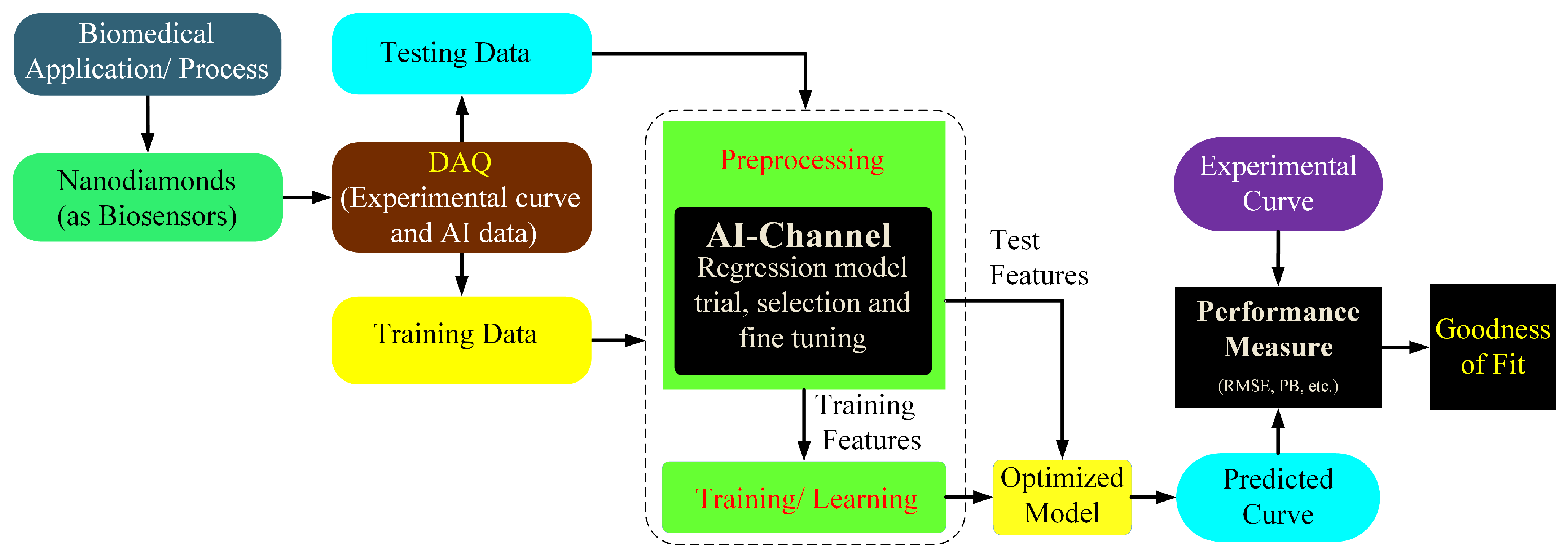
Currently, the main challenge is processing an extensive database for reliable results, demanding high computational resources and efficient algorithms providing accurate results. The fabrication of standard medicine is challenging and requires expert manpower, financial resources, and an unexpected time scale. In this regard, AI can significantly reduce the time and laborious effort by identifying the possible combinations of proteins and biomolecules, which could have better results and the least side effects. This idea can be further extended toward discovering appropriate biomarkers, which can then be utilized for disease diagnosis at different stages of progression and personalized drugs for individual cases. An example of this application where medical systems employing AI facilitated finding the optimized contents of a multi-drug conjugated with FNDs for curing human breast cancer was briefly examined in [117][16]. The efficiency and accuracy of this technique can be improved by using multiple data sources and sensors. An illustration of the model of the AI-based diagnosis is shown in Figure 1. Some of the AI’s tested and verified bioimaging applications associate brain imaging for detecting various types of tumors, such as glioma and Meningioma [118][17].
Figure 1. Illustration of AI scheme for the assessment and prediction of a biological process (BP). The proposed model relies upon the signal acquired from nanodiamonds which act as sensors providing electrical signals and an interface between BP and the data acquisition process. The accuracy of this model relies upon the comparison of the goodness of fit from the experimental and predicted curve based on root mean squared error values.

References

  1. Ozawa, H.; Hatano, Y.; Iwasaki, T.; Harada, Y.; Hatano, M. Formation of perfectly aligned high-density nv centers in (111) cvd-grown diamonds for magnetic field imaging of magnetic particles. Jpn. J. Appl. Phys. 2019, 58, SIIB26.
  2. Balasubramanian, G.; Neumann, P.; Twitchen, D.; Markham, M.; Kolesov, R.; Mizuochi, N.; Isoya, J.; Achard, J.; Beck, J.; Tissler, J. Ultralong spin coherence time in isotopically engineered diamond. Nat. Mater. 2009, 8, 383–387.
  3. Stehlik, S.; Varga, M.; Ledinsky, M.; Jirasek, V.; Artemenko, A.; Kozak, H.; Ondic, L.; Skakalova, V.; Argentero, G.; Pennycook, T. Size and purity control of hpht nanodiamonds down to 1 nm. J. Phys. Chem. C 2015, 119, 27708–27720.
  4. Chen, L.; Miao, X.; Ma, H.; Guo, L.; Wang, Z.; Yang, Z.; Fang, C.; Jia, X. Synthesis and characterization of diamonds with different nitrogen concentrations under high pressure and high temperature conditions. CrystEngComm 2018, 20, 7164–7169.
  5. Smeltzer, B.; Childress, L.; Gali, A. 13c hyperfine interactions in the nitrogen-vacancy centre in diamond. New J. Phys. 2011, 13, 025021.
  6. Felton, S.; Edmonds, A.; Newton, M.; Martineau, P.; Fisher, D.; Twitchen, D.; Baker, J. Hyperfine interaction in the ground state of the negatively charged nitrogen vacancy center in diamond. Phys. Rev. B 2009, 79, 075203.
  7. Antonov, D.; Häußermann, T.; Aird, A.; Roth, J.; Trebin, H.-R.; Müller, C.; McGuinness, L.; Jelezko, F.; Yamamoto, T.; Isoya, J. Statistical investigations on nitrogen-vacancy center creation. Appl. Phys. Lett. 2014, 104, 012105.
  8. Orwa, J.; Santori, C.; Fu, K.; Gibson, B.; Simpson, D.; Aharonovich, I.; Stacey, A.; Cimmino, A.; Balog, P.; Markham, M. Engineering of nitrogen-vacancy color centers in high purity diamond by ion implantation and annealing. J. Appl. Phys. 2011, 109, 083530.
  9. Pezzagna, S.; Naydenov, B.; Jelezko, F.; Wrachtrup, J.; Meijer, J. Creation efficiency of nitrogen-vacancy centres in diamond. New J. Phys. 2010, 12, 065017.
  10. Chen, S.; Li, W.; Zheng, X.; Yu, P.; Wang, P.; Sun, Z.; Xu, Y.; Jiao, D.; Ye, X.; Cai, M. Immunomagnetic microscopy of tumor tissues using quantum sensors in diamond. Proc. Natl. Acad. Sci. USA 2022, 119, e2118876119.
  11. Cui, F.; Yue, Y.; Zhang, Y.; Zhang, Z.; Zhou, H.S. Advancing biosensors with machine learning. ACS Sens. 2020, 5, 3346–3364.
  12. Dolenko, T.A.; Burikov, S.A.; Vervald, A.M.; Vlasov, I.I.; Dolenko, S.A.; Laptinskiy, K.A.; Rosenholm, J.M.; Shenderova, O.A. Optical imaging of fluorescent carbon biomarkers using artificial neural networks. J. Biomed. Opt. 2014, 19, 117007.
  13. Burikov, S.A.; Vervald, A.M.; Vlasov, I.I.; Dolenko, S.A.; Laptinskiy, K.; Dolenko, T.A. Use of neural network algorithms for elaboration of fluorescent biosensors on the base of nanoparticles. Opt. Mem. Neural Netw. 2013, 22, 156–165.
  14. Loh, K.P.; Ho, D.; Chiu, G.N.C.; Leong, D.T.; Pastorin, G.; Chow, E.K.H. Clinical applications of carbon nanomaterials in diagnostics and therapy. Adv. Mater. 2018, 30, 1802368.
  15. Blasiak, A.; Khong, J.; Kee, T. Curate. Ai: Optimizing personalized medicine with artificial intelligence. SLAS Technol. 2020, 25, 95–105.
  16. Wang, H.; Lee, D.-K.; Chen, K.-Y.; Chen, J.-Y.; Zhang, K.; Silva, A.; Ho, C.-M.; Ho, D. Mechanism-independent optimization of combinatorial nanodiamond and unmodified drug delivery using a phenotypically driven platform technology. ACS Nano 2015, 9, 3332–3344.
  17. Qureshi, S.A.; Raza, S.E.A.; Hussain, L.; Malibari, A.A.; Nour, M.K.; Rehman, A.u.; Al-Wesabi, F.N.; Hilal, A.M. Intelligent ultra-light deep learning model for multi-class brain tumor detection. Appl. Sci. 2022, 12, 3715.
More
Video Production Service