Wound healing continues to pose a challenge in clinical settings. Moreover, wound management must be performed properly and efficiently. Acute wound healing involves multiple cell divisions, a new extracellular matrix, and the process of formation, such as growth factors and cytokines, which are released at the site of the wound to regulate the process. Any changes that disrupt the healing process could cause tissue damage and prolong the healing process. Various factors, such as microbial infection, oxidation, and inflammation, can delay wound healing. In order to counter these problems, utilizing natural products with wound-healing effects has been reported to promote this process. A natural product with medicinal properties, which contribute to alleviating these factors, can facilitate the wound-healing process and be developed as a future drug. NOver the last few years, numerous research has investigated the wound-healing properties of natural products that contain antioxidant, anti-inflammatory, collagen promotion, and antibacterial properties. Various phytochemicals, including alkaloids, tannins, flavonoids, terpenoids, phenolic, essential oils, and saponin compounds, may contribute to the medicinal effects. Natural products, including phytochemicals, play an important role in wound healing due to these properties.
1. Biosurfactant
The ability of biosurfactants to interact with modifying surfaces makes them surface-active compounds (SACs). In nature and biotechnology, these biomolecules serve different functions due to their physiological roles and physicochemical properties due to their amphiphilic nature and their production by different microorganisms. The benefits of SACs over synthetic surfactants include low toxicity, increased biodegradability, low critical micelle concentrations, and environmental acceptability
[1][2][3][13,14,15].
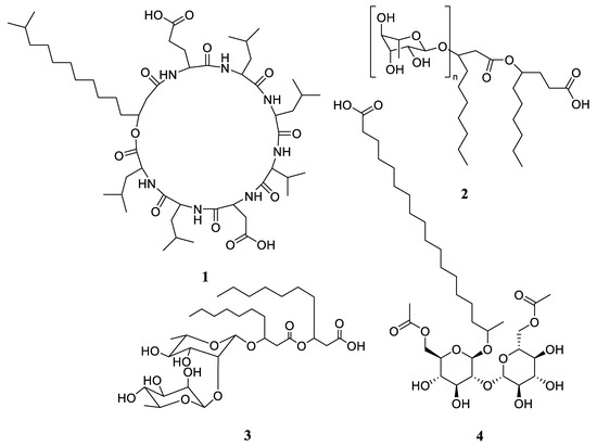
Surfactin A (
1,
Figure 1) isolated from
Bacillus subtilis was reported to promote wound healing and the inhibition effects of scars. Amid the healing process,
1 upregulated expression of hypoxia-inducible factor-1α (HIF-1α) and vascular endothelial growth factor. Moreover, it accelerated keratinocyte migration via the mitogen-activated protein kinase and factor nuclear-κB (NF-κB) signaling pathways, followed by the regulation of pro-inflammatory cytokine secretion and macrophage phenotypic exchange. In addition,
1 could heal the wound due to its antioxidant properties, with an IC
50 = 0.55 mg/mL
[4][5][25,26]. It also prevented scar tissue formation by inhibiting the expression of α-smooth muscle actin (α-SMA) and transforming growth factor (TGF-β).
Figure 1.
Chemical structures of surfactin A (
1
), BS glycolipid (
2
), rhamnolipid (
3
), and sophorolipid (
4
).
An in vitro study of a biosurfactant (BS) of a glycolipid nature (
2,
Figure 1), isolated from
Bacillus licheniformis SV1, exhibited adequate cytocompatibility and increased 3T3/NIH fibroblast proliferation. An approach using a BS ointment in vivo stimulated re-epithelialization, fibroblast proliferation, and the faster deposition of collagen in skin excision wounds in rats, thereby demonstrating its potential to improve wound healing through transdermal delivery
[6][27].
In a previous study, mice were treated with an ointment containing rhamnolipid (
3,
Figure 1) after creating severe wounds on their backs. The results of histopathological studies showed that
3 improved wound closure and collagenases, and it reduced inflammation by reducing the level of TNF-α without causing any negative skin reactions. There is evidence that dirhamnolipid therapy can alleviate scarring on the skin, as it has shown effects in rabbit ear hypertrophic scar models, with a depletion in α-SMA expression, type I collagen fibers, and scar elevation index scores
[7][28].
By substituting human skin with an in vitro model of human dermal fibroblasts, a cell culture model was utilized to demonstrate the wound-healing capacity of sophorolipid (
4,
Figure 1), revealing that
4 affected the human skin fibroblast expression of elastase inhibition collagen I mRNA, with IC
50 = 38.5 μg/mL. Additionally, an in vitro wound-healing evaluation in the human colorectal adenocarcinoma (HT-29) cell line showed a significant increase in collagenase-1 expression in HT-29 colorectal adenocarcinoma cells after treatment for 48 h with
4 [8][9][29,30].
2. Flavonoids
The flavonoid class of compounds possesses various biological functions, making them important sources of new pharmaceuticals, including those for treating skin wounds. The structure–activity relationship is one of the main factors that contributes to the pharmaceutical properties of flavonoids
[10][11][12][31,32,33]. Their anti-inflammatory
[13][34], antibacterial
[14][35], antioxidant
[15][36], and antifibrotic
[16][37] properties depend largely on the presence of hydroxyl groups due to high hydroxylation.
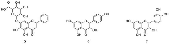
Baicalin (
5,
Figure 2) is a flavone glycoside with anti-inflammatory, antiviral, and photoprotective properties
[17][42]. Compound
5 blocks the pathological keratinocyte changes in psoriatic patients
[18][43]. An antioxidant and anti-inflammatory effect may be responsible for the wound-healing properties of the baicalin nanohydrogel preparation. Moreover,
5 inhibits nitric oxide and tumor necrosis factor-α (TNF-α), which are both important elements in the inflammatory process
[19][44]. Finally,
5 was found to exist in the endophytic fungus
Spiropes sp. and played a major role in the pharmacological action of
Scutellaria baicalensis [17][20][42,45].
Figure 2.
Chemical structures of baicalin (
5
), kaempferol (
6
), and quercetin (
7
).
Kaempferol (
6,
Figure 2) is known to inhibit cancer growth; reduce inflammation; promote antioxidant activity; and protect the heart, liver, and brain
[21][46]. With a 1%
w/
w ointment concentration,
6 exhibited wound-healing activity in diabetic and non-diabetic rats. This wound-healing activity was studied utilizing incision and excision wound models
[22][47]. According to both models, kaempferol showed crucial wound-healing activity, as indicated by the increase in the granulation tissue weight and hydroxyproline content in the incision wound model, as well as a reduction in the wound area, faster epithelialization, increased dry weight of the tissue, and increased hydroxyproline content in the excision wound model
[23][48]. Moreover,
6 was found in many edible vegetables and the extract of the endophytic fungi
Fusarium chlamydosporum from
Tylophora indica [24][25][26][49,50,51].
Bioflavonoids such as quercetin (
7,
Figure 2) are known for their anti-atherosclerotic, anti-hypercholesterolemic, anti-hypertensive, anti-inflammatory, and anti-obesity properties
[27][52]. Preclinical research has shown
7 to possess wound-healing properties
[28][29][53,54]. A study by Jangde et al. evaluated quercetin’s wound-healing properties in vitro and in vivo. The results indicated that wound healing was significantly accelerated, and the wound termination time significantly decreased compared to the regular dosage forms. These results suggested that connectivity tissue disorders can be treated effectively as wound-healing approaches
[29][54]. Quercetin was found in more than 20 plants
[17][42]. Moreover, in terms of obtaining quercetin from bacteria, it was isolated for the first time from a cyanobacterium,
Anabaena aequalis Borge
[30][55].
3. Quinones
Biochemical pigments called quinones are established in several living organisms, including fungi, bacteria, a few animals, and higher plants. There are many forms of quinones in nature, such as naphthoquinones, benzoquinones, polycyclic quinones, and anthraquinones.
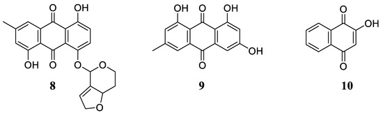
A new antibiotic, MT81 (
8,
Figure 3), was isolated and purified from
Penicillium nigricans culture media. It was found that
8 was a polyhydroxy anthraquinone, which exhibited antibacterial properties, along with an antiprotozoal effect toward
Leishmania donovani promastigotes in vitro. Moreover,
8 was investigated for its wound-healing effect in mice. The result exhibited the healing effects of an
8 ointment on mice infected with pathogenic organisms, and it was comparable to the positive control, nitrofurazone. This indicates that the antimicrobial effect of
8 helps to heal infected wounds by inhibiting microorganisms
[31][58].
Figure 3.
Chemical structures of MT81 (
8
), emodin (
9
), and hennotanic acid (
10
).
Emodin (
9,
Figure 3) is a natural anthraquinone derivative found in a wide variety of higher plants. Emodin obtained from microorganisms was first described as frangula-emodin, which was isolated from the fungus
Dermocybe sanguinea Wulf
[32][33][34][59,60,61]. In addition,
9 was identified as one of the pigmented products in the culture extracts of
Penicillium brunneum Udagawa,
Cladosporium fulvum Cooke,
Penicillium avellaneum,
Penicilliopsis clavariaeformis,
Penicillium islandicum Sopp,
Aspergillus wentii Wehmer,
Aspergillus ochraceus Wilhelm, and
Aspergillus cristatus [35][36][37][38][39][40][41][42][62,63,64,65,66,67,68,69]. Neuroprotective, anti-inflammatory, anticancer, antibacterial, anti-osteoporotic, hepatoprotective, antiviral, anti-allergic, and immunosuppressive properties are known to be associated with
9 [43][70]. Furthermore, the wound-healing activity of
9 using the excisional wound model in rats has been reported at dose levels of 100, 200, and 400 μg/mL
[44][71].
Hennotannic acid (
10,
Figure 3), also known as lawsone, is an orange-red dye extracted from the leaves of
Lawsonia inermis L., often known as the henna tree. However, Sarang et al. reported the production of 10 from an endophytic fungus,
Gibberella moniliformis, isolated from the leaf tissues of
Lawsonia inermis [45][72]. Moreover,
10 exhibited antibacterial, antifungal, antiparasitic, antitumor, and antiviral properties
[46][73]. By using an excision and incision model, Mandawgade and Patil investigated the wound-healing effects of
10 at dose levels of 50 mg/kg (per oral) and 0.1 mg/kg (topical/ointment). Based on the result,
10 exhibited significant wound-healing activity in both models
[47][74]. Another study on
10 also demonstrated crucial wound-healing activity in rodents
[48][75].
4. Phenolic Acids
Polyphenol is usually found in plants and marine organisms. Since polyphenols possess antimicrobial, regenerative, and antioxidant properties, they have significant potential for application in wound treatment
[49][50][51][76,77,78]. Besides their antimicrobial and antioxidant activity, polyphenols are also considered to be highly bioactive agents in wound dressings for acute wound treatment, thereby playing an important role in wound healing
[49][52][76,79].
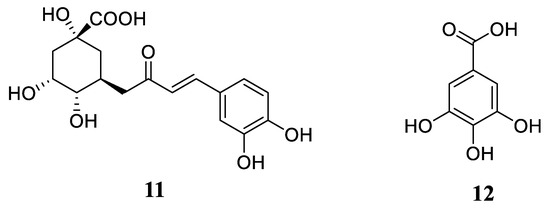
Chlorogenic acid (
11,
Figure 4) is an ester of caffeic acid with phenolic acid (quinic acid), established in twenty medicinal plants and twenty-nine fungal taxa, including
Phomopsis,
Colleterotrichum,
Phoma,
Alternaria, and
Xylariales [17][20][42,45]. Moreover,
11 showed significant wound-healing activity in both in vivo and in vitro investigations
[53][54][80,81]. A recent study demonstrated that
11 promotes wound closure and capillary tube formation, as well as enhanced wound closure with keratinocytes in vitro
[53][80]. In addition,
11 has been shown to enhance collagen synthesis by upregulating TNF-α and TGF-β during wound healing. It may promote wound healing in excision wounds due to its antioxidative properties
[54][81].
Figure 4.
Chemical structures of chlorogenic acid (
11
) and gallic acid (
12
).
Gallic acid (
12,
Figure 4) is a phenolic compound that can be found in plants, as well as nineteen endophytic fungal taxa
[20][45];
12 exhibits strong anti-inflammatory and antioxidant properties. During in vitro experiments,
12 accelerated keratinocyte and fibroblast migration and wound healing
[55][82]. In addition,
12 enhanced the expression of TGF-β and inhibited the nuclear factor κB (NF-κB), along with the proliferation and maturation phase of wound healing
[56][83].
5. Peptides
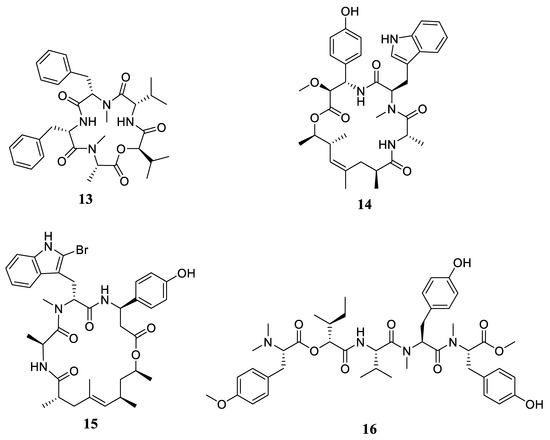
Acremonamide (
13,
Figure 5), a new cyclic depsipeptide obtained from
Acremonium sp., was isolated in an ongoing effort to discover new marine-derived natural products with wound-healing effects
[57][88]. It is known that species of the genus
Acremonium produce various secondary metabolites, such as hydroquinones, diterpenes, isocoumarins, sesquiterpenes, peptides, benzophenones, and butanolides
[58][89]. These fungal strains also produce cyclic depsipeptides, an interesting bioactive natural product
[58][59][60][61][89,90,91,92]. In in vivo experiments using the human wound healing RT
2 Profiler PCR array, adjustments in the wound-healing genes’ expression were screened to determine the mechanisms behind the wound-healing properties of
13. The result demonstrated, for the first time, that
13 increased keratinocyte and fibroblast motility, as well as COL1A2 and ACTC1 expression, thereby enhancing the wound-healing process
[57][88].
Figure 5.
Chemical structures of acremonamide (
13
), chondramide A (
14
), jasplakinolide (
15
), and apratyramide (
16
).
Chondramide A (
14,
Figure 5) and its analogs isolated from a terrestrial myxobacterium,
Chondramyces crocatus, were examined for the growth-inhibiting activity of actin cytoskeletons
[62][93]. The actin cytoskeleton plays a significant role in wound healing—it allows actomyosin to contract, recruits repair machinery, and migrates cells
[63][94]. By using highly purified recombinant actin from
T. gondii, in vitro polymerization assays confirmed that both synthetic and natural products target the actin cytoskeleton, with EC
50 values ranging from 0.3 to 1.3 μM. The results indicate that the chondramide treatment can prevent parasitic invasion and generate faster results than standard therapeutic agents such as pyrimethamine
[64][95].
Other studies reported the wound-healing activity of jasplakinolide (
15,
Figure 5) and apratyramide (
16,
Figure 5)
[65][66][96,97]. By binding to F-actin,
15 stabilized the actin filaments in vivo, leading to actin lumps and polynucleation, which are important in wound healing
[65][96]. Moreover,
16, a natural product isolated from a cyanobacterium, has been described to exert wound-healing effects by inducing vascular endothelial growth factor A
[66][97].
6. Triterpenoids
Terpenoids are plant-derived phytochemicals with a large variety of chemical structures derived from isoprene and usually have polycyclic structures. They are classified into monoterpenes, diterpenes, triterpenes, tetraterpenes, sesquiterpenes, and hemiterpenes, depending on the number of isoprene units in their structures. Triterpenes either have a tetracyclic or pentacyclic structure
[67][68][69][98,99,100]. Among pentacyclic triterpenes, oleanane, lupane, and ursane derivatives exhibit anti-inflammatory, anticancer, antioxidant, antiviral, and cardioprotective activities. In contrast, tetracyclic triterpenes have mostly been studied for their cytotoxic and anticancer biological properties
[70][71][72][73][101,102,103,104]. In recent years, systematic studies have examined the efficacy of triterpenes as wound healers. Based on the results of these studies, these phytocompounds have been displayed to promote wound healing by accelerating epithelialization and collagen formation and deposition, regardless of the wound type. Furthermore, their integration into various medicinal formulations is an effective option for wound management through their long-term delivery of active compounds. In conclusion, triterpenoids have been identified as an emerging class of wound care therapies
[67][74][98,105].
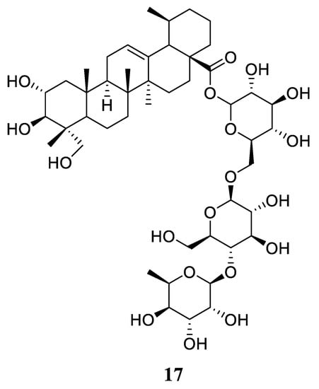
Asiaticoside (
17,
Figure 6) is a triterpenoid saponin, which is found in
Centella asiatica (L.), as well as in an endophytic fungus,
Colletotrichum gloeosporioides, obtained from
Centella asiatica (L.)
[75][76][106,107]. Based on the ability of
17 to induce the development of granulation tissue and collagenase-induced epithelialization in rabbits, it was proven that
17 exhibits wound-healing properties
[77][108]. In addition,
17 also induces type I collagen synthesis in human dermal fibroblast cells
[78][109].
Figure 6.
Chemical structures of asiaticoside (
17
).