Role of Endoscopy in Upper Gastrointestinal Cancers: Comparison
Please note this is a comparison between Version 2 by Jessie Wu and Version 3 by Jessie Wu.

Upper gastrointestinal (GI) malignancy is a leading cause of cancer-related morbidity and mortality. Upper endoscopy has an established role in diagnosing and staging upper GI cancers, screening for pre-malignant lesions, and providing palliation in cases of advanced malignancy. New advances in endoscopic techniques and technology have improved diagnostic accuracy and increased the therapeutic potential of upper endoscopy.

  • upper gastrointestinal cancer
  • endoscopy
  • cancer

1. Introduction

Since the inception of the flexible endoscope in the 1950s, gastrointestinal (GI) endoscopy has played an ever-increasing role in the diagnosis of upper GI tract cancers and screening of pre-cancerous lesions [1]. Modern advances in endoscopic technology and technique have led to significant improvements in diagnostic accuracy and expanded the role of endoscopy in cancer staging and treatment [2][3].
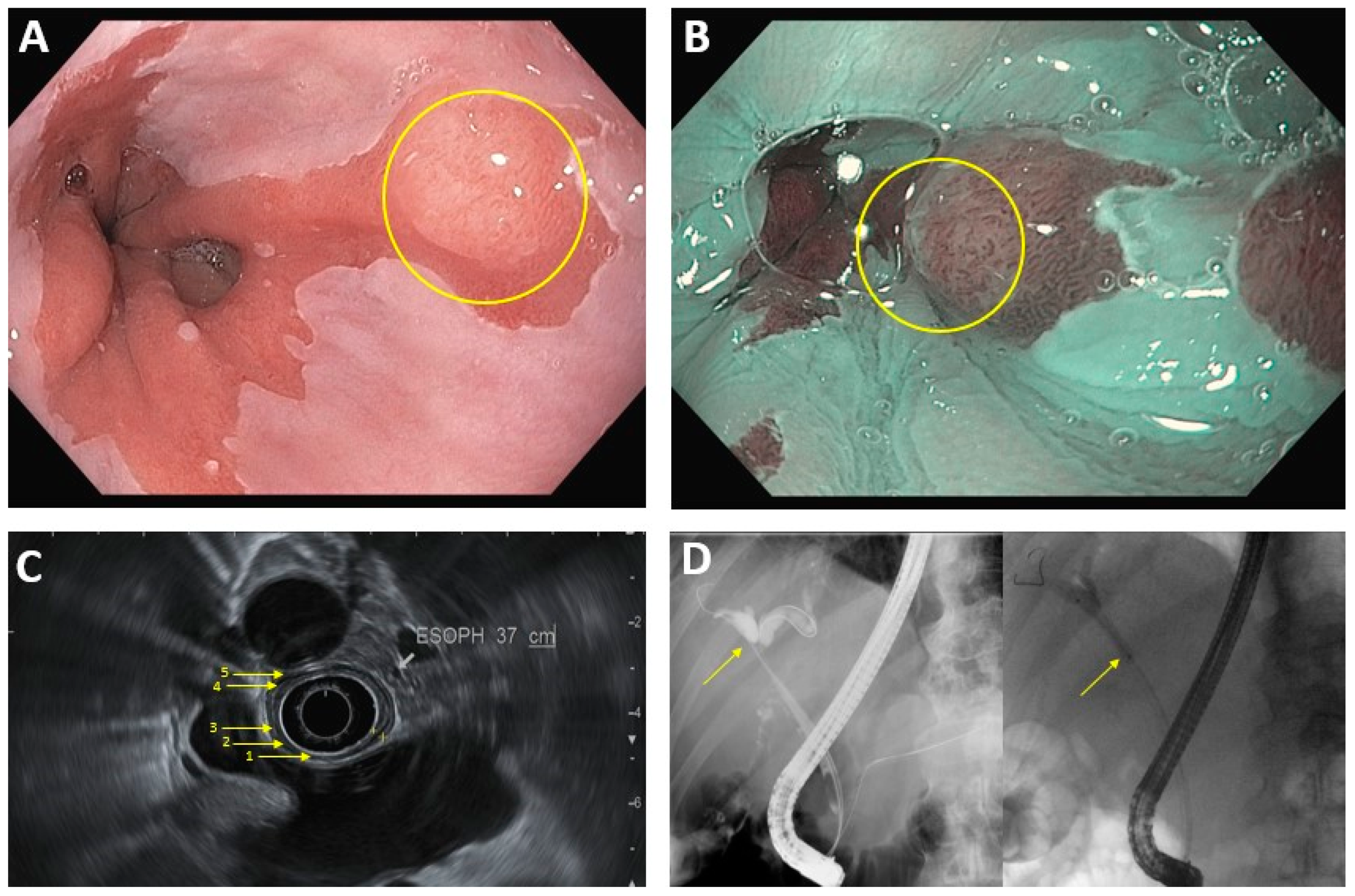
Conventional video endoscopy is the gold standard for diagnosing a wide range of upper GI malignancies. Chromoendoscopy, the intra-procedural application of stains and dyes to highlight abnormal mucosa, has traditionally been used to supplement video endoscopy and improve diagnostic yield [4]. Advances in HD imaging have led to the development of high-resolution endoscopy, allowing for closer magnification and clearer visualization of the mucosa [5]. Additionally, techniques such as narrow band imaging (NBI), which use camera filters or filter algorithms to identify malignant lesions, function as a modern “computerized virtual chromoendoscopy” that have lower interobserver variability compared to traditional chromoendoscopy while possibly providing similar (if not better) diagnostic accuracy (Figure 1) [3]. Autofluorescence imaging, which relies on detecting differences in light absorption/emission between dysplastic and normal tissue, is another form of image-enhancement endoscopy undergoing significant research, although its current utility is limited by a high false-positive rate [3][6]. Confocal laser endomicroscopy and endocytoscopy are newer, promising technologies that aim to visualize the mucosa at the cellular level in real-time, and additional studies are needed to better determine their optimal clinical utility [2][3][4][7][8].
Figure 1. Common endoscopic modalities. (A): Conventional HD endoscopy showing Barrett’s esophagus with lesion (circled). (B): Image in 1A under NBI. Area of abnormal vascular and mucosal patterns (circled) is concerning for malignancy. (C): EUS of the esophagus. Arrows denote 5 alternating hyperechoic/hypoechoic layers: (1) mucosal surface, (2) muscularis mucosa, (3) submucosa, (4) muscularis propia, (5) serosa. (D): ERCP in hilar cholangiocarcinoma. Left image: dilation of intrahepatic bile ducts because of complete blockage of the common hepatic duct (arrow). Right image: improved flow of bile/contrast after deployment of plastic biliary stent across the malignant obstruction.
In addition to direct visualization of luminal masses, evaluation of suspicious lesions in the gastrointestinal wall and extraluminal structures such as the pancreas can be performed via endoscopic ultrasound (EUS), and direct access to the biliary or pancreatic ducts for biopsy and palliative stent placement can be performed using endoscopic retrograde cholangiopancreatography (ERCP) [9][10][11][12]. Techniques such as fine-needle aspiration (FNA) can be used in conjunction with EUS to improve diagnostic accuracy, and technology such as elastography and contrast-enhanced ultrasound have been shown to improve sensitivity, specificity, and diagnostic yield when used to complement EUS-FNA [9][13][14]. Additionally, the function of EUS in visualizing and sampling the gastrointestinal wall layers, peri-intestinal lymph nodes, and surrounding structures gives it a crucial role in locoregional staging of malignancies [9][13][14]. Finally, there is an ever-growing therapeutic role for EUS, from supplementing ERCP in stent placement for malignant obstruction, to delivering anti-tumor agents via fine needle injection (FNI) in pancreatic cancer [15].

2. Diagnosis and Staging

2.1. Luminal Upper GastrointestinalI Cancer

EGD is indicated for patients with risk factors and symptoms concerning for upper GI malignancy, such as persistent dyspepsia, esophageal dysphagia or vomiting in patients older than 50 years, anorexia, weight loss, early satiety, and unexplained iron deficiency anemia [16]. Endoscopy with biopsy is required to definitively diagnose malignancy of the upper GI tract [17][18]. Newer studies that incorporate confocal light endomicroscopy have found a sensitivity and specificity of greater than 90%, suggesting a role for this technique in identifying targets to biopsy or delineating tumor margins prior to resection [19][20][21].
Tumors are most common staged according to the TNM (Tumor depth or size, Nodal metastasis, and Metastatic disease) system. This evaluation begins with CT imaging, since the presence of metastatic disease precludes curative tumor resection [18][22]. In patients without evidence of distant metastasis, EUS is the first-line method for T (tumor depth) and N (nodal metastasis) staging due to its minimally invasive nature (Table 1) [17][23][24][25]. It has been shown to be effective in distinguishing T1/T2 from T3/T4 cancer, and tends to be more sensitive for identifying advanced cancers (sensitivity greater than 90% for T3 and T4 cancers) than earlier stage ones (sensitivity approximately 81% for T1 and T2) [17][23][26][27]. However, the accuracy of EUS may be limited by inter-observer variability, proceduralist experience, and the anatomic location of the tumor [23][25]. Additionally, EUS cannot reliably distinguish T1a from T1b tumors [17][27][28][29][30]. This is an important distinction, because T1b tumors carry a significantly higher risk of nodal metastasis and are generally treated surgically, whereas T1a lesions may be managed with endoscopic resection [31][32]. In these instances, T-staging may require endoscopic resection, which has been shown to have better accuracy and lower inter-observer variability [23][28].
Endoscopic ultrasound (EUS) can inform N-staging by visualization locoregional lymph nodes or by lymph node biopsy via EUS-FNA. Four characteristics of lymph nodes seen on EUS—size > 10 mm, round shape, sharp borders, and absence of central intranodal vessels—are traditionally associated with a higher likelihood of malignancy, although its accuracy is poor (approximately 70% in esophageal cancer, and as low as 30% in gastric cancer) [17][23]. Studies have shown that accounting for three additional features—number of lymph nodes (≥5), involvement of the celiac nodes, and advanced primary tumor (T3 or greater)—improves the diagnostic accuracy for esophageal cancer (86% when ≥ 3 out of 7 features are present) [17][23]. When EUS findings are equivocal, EUS-FNA can be used to sample lymph nodes for histologic evaluation. However, EUS-FNA can yield false-positive results if the lymph nodes are accessed through the tumor site, and additional studies are needed to establish the definitive role of EUS-FNA in cancer staging [17][23][25].

2.2. Pancreaticobiliary Cancer

Pancreatic cancer is most commonly diagnosed via initial CT imaging; equivocal imaging results can often be clarified with EUS-FNA, which has been shown to have sensitivity and specificity greater than 90% [33]. Studies comparing EUS-FNA to cross-sectional imaging found that the former is especially accurate for diagnosing smaller pancreatic masses (<2 cm) [15][33].
Cholangiocarcinoma is traditionally diagnosed with cross-sectional imaging (MRCP is preferred), although distal extrahepatic neoplasms may be diagnosed via EUS-FNA or endoscopic retrograde cholangiography (ERC). Fluoroscopy-guided shaped endobiliary biopsy (FSEB) is a newer technique that involves manually re-shaping the endoscopy forceps to permit easier access to a biliary stricture, and has been shown to have high sensitivity for diagnosing both proximal and distal biliary neoplasms [34]. Intraductal cholangiocarcinoma is not easily accessible, and is typically diagnosed via multi-phase contrast MRI [35].
Gallbladder cancer is a rare entity that is diagnosed incidentally after cholecystectomy in approximately 50% of cases [36]. Individuals with a gallbladder polyp >1 cm seen on transabdominal ultrasound (TUS) are advised to undergo surgical resection. For patients with additional risk factors—such as primary sclerosing cholangitis (PSC), age > 50, focal wall thickening, or Indian ethnicity—cholecystectomy is recommended for polyps >5 cm [37]. EUS can be helpful in clarifying the diagnosis in less definitive cases [38][39]. EUS is more sensitive and specific than TUS in distinguishing neoplastic from non-neoplastic lesions; additionally, EUS-FNA can be performed to directly sample gallbladder lesions, with a reported accuracy between 80–100% [40].
Staging of pancreaticobiliary cancers generally begins with cross-sectional imaging, similar to the case with intraluminal cancers [35][38][41]. Surgical resection and staging laparoscopy are the most accurate modalities for evaluating gallbladder cancer and cholangiocarcinoma, respectively [35][38]. Pancreatic cancer may be accurately staged via endoscopy and imaging [41]. Studies have shown that EUS has a sensitivity of up to 94% for T staging and 82% for N staging [41]. It has also been shown to have high sensitivity and specificity for detecting portal venous invasion, but has poor accuracy for diagnosing arterial involvement of the tumor. This is an important distinction, since vascular involvement in pancreaticobiliary cancer makes the tumor non-resectable [41]. Helical CT remains the first-line technique for evaluating the vasculature; when these findings are non-diagnostic, supplementing with EUS has been shown to predict non-resectability with >90% accuracy [41].

3. Treatment of Cancer

3.1. Esophageal Squamous Cell CarcinomaC/Adenocarcinoma
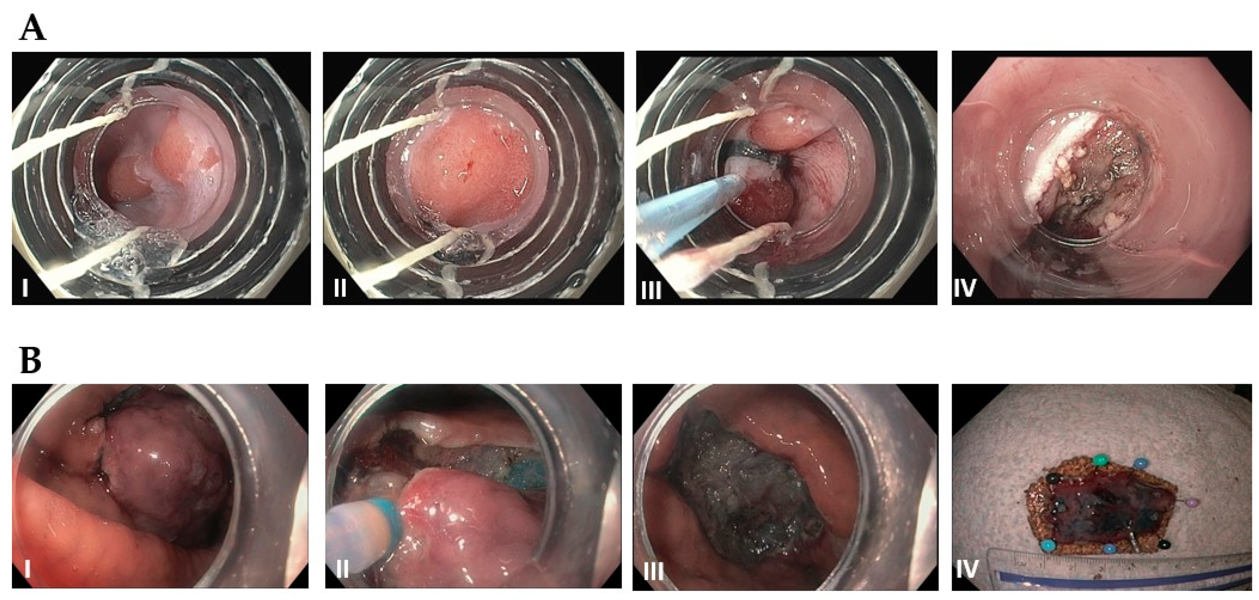
As mentioned above, stage T1a esophageal carcinoma in the absence of metastasis can be treated endoscopically, with the goal of achieving R0 resection (negative horizontal and vertical margins on histology) [31][42]. Studies have shown similar survival outcomes between endoscopic resection and esophagectomy, and lower morbidity, faster recovery, and decreased length of hospital stay with endoscopic treatment [43]. EMR has become the most common treatment technique for T1a esophageal cancer in the US. The ideal candidates are solitary, small (<1.5–2 cm), flat-type mucosal lesions without evidence of lymphovascular invasion [42][43][44]. While piecemeal resection may be performed for larger tumors, the excised samples cannot be accurately evaluated for negative margins [42]. ESD, is a newer technique that permits en bloc resection of larger tumors, and is associated with a lower risk of recurrence, but a higher rate of complications (Figure 2 for comparison of EMR and ESD technique) [42]. Endoscopic resection should always be followed up with ablative therapy of concomitant Barrett’s esophagus [43][44]. Patients who undergo curative resection still require interval surveillance, as the rate of developing metachronous tumors is high [44].
Figure 2. (A): Endoscopic mucosal resection (EMR) by band and snare technique: (I) Initial 1 cm, slightly raised esophageal lesion with surrounding Barrett’s esophagus. (II) Abnormal mucosal area is being capture completely into the cap with a cuff of normal surrounding mucosa. (III) Snare deployed around the band and resected using blended current. (IV) Resection base after cautery of underlying vessels. (B): Endoscopic Submucosal Dissection (ESD): (I) Initial large, well-demarcated lesion of the stomach. Lesions of the border are usually marked in the beginning and methylene bue or other lifting agents are injected to elevate the lesion (not performed here). (II) The lesion is resected with a needle-knife using electrocautery, starting at the previously demarcated margins. (III) Base of the lesion that has been fully resected. (IV) 3 cm resected lesion.

3.2. Gastric Cancer

Gastric adenocarcinoma makes up the overwhelming majority of gastric cancer cases, and most of the endoscopic treatment guidelines come from countries with a high prevalence, such as Japan and Korea. In general, well-differentiated tumors that are limited to the mucosa without ulceration, ≤2 cm without evidence of nodal or distant metastasis should be treated with EMR [44][45]. Well-differentiated non-ulcerated tumors >2 cm or ulcerated tumors ≤3 cm may be treated with ESD. Recent studies suggest that undifferentiated/poorly differentiated adenocarcinoma ≤2 cm are at low risk for developing nodal metastasis and can reasonably be treated with endoscopic resection [44]. However, undifferentiated adenocarcinomas often have indistinct borders that make achieving negative margins difficult, and may exhibit different behavior depending on histologic features (i.e., predominantly tubular vs. signet ring); therefore, additional studies are needed to specify which cases are most suitable for endoscopic treatment [44][46].
Patients found to have lympho-vascular invasion, positive vertical margins, or greater than 500μm tumor extension into the submucosa are at high rate for nodal metastasis and should be treated with gastrectomy [44][45]. In contrast, well-differentiated tumors with positive horizontal margins are at low risk for lymph node metastasis, and may be treated further with endoscopic resection [44][45]. Patients with negative tumor margins should undergo routine surveillance with endoscopy and CT imaging every 6–12 months [44][45].
In addition to adenocarcinoma, gastric neuroendocrine tumor (G-NET) is another type of gastric cancer that is being detected at increasing frequency [47][48][49]. G-NETs are traditionally classified into three types (gastric adenocarcinoma with neuroendocrine features on histology has been recognized as a fourth type in recent years), which guide management [47][48]. Type 1 comprises 70–80% of all G-NET cases, is caused by hypergastrinemia in the setting of chronic gastritis, and is generally superficial and sub-centimeter in size; in contrast, Type 2 G-NETs are less common, caused by hypergastrinemia related to Zollinger-Ellison Syndrome, and tend to be slightly larger in size [48]. The National Comprehensive Cancer Network (NCCN) guidelines recommend endoscopic treatment for Type 1 tumors that have not spread beyond the submucosa, and surgical resection of Type 2 G-NETs due to a higher risk for metastasis [48][49][50][51]. Type 3 G-NETs are sporadic, tend to be larger (>2 cm) on presentation, and have a >50% risk for metastasis; therefore, these are classically treated with gastrectomy and chemotherapy [47][48][49]. However, a small study from Kwon et al. showed that Type 3 G-NETs <2 cm without lymphovascular invasion may be safely treated with endoscopic resection [52]. The most recent NCCN guidelines recommend that localized, Type 3 G-NETs <1 cm can be resected endoscopically [51].

3.3. Gastrointestinal Stromal Tumor

Surgical resection was traditionally preferred over endoscopic therapy as the first line treatment for patients with resectable gastrointestinal stromal tumors (GIST) of the stomach, since these malignancies originate in the deeper muscularis propria layer [53]. However, advances in EMR technique have led to the development of endoscopic full-thickness resection (EFTR) [54]. This technique can be performed in an “exposed” manner, which involves dissection of the tumor followed by endoscopic closure of the serosa layer via Endoclip or suture (so the peritoneal cavity is briefly exposed to the intraluminal space), or via the “unexposed” approach by appositioning the serosal layers below the tumor prior to resection [54]. Submucosal lesions can also be accessed via the “tunneled” approach—the endoscope/resection device is passed into the submucosal layer, the tumor is removed, and the endoscope is withdrawn followed by closure of the tunneled tract [54]. A recent study comparing EFTR to laparoscopic surgery in treating small, focal GIST found complete tumor resection with no recurrence after 6 years in all cases, but EFTR was associated with a shorter procedure time and hospital length of stay [53]. Limitations of EFTR include reduced efficacy for lesions larger than 4–5 cm due to difficulty with closure, and inability to treat cases with metastatic lymphadenopathy [53][54]. Certain locations, such as the fornix of the gastric fundus, are also more challenging targets for endoscopic closure; however, techniques such as over-the-scope clip (OTSC) have been shown to be effective for supplementing EFTR in locations that are more difficult to access [55]. The role of EFTR will likely continue to grow with more advanced endoscopic technology and development of resection techniques.

3.4. Pancreatic and Ampullary Cancer

Pancreatic cysts are normally treated with surgical resection, and adenocarcinoma (PDAC) or neuroendocrine tumors (PNET) are treated with surgery (if resectable) or systemic chemoradiation. In recent years, endoscopic ablative procedures have been used with increasing frequency [56][57]. A small study by Park et al. examined the results of EUS-guided ethanol ablation in patients with small PNETs who were poor surgical candidates, and found that ≥60% of patients had a complete response after multiple treatments [58]. Similar levels of technical success and treatment efficacy were found when ethanol ablation followed by paclitaxel injection was used to treat pancreatic cysts [59]. RFA for non-resectable cancer has been shown to reduce chemotherapy requirement, and small studies have shown a good safety profile [57][60]; however, additional studies are needed to determine whether RFA provides any benefit to mortality or quality of life.
Similar to pancreatic cancer, resectable ampullary adenocarcinoma is typically treated surgically, and ampullectomy or pancreaticoduodenectomy currently remain the standard of care [61]. However, a few recent studies suggests that endoscopic papillectomy may be an appropriate treatment in cases of carcinoma in situ without intraductal extension [62][63][64]. Further investigation with a larger patient population and longer follow-up is warranted to determine the efficacy of endoscopic resection compared to surgery.

3.5. Extrahepatic Cholangiocarcinoma

Endoscopic radiofrequency ablation (RFA) is a relatively new technique that promising outcomes in patients with malignant biliary obstruction who are not candidates for surgery [65]. When used with biliary stent placement, endoscopic RFA has been shown to prolong stent patency and may prolong survival [65]. Additionally, RFA may be effective for clearing occluded metal stents [65]. Adverse effects are rare, with cholecystitis being one of the most commonly reported [65]. RFA can also be applied in conjunction with local chemotherapy—a study by Yang et al. found that treatment with endoscopic RFA and local administration of 5-fluorouracil is associated with a median 5-month improvement in survival and a 1-month improvement in biliary stent patency compared to treatment with RFA alone (all patients in the study were receiving concomitant systemic chemotherapy) [66]. Using RFA with chemotherapy may produce a synergistic benefit by improving stent patency (thereby prolonging duration of localized chemotherapy delivery to the malignant stricture) and tumor sensitization [65][66]. Additional studies are needed to better evaluate the benefits and adverse effects of RFA and define its role in cancer therapy.

4. Conclusion

Recent advancements have made endoscopic treatments the preferred technique for resection of small focal tumors. Additionally, early studies in the areas of endoscopic intra-tumoral injections and EUS-guided brachytherapy seed placement have shown promising results. While further studies are needed to determine the clinical benefit of these procedures, these developments demonstrate the potential to supplement existing treatments and expand the ever increasing role for the endoscopic oncologist. 

References

  1. Axon, A.T.R. Fifty years of digestive endoscopy: Successes, setbacks, solutions and the future. Dig. Endosc. 2019, 32, 290–297.
  2. Teh, J.-L.; Shabbir, A.; Yuen, S.; So, J.B.-Y. Recent advances in diagnostic upper endoscopy. World J. Gastroenterol. 2020, 26, 433–447.
  3. Subramanian, V.; Ragunath, K. Advanced Endoscopic Imaging: A Review of Commercially Available Technologies. Clin. Gastroenterol. Hepatol. 2014, 12, 368–376.e1.
  4. Akarsu, M. Evaluation of New Technologies in Gastrointestinal Endoscopy. JSLS J. Soc. Laparoendosc. Surg. 2018, 22, e2017.00053.
  5. Shin, D.; Protano, M.-A.; Polydorides, A.D.; Dawsey, S.M.; Pierce, M.C.; Kim, M.K.; Schwarz, R.A.; Quang, T.; Parikh, N.; Bhutani, M.S.; et al. Quantitative Analysis of High-Resolution Microendoscopic Images for Diagnosis of Esophageal Squamous Cell Carcinoma. Clin. Gastroenterol. Hepatol. 2014, 13, 272–279.e2.
  6. Song, L.-M.W.K.; Banerjee, S.; Desilets, D.; Diehl, D.L.; Farraye, F.A.; Kaul, V.; Kethu, S.R.; Kwon, R.S.; Mamula, P.; Pedrosa, M.C.; et al. Autofluorescence imaging. Gastrointest. Endosc. 2011, 73, 647–650.
  7. Jang, J.-Y. The Past, Present, and Future of Image-Enhanced Endoscopy. Clin. Endosc. 2015, 48, 466–475.
  8. Shukla, R.; Abidi, W.M.; Richards-Kortum, R.; Anandasabapathy, S. Endoscopic imaging: How far are we from real-time histology? World J. Gastrointest. Endosc. 2011, 3, 183–194.
  9. Bhutani, M.S.; Cazacu, I.M.; Chavez, A.A.L.; Saftoiu, A.; Vilmann, P. A quarter century of EUS-FNA: Progress, milestones, and future directions. Endosc. Ultrasound 2018, 7, 141–160.
  10. Bang, J.Y.; Navaneethan, U.; Hasan, M.; Hawes, R.; Varadarajulu, S. Stent placement by EUS or ERCP for primary biliary decompression in pancreatic cancer: A randomized trial (with videos). Gastrointest. Endosc. 2018, 88, 9–17.
  11. Yousaf, M.N.; Ehsan, H.; Wahab, A.; Muneeb, A.; Chaudhary, F.S.; Williams, R.; Haas, C.J. Endoscopic retrograde cholangiopancreatography guided interventions in the management of pancreatic cancer. World J. Gastrointest. Endosc. 2020, 12, 323–340.
  12. Hanada, K.; Minami, T.; Shimizu, A.; Fukuhara, M.; Yano, S.; Sasaki, K.; Koda, M.; Sugiyama, K.; Yonehara, S.; Yanagisawa, A. Roles of ERCP in the Early Diagnosis of Pancreatic Cancer. Diagnostics 2019, 9, 30.
  13. Friedberg, S.R.; Lachter, J. Endoscopic ultrasound: Current roles and future directions. World J. Gastrointest. Endosc. 2017, 9, 499–505.
  14. Yeo, S.T.; Bray, N.; Haboubi, H.; Hoare, Z.; Edwards, R.T. Endoscopic ultrasound staging in patients with gastro-oesophageal cancers: A systematic review of economic evidence. BMC Cancer 2019, 19, 900.
  15. Bhutani, M.; Cazacu, I.; Singh, B.; Saftoiu, A. Recent developments in hepatopancreatobiliary EUS. Endosc. Ultrasound 2019, 8, 146–150.
  16. Early, D.S.; Ben-Menachem, T.; Decker, G.A.; Evans, J.A.; Fanelli, R.D.; Fisher, D.A.; Fukami, N.; Hwang, J.H.; Jain, R.; Jue, T.L.; et al. Appropriate use of GI endoscopy. Gastrointest. Endosc. 2012, 75, 1127–1131.
  17. Polkowski, M. Endosonographic staging of upper intestinal malignancy. Best Pract. Res. Clin. Gastroenterol. 2009, 23, 649–661.
  18. Boniface, M.; Wani, S.; Schefter, T.; Koo, P.; Meguid, C.; Leong, S.; Kaplan, J.; Wingrove, L.; McCarter, M. Multidisciplinary management for esophageal and gastric cancer. Cancer Manag. Res. 2016, 8, 39–44.
  19. Pilonis, N.D.; Januszewicz, W.; di Pietro, M. Confocal laser endomicroscopy in gastro-intestinal endoscopy: Technical aspects and clinical applications. Transl. Gastroenterol. Hepatol. 2022, 7, 7.
  20. Gaddam, S.; Mathur, S.C.; Singh, M.; Arora, J.; Wani, S.B.; Gupta, N.; Overhiser, A.; Rastogi, A.; Singh, V.; Desai, N.; et al. Novel Probe-Based Confocal Laser Endomicroscopy Criteria and Interobserver Agreement for the Detection of Dysplasia in Barrett’s Esophagus. Am. J. Gastroenterol. 2011, 106, 1961–1969.
  21. Yoon, H. New approaches to gastric cancer staging: Beyond endoscopic ultrasound, computed tomography and positron emission tomography. World J. Gastroenterol. 2014, 20, 13783–13790.
  22. Zhu, Z.; Gong, Y.; Xu, H. Clinical and pathological staging of gastric cancer: Current perspectives and implications. Eur. J. Surg. Oncol. (EJSO) 2020, 46, e14–e19.
  23. Papanikolaou, I.S.; Triantafyllou, M.; Triantafyllou, K.; Rösch, T. EUS in the management of gastric cancer. Ann. Gastroenterol. 2011, 24, 9–15.
  24. Valero, M.; Robles-Medranda, C. Endoscopic ultrasound in oncology: An update of clinical applications in the gastrointestinal tract. World J. Gastrointest. Endosc. 2017, 9, 243–254.
  25. Thakkar, S.; Kaul, V. Endoscopic Ultrasound Stagingof Esophageal Cancer. Gastroenterol. Hepatol. 2020, 16, 14–20.
  26. Thosani, N.; Singh, H.; Kapadia, A.; Ochi, N.; Lee, J.H.; Ajani, J.; Swisher, S.G.; Hofstetter, W.L.; Guha, S.; Bhutani, M.S. Diagnostic accuracy of EUS in differentiating mucosal versus submucosal invasion of superficial esophageal cancers: A systematic review and meta-analysis. Gastrointest. Endosc. 2012, 75, 242–253.
  27. Krill, T.; Baliss, M.; Roark, R.; Sydor, M.; Samuel, R.; Zaibaq, J.; Guturu, P.; Parupudi, S. Accuracy of endoscopic ultrasound in esophageal cancer staging. J. Thorac. Dis. 2019, 11, S1602–S1609.
  28. Pouw, R.E.; Heldoorn, N.; Herrero, L.A.; Kate, F.J.T.; Visser, M.; Busch, O.R.; Henegouwen, M.I.V.B.; Krishnadath, K.K.; Weusten, B.L.; Fockens, P.; et al. Do we still need EUS in the workup of patients with early esophageal neoplasia? A retrospective analysis of 131 cases. Gastrointest. Endosc. 2011, 73, 662–668.
  29. Young, P.E.; Gentry, A.B.; Acosta, R.D.; Greenwald, B.D.; Riddle, M. Endoscopic Ultrasound Does Not Accurately Stage Early Adenocarcinoma or High-Grade Dysplasia of the Esophagus. Clin. Gastroenterol. Hepatol. 2010, 8, 1037–1041.
  30. He, L.-J. Endoscopic ultrasonography for staging of T1a and T1b esophageal squamous cell carcinoma. World J. Gastroenterol. 2014, 20, 1340–1347.
  31. Prasad, G.A.; Wu, T.T.; Wigle, D.A.; Buttar, N.S.; Wongkeesong, L.; Dunagan, K.T.; Lutzke, L.S.; Borkenhagen, L.S.; Wang, K.K. Endoscopic and Surgical Treatment of Mucosal (T1a) Esophageal Adenocarcinoma in Barrett’s Esophagus. Gastroenterology 2009, 137, 815–823.
  32. Ngamruengphong, S.; Ferri, L.; Aihara, H.; Draganov, P.V.; Yang, D.J.; Perbtani, Y.B.; Jue, T.L.; Munroe, C.A.; Boparai, E.S.; Mehta, N.A.; et al. Efficacy of Endoscopic Submucosal Dissection for Superficial Gastric Neoplasia in a Large Cohort in North America. Clin. Gastroenterol. Hepatol. 2020, 19, 1611–1619.e1.
  33. Saftoiu, A.; Vilmann, P. Role of endoscopic ultrasound in the diagnosis and staging of pancreatic cancer. J. Clin. Ultrasound 2009, 37, 1–17.
  34. Wang, B.-C.; Wang, K.K.; Paul, N.; Jayaraman, V.; Wang, Q.; Abboud, Y.; Jamil, L.H.; Gaddam, S.; Lo, S.K. Fluoroscopy-guided shaped endobiliary biopsy at endoscopic retrograde cholangiography can accurately diagnose biliary neoplasia: Results from a large cohort. Endosc. Int. Open 2021, 9, E1039–E1048.
  35. Brindley, P.J.; Bachini, M.; Ilyas, S.I.; Khan, S.A.; Loukas, A.; Sirica, A.E.; Teh, B.T.; Wongkham, S.; Gores, G.J. Cholangiocarcinoma. Nat. Rev. Dis. Prim. 2021, 7, 1–17.
  36. Duffy, A.; Capanu, M.; Abou-Alfa, G.; Huitzil, D.; Jarnagin, W.; Fong, Y.; D’Angelica, M.; DeMatteo, R.; Blumgart, L.; O’Reilly, E. Gallbladder cancer (GBC): 10-year experience at Memorial Sloan-Kettering Cancer Centre (MSKCC). J. Surg. Oncol. 2008, 98, 485–489.
  37. Gurusamy, K.S.; Abu-Amara, M.; Farouk, M.; Davidson, B.R. Cholecystectomy for gallbladder polyp. Cochrane Database Syst. Rev. 2009, 2009, CD007052.
  38. Ito, H.; Ito, K.; D’Angelica, M.; Gonen, M.; Klimstra, D.; Allen, P.; DeMatteo, R.P.; Fong, Y.; Blumgart, L.H.; Jarnagin, W.R. Accurate Staging for Gallbladder Cancer: Implications for Surgical Therapy and Pathological Assessment. Ann. Surg. 2011, 254, 320–325.
  39. Azuma, T.; Yoshikawa, T.; Araida, T.; Takasaki, K. Differential diagnosis of polypoid lesions of the gallbladder by endoscopic ultrasonography. Am. J. Surg. 2001, 181, 65–70.
  40. Hijioka, S.; Hara, K.; Mizuno, N.; Imaoka, H.; Ogura, T.; Haba, S.; Mekky, M.A.; Bhatia, V.; Hosoda, W.; Yatabe, Y.; et al. Diagnostic yield of endoscopic retrograde cholangiography and of EUS-guided fine needle aspiration sampling in gallbladder carcinomas. J. Hepato-Biliary-Pancreatic Sci. 2011, 19, 650–655.
  41. Appel, B.L.; Tolat, P.; Evans, D.B.; Tsai, S. Current Staging Systems for Pancreatic Cancer. Cancer J. 2012, 18, 539–549.
  42. Noordzij, I.C.; Curvers, W.L.; Schoon, E.J. Endoscopic resection for early esophageal carcinoma. J. Thorac. Dis. 2019, 11, S713–S722.
  43. Shah, P.M.; Gerdes, H. Endoscopic options for early stage esophageal cancer. J. Gastrointest. Oncol. 2015, 6, 20–30.
  44. Park, C.H.; Yang, D.-H.; Kim, J.W.; Kim, J.-H.; Min, Y.W.; Lee, S.H.; Bae, J.H.; Chung, H.; Choi, K.D.; Park, J.C.; et al. Clinical Practice Guideline for Endoscopic Resection of Early Gastrointestinal Cancer. Clin. Endosc. 2020, 53, 142–166.
  45. Japanese Gastric Cancer Association. Japanese gastric cancer treatment guidelines 2018 (5th edition). Gastric Cancer 2021, 24, 1–21.
  46. Ahn, J.Y.; Park, H.J.; Park, Y.S.; Lee, J.H.; Choi, K.-S.; Jeong, K.W.; Kim, D.H.; Choi, K.D.; Song, H.J.; Lee, G.H.; et al. Endoscopic Resection for Undifferentiated-Type Early Gastric Cancer: Immediate Endoscopic Outcomes and Long-Term Survivals. Am. J. Dig. Dis. 2015, 61, 1158–1164.
  47. Ahmed, M. Gastrointestinal neuroendocrine tumors in 2020. World J. Gastrointest. Oncol. 2020, 12, 791–807.
  48. Sato, Y.; Hashimoto, S.; Mizuno, K.-I.; Takeuchi, M.; Terai, S. Management of gastric and duodenal neuroendocrine tumors. World J. Gastroenterol. 2016, 22, 6817–6828.
  49. Scherübl, H.; Cadiot, G.; Jensen, R.; Rösch, T.; Stölzel, U.; Klöppel, G. Neuroendocrine tumors of the stomach (gastric carcinoids) are on the rise: Small tumors, small problems? Endoscopy 2010, 42, 664–671.
  50. Sivandzadeh, G.R.; Ejtehadi, F.; Shoaee, S.; Aminlari, L.; Niknam, R.; Taghavi, A.R.; Geramizadeh, B.; Hormati, A.; Safarpour, A.R.; Lankarani, K.B. Endoscopic mucosal resection: Still a reliable therapeutic option for gastrointestinal neuroendocrine tumors. BMC Gastroenterol. 2021, 21, 1–6.
  51. Shah, M.H.; Goldner, W.S.; Benson, A.B.; Bergsland, E.; Blaszkowsky, L.S.; Brock, P.; Chan, J.; Das, S.; Dickson, P.V.; Fanta, P.; et al. Neuroendocrine and Adrenal Tumors, Version 2.2021, NCCN Clinical Practice Guidelines in Oncology. J. Natl. Compr. Canc. Netw. 2021, 19, 839–868.
  52. Kwon, Y.H. Long-term follow up of endoscopic resection for type 3 gastric NET. World J. Gastroenterol. 2013, 19, 8703–8708.
  53. Wang, H.; Feng, X.; Ye, S.; Wang, J.; Liang, J.; Mai, S.; Lai, M.; Feng, H.; Wang, G.; Zhou, Y. A comparison of the efficacy and safety of endoscopic full-thickness resection and laparoscopic-assisted surgery for small gastrointestinal stromal tumors. Surg. Endosc. 2015, 30, 3357–3361.
  54. Andalib, I.; Yeoun, D.; Reddy, R.; Xie, S.; Iqbal, S. Endoscopic resection of gastric gastrointestinal stromal tumors originating from the muscularis propria layer in North America: Methods and feasibility data. Surg. Endosc. 2017, 32, 1787–1792.
  55. Wang, W.; Liu, C.-X.; Niu, Q.; Wang, A.-L.; Shi, N.; Ma, F.-Z.; Hu, Y.-B. OTSC assisted EFTR for the treatment of GIST: 40 cases analysis. Minim. Invasive Ther. Allied Technol. 2020, 31, 238–245.
  56. Hadjicostas, P.; Malakounides, N.; Varianos, C.; Kitiris, E.; Lerni, F.; Symeonides, P. Radiofrequency ablation in pancreatic cancer. Hpb 2006, 8, 61–64.
  57. Yousaf, M.N.; Ehsan, H.; Muneeb, A.; Wahab, A.; Sana, M.K.; Neupane, K.; Chaudhary, F.S. Role of Radiofrequency Ablation in the Management of Unresectable Pancreatic Cancer. Front. Med. 2021, 7, 1070.
  58. Park, D.H.; Choi, J.-H.; Oh, D.; Lee, S.S.; Seo, D.-W.; Lee, S.K.; Kim, M.-H. Endoscopic Ultrasonography-Guided Ethanol Ablation for Small Pancreatic Neuroendocrine Tumors: Results of a Pilot Study. Clin. Endosc. 2015, 48, 158–164.
  59. Oh, H.-C.; Seo, D.W.; Song, T.J.; Moon, S.; Park, D.H.; Lee, S.S.; Lee, S.K.; Kim, M.; Kim, J. Endoscopic Ultrasonography-Guided Ethanol Lavage With Paclitaxel Injection Treats Patients With Pancreatic Cysts. Gastroenterology 2011, 140, 172–179.
  60. Song, T.J.; Seo, D.W.; Lakhtakia, S.; Reddy, N.; Oh, D.W.; Park, D.H.; Lee, S.S.; Lee, S.K.; Kim, M.-H. Initial experience of EUS-guided radiofrequency ablation of unresectable pancreatic cancer. Gastrointest. Endosc. 2015, 83, 440–443.
  61. Chathadi, K.V.; Khashab, M.A.; Acosta, R.D.; Chandrasekhara, V.; Eloubeidi, M.A.; Faulx, A.L.; Fonkalsrud, L.; Lightdale, J.R.; Saltzman, J.R.; Shaukat, A.; et al. The role of endoscopy in ampullary and duodenal adenomas. Gastrointest. Endosc. 2015, 82, 773–781.
  62. Seewald, S.; Omar, S.; Soehendra, N. Endoscopic resection of tumors of the ampulla of Vater: How far up and how deep down can we go? Gastrointest. Endosc. 2006, 63, 789–791.
  63. Alali, A.; Espino, A.; Moris, M.; Martel, M.; Schwartz, I.; Cirocco, M.; Streutker, C.; Mosko, J.; Kortan, P.; Barkun, A.; et al. Endoscopic Resection of Ampullary Tumours: Long-term Outcomes and Adverse Events. J. Can. Assoc. Gastroenterol. 2019, 3, 17–25.
  64. Pérez-Cuadrado-Robles, E.; Piessevaux, H.; Moreels, T.G.; Yeung, R.; Aouattah, T.; Komuta, M.; Dano, H.; Jouret-Mourin, A.; Deprez, P.H. Combined excision and ablation of ampullary tumors with biliary or pancreatic intraductal extension is effective even in malignant neoplasms. United Eur. Gastroenterol. J. 2019, 7, 369–376.
  65. Sofi, A.A.; Khan, M.A.; Das, A.; Sachdev, M.; Khuder, S.; Nawras, A.; Lee, W. Radiofrequency ablation combined with biliary stent placement versus stent placement alone for malignant biliary strictures: A systematic review and meta-analysis. Gastrointest. Endosc. 2018, 87, 944–951.e1.
  66. Yang, J.; Wang, J.; Zhou, H.; Wang, Y.; Huang, H.; Jin, H.; Lou, Q.; Shah, R.J.; Zhang, X. Endoscopic radiofrequency ablation plus a novel oral 5-fluorouracil compound versus radiofrequency ablation alone for unresectable extrahepatic cholangiocarcinoma. Gastrointest. Endosc. 2020, 92, 1204–1212.e1.
More
ScholarVision Creations