Features of Nucleic Acid Vaccines: Comparison
Please note this is a comparison between Version 2 by Sirius Huang and Version 1 by Antonio Carlos de Freitas.

Gene immunization comprises mRNA and DNA vaccines, which stand out due to their simple design, maintenance, and high efficacy. Several studies indicate promising results in preclinical and clinical trials regarding immunization against ebola, human immunodeficiency virus (HIV), influenza, and human papillomavirus (HPV). The efficiency of nucleic acid vaccines has been highlighted in the fight against COVID-19 with unprecedented approval of their use in humans. 

  • vaccines
  • nucleic acids
  • synthetic genes

1. Introduction

Throughout history, we have experienced how several disease outbreaks have caused health risks, many of them with pandemic potential, culminating in the deaths of millions of people worldwide. The emergence of new diseases accompanied by population growth and globalization indicates the need to obtain new tools capable of reducing the transmission of infectious agents and the risk of future pandemics [1,2][1][2]. In this context, vaccines represent a valuable measure for maintaining global health, offering protection, and contributing to the control and combat of several pathogens that threaten human and veterinary health [3]. More than two centuries after the creation of the first vaccine, the field of vaccinology has promoted the improvement of classic immunization techniques. These approaches include use of attenuated or inactivated pathogens, or even toxoids, and the creation and application of new strategies, such as live vectors and nucleic acids [4].

As seen in recent epidemic outbreaks, future pandemics are likely to require the continued development of new models and approaches for designing nucleic acid vaccines. Particularly with viral infections, there is a demand for rapid production and updating of vaccine platforms. These improvements are essential not only for diseases not controlled through vaccination, but also in the context of the appearance of mutations that lead to the emergence of variants or the establishment of new serotypes. In addition, it is also essential to invest in the vaccine targets’ presentation, which can be whole genes or constructs based on epitopes predicted in silico, besides the development of adjuvants and immunomodulators [6,7][5][6]. Therefore, this research field is continuously expanding, especially regarding third-generation vaccines.

2. New Technologies: Gene-bBased Vaccines

Genetic vaccines consist of immunizing or immunotherapeutic approaches that employ DNA or RNA plasmids as antigen precursors. The gene sequence (of one or more genes) encoding the antigen of interest is taken up and translated into protein by host cells. This so-called third-generation vaccine technology, considered innovative among vaccine platforms, was widely used in the COVID-19 immunization program and has great potential to become increasingly common. In addition, these vaccines can trigger cellular and humoral responses and can be improved through in silico tools, which allow the selection of antigenic epitopes, called synthetic antigen vaccines.
The manufacture of vaccines that have RNA or DNA molecules in their composition dispenses with the large-scale cultivation of pathogenic microorganisms in a laboratory with a high level of biosafety, such as BSL3, to the detriment of conventional vaccine strategies, such as those using attenuated or inactivated vaccines. In addition, the absence of the pathogen prevents the virus reactivation. This aspect favors the vaccination of immunocompromised people [8][7].
Indeed, nucleic acid vaccines also exhibit significant advantages over traditional vaccines regarding their ability to induce CD4+ and CD8+ T cell responses. The inherent immunostimulatory nature of the mRNA molecule and its functionality as an immunoadjuvant is regarded as a strength that can be exploited in vaccine strategies. Transforming these characteristics into a safe and effective clinical product presents the challenge of balancing immune stimulation with the expression of the encoded antigen. Lately, mRNA vaccines have received special attention and exhibit some advantages over DNA vaccines, such as targeting delivery only to the cell cytoplasm, nullifying the risk of genomic integration, and performing its function independently of cell division. They have a transient and controlled expression of the encoded antigen due to a relatively short half-life, and the absence of additional foreign genes ensures their safety. Besides, cell-free manufacture reduces the chances of contamination with bacterial components and facilitates their production under good manufacturing practices. However, RNA vaccines require additional steps in their production and are susceptible to degradation ex vivo and in vivo, while DNA vaccines are more thermostable, facilitating their storage [9,10,11][8][9][10].
One of the main advantages of nucleic acid vaccines over predecessor vaccine platforms is the improvement of the immune response targeting. Besides, this approach allows the addition of antigens from two or more variants in the same vaccine, rapid production, and subsequent modifications to include new variants. Despite initial concerns regarding the possibility of integrating the vaccine plasmid integration into the host’s genome, DNA vaccines have shown remarkable safety, and no significant evidence of integration has been demonstrated [12][11]. DNA vaccines are composed of synthetic DNA sequences coding an antigen of the targeted pathogen, cloned in expression vectors. After in vivo transfection, the vaccine plasmid needs to reach the nucleus, where the transcription in mRNA will take place, followed by the translation of vaccine antigen peptides in the cytoplasm.
After translation, these intracellular antigens are processed inside the proteasome, generating the vaccine epitopes. Then, peptides are transported to the endoplasmic reticulum by the TAP transporter and are linked to MHC-I molecules to be presented to the T cell surface receptors and activate cytotoxic responses. Another pathway of immune response activation can occur from the production and secretion of vaccine antigens from transfected cells, like myocytes. These secreted products are phagocytized by antigen-presenting cells (APCs) and can activate helper responses when presented via MHC-II. The helper response is important because it allows the cross-activation of other cells in the immune system, such as B and TCD8 lymphocytes [13][12]. More details concerning the activation of immunological pathways by gene-based vaccines are in Figure 1.
Figure 1. Activation of immunological pathways generated by nucleic acid vaccines. After the vaccine administration, the nucleic acids can be introduced into the dendritic cell through delivery mechanisms such as electroporation and lipidic nanoparticles. (1) Represented by the electric ray, the DNA electroporation facilitates the vaccine entry into the cell through transmembrane destabilization and favors the access of the genetic material to the nucleus and its subsequent transcription. (2) After that, the mRNA is formed and undergoes post-transcriptional modifications, allowing it to escape the nucleus and reach the cytoplasm. (3) The routes for DNA and mRNA vaccines are the same, with the translation of antigen occurring after the endocytosis of the mRNA vaccine. (4) The antigenic proteins processed by proteasomes generate epitopes that are associated with antigen processing (TAP), transported to endoplasmic reticulum, and carried in MHC-I molecules through the Golgi vesicles to be displayed on the cell surface. (5) Thus, MHC-I presenting antigen epitopes and costimulation signals activate naive CD8+ T lymphocytes leading to the production of effector cytotoxic cells, and the induction of immunological memory. (6) Furthermore, exogenous proteins released by transfected cells such as keratinocytes and myocytes can be recognized directly by B cells or phagocytosed by DCs, processed, and presented by MHC-II. (7) In this case, they can activate antigen-specific CD4+ T lymphocytes that expand into differentiated subtypes, release cytokines, and interact with B lymphocytes, leading to a strong humoral response. After antigen stimulus, some lymphocytes migrate to the different lymph nodes as memory cells (or sentinel cells) and are ready for an eventual infection.

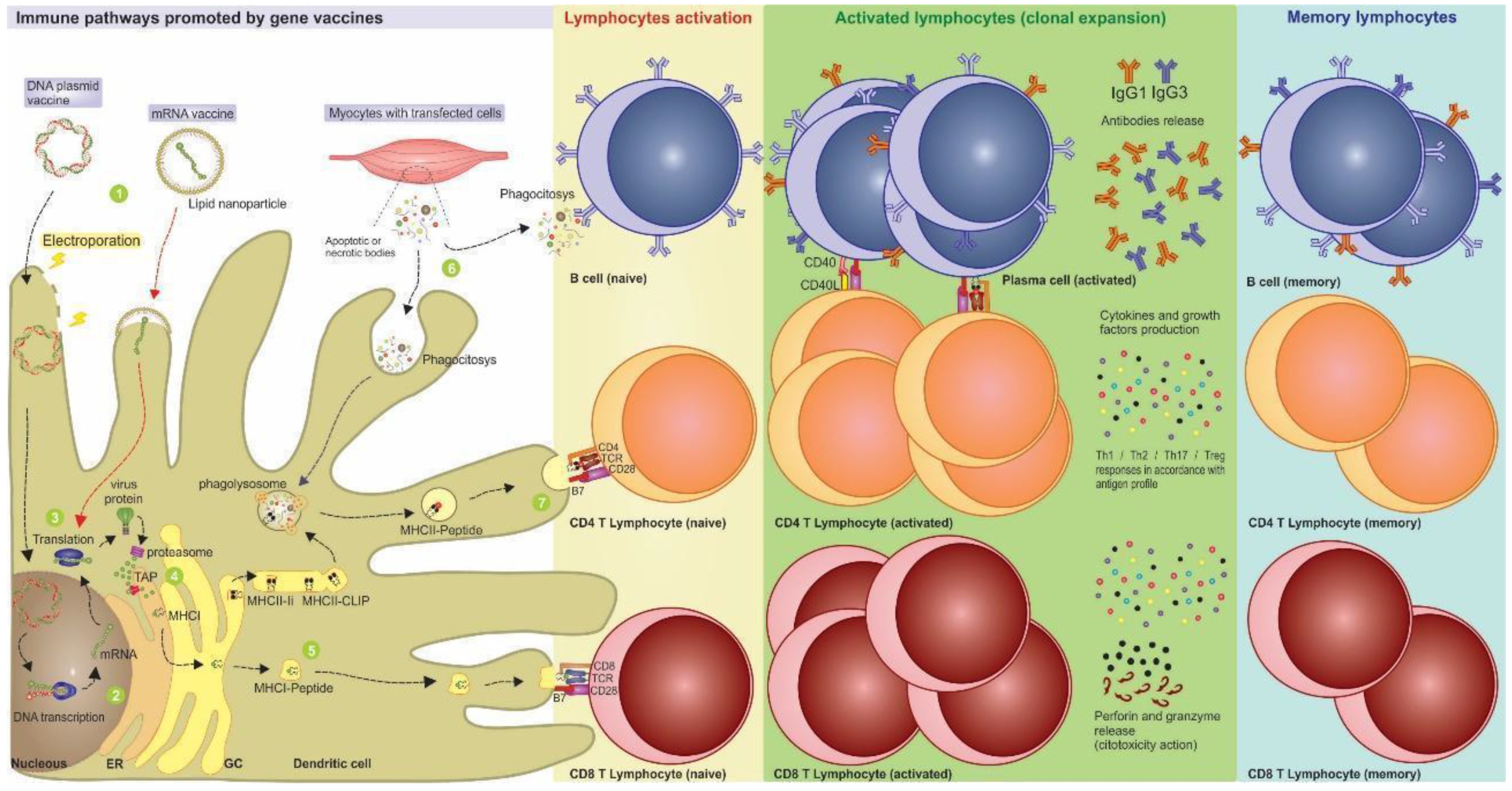
3. Nucleic Acid Vaccines Allow Better Immune Response Directing

This versatility in activating different immunological response pathways makes nucleic acid vaccines useful for both prophylactic and therapeutic purposes. In this sense, since the main objective of prophylactic vaccines is to promote immunological memory, these strategies demand a strong humoral response through specific CD4+ T cells. A vaccine for therapeutic purposes, on the other hand, requires primarily cytotoxic CD8+ T cell responses to recognize and cause apoptosis of chronically infected or tumoral cells [14,15][13][14].
As an example of a prophylactic study, the effects of the mRNA-1273 vaccine against SARS-CoV-2 demonstrated high neutralization and Th1-shifted CD4+ T cell responses in humans. This response profile involved a reduction in the risk of increased vaccine-associated respiratory disease or increasing antibody-dependent replication. Furthermore, these characteristics of mRNA composition and formulation have been associated with prolonged protein expression, induction of antigen-specific follicular T helper cells, and activation of germinal center B cells [16,17][15][16].
Regarding the therapeutic approach, Rittig et al. [18][17] carried out a phase I/II mRNA-based vaccine trial in patients with stage IV renal cell cancer. Direct injection of naked mRNA induced a safe and efficient immune response with specific antitumor immunity promoted by CD4+ T and CD8+ T effector cells. In the study by Cafri et al. [19][18], an mRNA vaccine for patients with metastatic GI cancer was proven as safe and induced mutation-specific T-cell responses against predicted neoepitopes not detected before vaccination.
Regarding DNA vaccines, two clinical studies tested the VGX-3100 vaccine (NCT01304524) [20][19] and the GX-188E vaccine (NCT01634503) [21][20], both encoding immunogenic peptides based on E6 and E7 genes of HPV-16 and 18, in patients with CINs II and III. In the first study, the histopathological results showed regression of lesions in 49.5% of patients (n = 53), whereas in placebos, spontaneous regression of lesions occurred in 30.6% (n = 11). Furthermore, in this study, the immunological analysis demonstrated significantly greater specific activation of cytotoxic T lymphocytes and increased humoral response in the vaccinated patients [22][21]. Meanwhile, in the study by Kim et al. [21][20], 8 out of 9 patients exhibited a polyfunctional response of specific cytotoxic T cells, and 7 out of 9 patients showed complete regression of the lesion, with no viral detection after 36 weeks of follow-up. Due to the COVID-19 pandemic, different platforms have stood out in the face of the public health emergency. DNA vaccines such as the ZyCOV-D vaccine [23][22] were first licensed in India for emergency use in humans. Others, such as AG0302-COVID19 (NCT04655625), GX-19N (NCT05067946), and INO-4800 (NCT04642638), are currently in phase II/III trials and consist of plasmid vaccines encoding SARS-CoV-2 proteins.
The mechanism by which mRNA vaccines act is like that of DNA vaccines. The main difference is that after immunization, the mRNA vaccines are transported to the cell cytoplasm ready for translation, without the need to reach the nucleus. Besides, the mRNA molecule is less stable and needs to undergo structural changes, such as the addiction of modified nucleosides. Among these modifications is the addition of a synthetic cap on the 5′ region and the poly(A) tail on the 3′ region, needing at least 120 bases to form a mature mRNA sequence. Together, these increments are responsible for increase of translation efficiency, avoiding molecule degradation by cytoplasmic nucleases [11,22,24][10][21][23]. There are two types of mRNA vaccines: non-replicating mRNA vaccines, that encode only the target antigen, and self-replicating RNA vaccines that, besides the antigen of interest, have the replication machinery of positive-stranded RNA viruses such as alphavirus, flavivirus, measles virus, and rhabdovirus, enabling intracellular replication of the vaccine [25][24].
During the design of a self-replicating mRNA vaccine, the coding sequences of the viral RNA replicase are conserved, while the coding regions of viral structural proteins are replaced by the antigen sequence, preventing the formation of virions in the host. In addition, eukaryotic promoters, such as the CMV promoter, are inserted into the vaccine sequence for recognition by the host’s translation machinery. The action of self-replicating vaccines is like that of conventional mRNA vaccines, except for the fact that after the vaccine transfection, the alphavirus replicase is translated and allows the subsequent replication of more vaccine mRNA molecules. Thus, much lower vaccine doses of self-replicating mRNA vaccines are required to achieve immunizing potential compared to with conventional mRNA vaccines [24,25][23][24].
DNA vaccines have been broadly tested in human clinical trials where the immunogenicity, the lack of significant reactions, and the tolerance for doses between 20 µg and 2500 µg have been demonstrated [24,26][23][25]. This platform also has high stability at room temperature without the demand for an uninterrupted cold chain for transport and storage, facilitating worldwide access, especially in poor rural areas and tropical countries. Meanwhile, mRNA vaccines have become the focus of different studies, particularly in cancer immunotherapy research, mainly those which use ex vivo modification of antigen-presenting cells [11,27][10][26]. Nowadays, this platform has received significant visibility due to the promising results obtained in assays against ebola and H1N1 influenza pathogens [28][27] and in the face of its extensive use during the COVID-19 pandemic. Furthermore, the first licensed emergency vaccine strategy in the SARS-CoV-2 pandemic was the mRNA vaccine, which retained the highest level of efficacy even after the approval of other vaccine platforms.
Optimizations in the formulation of mRNA vaccines have been sought to maximize their thermostability. An example is the protamine-encapsulated conventional mRNA-based rabies vaccine developed by Sitiz et al. [29][28]. This study showed the maintenance of vaccine immunogenicity and protective effects through temperature oscillation between 4 and 56 °C per 20 cycles and after prolonged storage (from −80 °C to 70 °C) for several months.
Inside the cell, nucleic acid vaccines can simulate a natural viral infection because they act as an intracellular antigen that can generate specific cellular responses after endogenous production and induce antibody production. Furthermore, the cell transfected with the DNA or mRNA vaccines does not need to be a professional APC to produce the protein antigen capable of stimulating a B or T cell. For example, once expressed by neighboring myocytes, the vaccine antigens can be phagocytosed by APCs and undergo immune cross-presentation [30][29].
The importance of B cells in the efficacy of prophylactic vaccines should not underestimate the role of T-cell responses that are essential for the induction of high-affinity/avidity neutralizing antibodies and memory cells. This role can be explained because the follicular helper cells (Tfh) provide support for the B cell maturation within the germinal centers of secondary lymphoid organs, which can produce high titers of high-affinity and neutralizing antibodies [31][30]. In addition, activation of helper Th1 response stimulates the secretion of interleukin (IL-2), interferon (IFN-γ), and tumor necrosis factor (TNF-β), with direct antiviral functions and support for cytotoxic T cells and macrophages [32][31]. In contrast, the Th2 response is suggested as a key factor for the development of vaccine-associated disease enhancement through the production of low-affinity antibodies [33][32].
The complete activation of both arms of the immune system (humoral and cellular responses) is vital to avoid the lack of affinity maturation of the antibodies. This factor is especially important in the context of COVID-19, since studies have demonstrated the occurrence of vaccine-associated disease enhancement following viral challenge with SARS-CoV, a virus related to SARS-CoV-2 [34][33]. This issue can be avoided through the careful choice of vaccine antigens, predicted in silico, which must be highly immunogenic and contain MHC-I and MHC-II ligands to activate the cellular response.
Nucleic acid vaccines, especially those with synthetic antigens, allow for the direction of immune response reached by including epitopes recognized by B lymphocytes, MHC-I ligands (cytotoxic response), and MHC-II (helper response) or preferably, all of them simultaneously, in the synthetic construction. One advantage of including T cell epitopes in the vaccine construction is that they can be from any region in the viral antigen, either localized internally or on the protein surface. The recognition of B cell antigens, however, is limited to conformational determinants composed of amino acids located on the surface of the viral antigen.

References

  1. Domínguez-Andrés, J.; van Crevel, R.; Divangahi, M.; Netea, M.G. Designing the Next Generation of Vaccines: Relevance for Future Pandemics. mBio 2020, 11, e02616–e02620.
  2. Wang, H.; Naghavi, M.; Allen, C.; Barber, R.M.; Bhutta, Z.A.; Carter, A.; Casey, D.C.; Charlson, F.J.; Chen, A.Z.; Coates, M.M.; et al. Global, Regional, and National Life Expectancy, All-Cause Mortality, and Cause-Specific Mortality for 249 Causes of Death, 1980–2015: A Systematic Analysis for the Global Burden of Disease Study 2015. Lancet 2016, 388, 1459–1544.
  3. Plotkin, S.A. Vaccines: Past, Present and Future. Nat. Med. 2005, 11, S5–S11.
  4. Francis, M.J. Recent Advances in Vaccine Technologies. Vet. Clin. North Am. Small Anim. Pract. 2018, 48, 231–241.
  5. Rappuoli, R.; De Gregorio, E.; Del Giudice, G.; Phogat, S.; Pecetta, S.; Pizza, M.; Hanon, E. Vaccinology in the Post−COVID-19 Era. Proc. Natl. Acad. Sci. USA 2021, 118, e2020368118.
  6. González-Romo, F.; Picazo, J.J. El desarrollo de nuevas vacunas. Enferm. Infecc. Y Microbiol. Clínica 2015, 33, 557–568.
  7. Carter, C.; Houser, K.V.; Yamshchikov, G.V.; Bellamy, A.R.; May, J.; Enama, M.E.; Sarwar, U.; Larkin, B.; Bailer, R.T.; Koup, R.; et al. Safety and Immunogenicity of Investigational Seasonal Influenza Hemagglutinin DNA Vaccine Followed by Trivalent Inactivated Vaccine Administered Intradermally or Intramuscularly in Healthy Adults: An Open-Label Randomized Phase 1 Clinical Trial. PLoS ONE 2019, 14, e0222178.
  8. Barbier, A.J.; Jiang, A.Y.; Zhang, P.; Wooster, R.; Anderson, D.G. The Clinical Progress of MRNA Vaccines and Immunotherapies. Nat. Biotechnol. 2022, 40, 840–854.
  9. Tombácz, I.; Weissman, D.; Pardi, N. Vaccination with Messenger RNA: A Promising Alternative to DNA Vaccination. In DNA Vaccines; Sousa, Â., Ed.; Methods in Molecular Biology; Springer: New York, NY, USA, 2021; Volume 2197, pp. 13–31. ISBN 978-1-07-160871-5.
  10. Wadhwa, A.; Aljabbari, A.; Lokras, A.; Foged, C.; Thakur, A. Opportunities and Challenges in the Delivery of MRNA-Based Vaccines. Pharmaceutics 2020, 12, 102.
  11. Sheets, R.L.; Stein, J.; Manetz, T.S.; Duffy, C.; Nason, M.; Andrews, C.; Kong, W.-P.; Nabel, G.J.; Gomez, P.L. Biodistribution of DNA Plasmid Vaccines against HIV-1, Ebola, Severe Acute Respiratory Syndrome, or West Nile Virus Is Similar, without Integration, despite Differing Plasmid Backbones or Gene Inserts. Toxicol. Sci. 2006, 91, 610–619.
  12. Wang, J.-C.E.; Livingstone, A.M. Cutting Edge: CD4+ T Cell Help Can Be Essential for Primary CD8 + T Cell Responses In Vivo. J. Immunol. 2003, 171, 6339–6343.
  13. Lee, S.-H.; Danishmalik, S.N.; Sin, J.-I. DNA Vaccines, Electroporation and Their Applications in Cancer Treatment. Hum. Vaccines Immunother. 2015, 11, 1889–1900.
  14. Qin, S.; Tang, X.; Chen, Y.; Chen, K.; Fan, N.; Xiao, W.; Zheng, Q.; Li, G.; Teng, Y.; Wu, M.; et al. MRNA-Based Therapeutics: Powerful and Versatile Tools to Combat Diseases. Signal Transduct. Target. Ther. 2022, 7, 166.
  15. Baden, L.R.; El Sahly, H.M.; Essink, B.; Kotloff, K.; Frey, S.; Novak, R.; Diemert, D.; Spector, S.A.; Rouphael, N.; Creech, C.B.; et al. Efficacy and Safety of the MRNA-1273 SARS-CoV-2 Vaccine. N. Engl. J. Med. 2021, 384, 403–416.
  16. Jackson, L.A.; Anderson, E.J.; Rouphael, N.G.; Roberts, P.C.; Makhene, M.; Coler, R.N.; McCullough, M.P.; Chappell, J.D.; Denison, M.R.; Stevens, L.J.; et al. An MRNA Vaccine against SARS-CoV-2—Preliminary Report. N. Engl. J. Med. 2020, 383, 1920–1931.
  17. Rittig, S.M.; Haentschel, M.; Weimer, K.J.; Heine, A.; Muller, M.R.; Brugger, W.; Horger, M.S.; Maksimovic, O.; Stenzl, A.; Hoerr, I.; et al. Intradermal Vaccinations With RNA Coding for TAA Generate CD8+ and CD4+ Immune Responses and Induce Clinical Benefit in Vaccinated Patients. Mol. Ther. 2011, 19, 990–999.
  18. Cafri, G.; Gartner, J.J.; Zaks, T.; Hopson, K.; Levin, N.; Paria, B.C.; Parkhurst, M.R.; Yossef, R.; Lowery, F.J.; Jafferji, M.S.; et al. MRNA Vaccine–Induced Neoantigen-Specific T Cell Immunity in Patients with Gastrointestinal Cancer. J. Clin. Investig. 2020, 130, 5976–5988.
  19. Trimble, C.L.; Morrow, M.P.; Kraynyak, K.A.; Shen, X.; Dallas, M.; Yan, J.; Edwards, L.; Parker, R.L.; Denny, L.; Giffear, M.; et al. Safety, Efficacy, and Immunogenicity of VGX-3100, a Therapeutic Synthetic DNA Vaccine Targeting Human Papillomavirus 16 and 18 E6 and E7 Proteins for Cervical Intraepithelial Neoplasia 2/3: A Randomised, Double-Blind, Placebo-Controlled Phase 2b Trial. Lancet 2015, 386, 2078–2088.
  20. Kim, T.J.; Jin, H.-T.; Hur, S.-Y.; Yang, H.G.; Seo, Y.B.; Hong, S.R.; Lee, C.-W.; Kim, S.; Woo, J.-W.; Park, K.S.; et al. Clearance of Persistent HPV Infection and Cervical Lesion by Therapeutic DNA Vaccine in CIN3 Patients. Nat. Commun. 2014, 5, 5317.
  21. Houseley, J.; Tollervey, D. The Many Pathways of RNA Degradation. Cell 2009, 136, 763–776.
  22. Khobragade, A.; Bhate, S.; Ramaiah, V.; Deshpande, S.; Giri, K.; Phophle, H.; Supe, P.; Godara, I.; Revanna, R.; Nagarkar, R.; et al. Efficacy, Safety, and Immunogenicity of the DNA SARS-CoV-2 Vaccine (ZyCoV-D): The Interim Efficacy Results of a Phase 3, Randomised, Double-Blind, Placebo-Controlled Study in India. Lancet 2022, 399, 1313–1321.
  23. van de Wall, S.; Ljungberg, K.; Ip, P.P.; Boerma, A.; Knudsen, M.L.; Nijman, H.W.; Liljeström, P.; Daemen, T. Potent Therapeutic Efficacy of an Alphavirus Replicon DNA Vaccine Expressing Human Papilloma Virus E6 and E7 Antigens. OncoImmunology 2018, 7, e1487913.
  24. Ljungberg, K.; Liljeström, P. Self-Replicating Alphavirus RNA Vaccines. Expert Rev. Vaccines 2015, 14, 177–194.
  25. Le, T.P.; Coonan, K.M.; Hedstrom, R.C.; Charoenvit, Y.; Sedegah, M.; Epstein, J.E.; Kumar, S.; Wang, R.; Doolan, D.L.; Maguire, J.D.; et al. Safety, Tolerability and Humoral Immune Responses after Intramuscular Administration of a Malaria DNA Vaccine to Healthy Adult Volunteers. Vaccine 2000, 18, 1893–1901.
  26. Palucka, K.; Banchereau, J. Cancer Immunotherapy via Dendritic Cells. Nat. Rev. Cancer 2012, 12, 265–277.
  27. Chahal, J.S.; Khan, O.F.; Cooper, C.L.; McPartlan, J.S.; Tsosie, J.K.; Tilley, L.D.; Sidik, S.M.; Lourido, S.; Langer, R.; Bavari, S.; et al. Dendrimer-RNA Nanoparticles Generate Protective Immunity against Lethal Ebola, H1N1 Influenza, and Toxoplasma Gondii Challenges with a Single Dose. Proc. Natl. Acad. Sci. USA 2016, 113, E4133–E4142.
  28. Stitz, L.; Vogel, A.; Schnee, M.; Voss, D.; Rauch, S.; Mutzke, T.; Ketterer, T.; Kramps, T.; Petsch, B. A Thermostable Messenger RNA Based Vaccine against Rabies. PLoS Negl. Trop. Dis. 2017, 11, e0006108.
  29. Liu A Comparison of Plasmid DNA and MRNA as Vaccine Technologies. Vaccines 2019, 7, 37.
  30. Hollister, K.; Chen, Y.; Wang, S.; Wu, H.; Mondal, A.; Clegg, N.; Lu, S.; Dent, A. The Role of Follicular Helper T Cells and the Germinal Center in HIV-1 Gp120 DNA Prime and Gp120 Protein Boost Vaccination. Hum. Vaccines Immunother. 2014, 10, 1985–1992.
  31. Holdsworth, S.R.; Kitching, A.R.; Tipping, P.G. Th1 and Th2 T Helper Cell Subsets Affect Patterns of Injury and Outcomes in Glomerulonephritis. Kidney Int. 1999, 55, 1198–1216.
  32. Delgado, M.F.; Coviello, S.; Monsalvo, A.C.; Melendi, G.A.; Hernandez, J.Z.; Batalle, J.P.; Diaz, L.; Trento, A.; Chang, H.-Y.; Mitzner, W.; et al. Lack of Antibody Affinity Maturation Due to Poor Toll-like Receptor Stimulation Leads to Enhanced Respiratory Syncytial Virus Disease. Nat. Med. 2009, 15, 34–41.
  33. Lee, W.S.; Wheatley, A.K.; Kent, S.J.; DeKosky, B.J. Antibody-Dependent Enhancement and SARS-CoV-2 Vaccines and Therapies. Nat. Microbiol. 2020, 5, 1185–1191.
More
Video Production Service