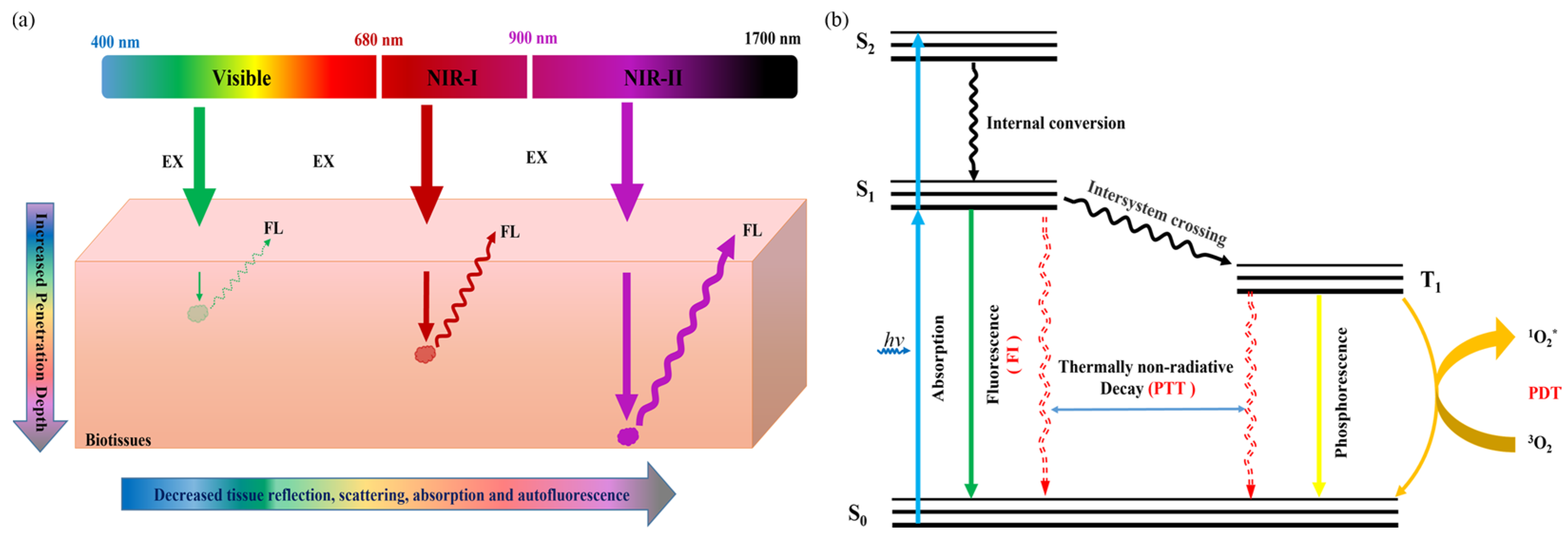
Much effort has been devoted to developing Pdots with emission bands located in the second near-infrared (NIR-II, 1000–1700 nm) region, which hold great advantages of higher spatial resolution, better signal-to-background ratios (SBR), and deeper tissue penetration for solid-tumor imaging in comparison with the visible region (400–680 nm) and the first near-infrared (NIR-I, 680–900 nm) window, by virtue of the reduced tissue autofluorescence, minimal photon scattering, and low photon absorption.
Herein, we will focus on the latest reported NIR-II Pdots for in vivo tumor imaging, and in particular on the molecular engineering to optimize the fluorescence quantum yields and surface functionalization to promote the ability of active tumor targeting. Some of the NIR-II theranostic Pdots used for integrated FI diagnosis and phototherapy are also included. Finally, we will discuss the future directions and challenges in this field.
- semiconducting polymer dots
- NIR-II
- fluorescence probes
- Tumor Theranostics
1. Introduction
2. Molecular Engineering of Efficient NIR-II Pdots for In Vivo Tumor FI
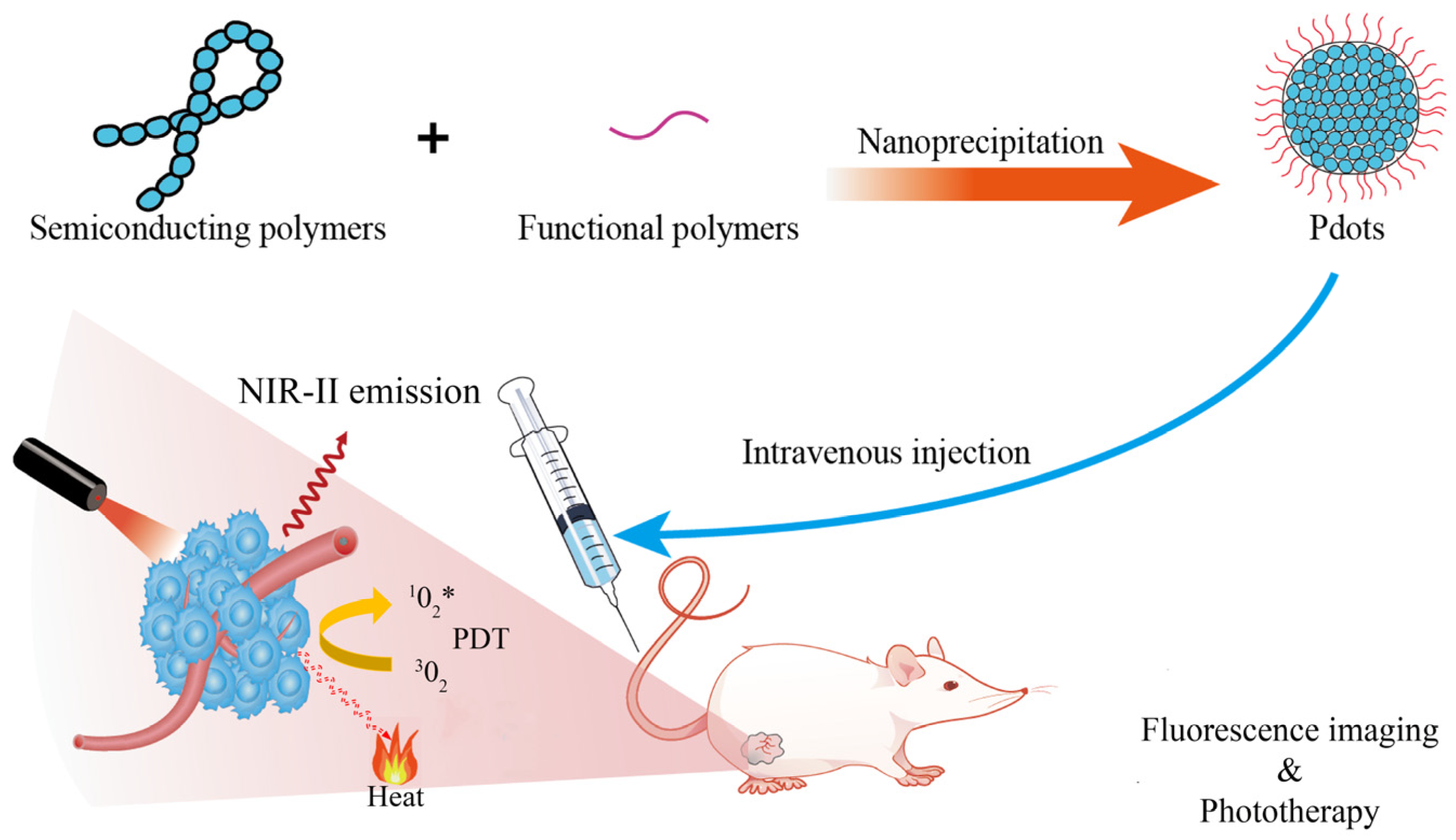
As mentioned above, light with wavelengths in the NIR-II region has a high development potential for in vivo imaging of tumors due to its small absorption and scattering in animal tissues, better tissue penetration and higher spatial resolution. However, NIR-II emissive Pdots usually exhibit low Φf due to an intrinsic small energy bandgap between the lowest unoccupied molecular orbital (LUMO) and the highest occupied molecular orbital (HOMO) of conjugated polymers, as well as the severe aggregation-caused quenching (ACQ) effect in the Pdot state, both of which facilitate non-radiative decay, resulting in low Φf of NIR-II Pdots [49,62,63][49][54][55]. Therefore, one important challenge is to design NIR-II Pdots with high Φf, to realize high brightness in vivo and hence better SBR of fluorescence imaging. To develop NIR-II Pdot probes with high Φf, Liu et al., proposed a fluorination strategy to design semiconducting polymers to optimize the Φf of corresponding Pdot probes [54][56]. Using benzodithiophene (BDT) as electron donor (D) and triazole [4,5-g]-quinoxaline (TQ) derivatives as electron acceptor (A), the team synthesized two sets of fluorine-substituted D-A type semiconducting polymers based on the two fluorine substitution modes, which are named as m-PBTQ, m-PBTQ2F, m-PBTQ4F, m-PBTQ4F, with alkoxy chains anchored at the meta site of benzene and p-PBTQ, p-PBTQ2F, and p-PBTQ4F, with alkoxy chains anchored to the para position of benzene. The semiconducting polymers and amphiphilic polystyrene polymer (PS-PEG-COOH) were prepared into Pdots by the classic nano-precipitation method. The Φf of obtained m-series Pdots (1.0%, 2.2%, and 3.2%) were consistently higher than those of p-series Pdots (0.6%, 0.9%, and 1.5%), mainly because the different alkoxy positions on TQ affected the hydrophobicity and steric hindrance of molecules. Additionally, the Φf of Pdots was increased with more fluorine atoms on the TQ acceptor. As a result, the tetrafluorinated m-PBTQ4F Pdots yielded the highest Φf of 3.2%, which was three times higher than that of the non-fluoridated counterpart and six times higher than that of IR26. Liu et al. attributed the fluorescence enhancement in the fluorinated Pdots to the nanoscale fluorous effect that increases the planarity of the conjugated backbone and minimizes the structure distortion between the excited-state and ground-state, thus decreasing the nonradiative decay rates. The fluorescence intensity of m-PBTQ4F Pdots remained at 80% of the initial state under 120 min laser irradiation, indicating good photostability. The authors suggest that fluorination of PBTQ polymers can effectively modulate the optical properties of the resulting Pdot in several ways, including energy-level reduction and fluorescence enhancement. Quantitative cranial and scalp-puncture imaging of brain tumor vasculature in vivo demonstrated that m-PBTQ4F Pdot images showed better imaging performance than non-fluoridated m-PBTQ images, and could distinguish normal, uniform, and orderly brain blood vessels from the images, and also clearly differentiate uneven and chaotic distribution. These results indicate that fluorinated Pdots have good photostability and high brightness, and have great development potential in the diagnosis and detection of brain tumors. To solve the low Φf problem of NIR-II probes arising from the ACQ effect, Li et al., proposed two strategies to develop efficient NIR-II Pdots, by incorporating an anti-ACQ unit or an aggregation-induced emission (AIE) segment into the polymer backbone [55][57]. The authors first designed a conjugated polymer skeleton using [1,2,5]thiadiazolo [3,4-g]quinoxaline (TQ) as a strong A unit and alkylthio-thiophene-substituted benzodithiophene as the D unit. A phenothiazines with Pttc (anti-ACQ), TPA (AIE), or TPE (AIE) unit was then added to the semiconducting polymer backbone. The amphiphilic lipids were subsequently made into Pdots by a nanoprecipitate with semiconducting polymers. The results showed that IR-PTTC Pdots had the lowest Φf of 4.9% among the three Pdots, while IR-TPA and IR-TPE Pdots had Φf of 6.7% and 14%, respectively. To the best of our knowledge, the IR-TPE Pdot is, to date, the most efficient NIR-II emissive Pdot. At the same time, IT-TPE has a strong absorption at 700 nm, and the maximum emission peak is 1010 nm. Due to the high Φf of IR-TPE Pdots, the authors used these Pdots for subsequent bioimaging. First, the authors functionalized the surface of IR-TPE Pdots with folic acid so that specific cancer cells with folate receptors could internalize them. Subsequently, Pdots were injected intravenously into mice through the tail vein, and the SBR reached 2.42 when equipped with a 1400 nm long-pass filter. At the same time, their fluorescence intensity remained more than 80% of their original intensity after 20 min of continuous UV irradiation. These results indicate that Pdots are very light resistant and suitable for long-term fluorescence imaging or tracking. The authors performed in vivo tumor imaging using IR-TPE Pdots and ICG in live mice bearing 4T1 tumors and compared the performance of these two probes. At the same time, 3D tumor mapping was performed in vivo in tumor-bearing mice 6 h after injection. The images in the film were reconstructed from a series of images with different rotation angles from −45° to 45°, from which wresearchers could easily identify the location of the tumor.References
- Liu, Y.; Bhattarai, P.; Dai, Z.; Chen, X. Photothermal therapy and photoacoustic imaging via nanotheranostics in fighting cancer. Chem. Soc. Rev. 2019, 48, 2053–2108.
- Fu, Q.; Zhu, R.; Song, J.; Yang, H.; Chen, X. Photoacoustic imaging: Contrast agents and their biomedical applications. Adv. Mater. 2019, 31, e1805875.
- Zhen, X.; Pu, K.; Jiang, X. Photoacoustic imaging and photothermal therapy of semiconducting polymer nanoparticles: Signal amplification and second near-infrared construction. Small 2021, 17, e2004723.
- Wang, S.; Ren, W.X.; Hou, J.T.; Won, M.; An, J.; Chen, X.; Shu, J.; Kim, J.S. Fluorescence imaging of pathophysiological microenvironments. Chem. Soc. Rev. 2021, 50, 8887–8902.
- Kim, T.H.; Schnitzer, M.J. Fluorescence imaging of large-scale neural ensemble dynamics. Cell 2022, 185, 9–41.
- Li, Y.; Younis, M.H.; Wang, H.; Zhang, J.; Cai, W.; Ni, D. Spectral computed tomography with inorganic nanomaterials: State-of-the-art. Adv. Drug Deliv. Rev. 2022, 189, 114524.
- Arif, W.M.; Elsinga, P.H.; Gasca-Salas, C.; Versluis, M.; Martínez-Fernández, R.; Dierckx, R.; Borra, R.J.H.; Luurtsema, G. Focused ultrasound for opening blood-brain barrier and drug delivery monitored with positron emission tomography. J. Control. Release 2020, 324, 303–316.
- Floresta, G.; Abbate, V. Recent progress in the imaging of c-Met aberrant cancers with positron emission tomography. Med. Res. Rev. 2022, 42, 1588–1606.
- Verger, A.; Grimaldi, S.; Ribeiro, M.J.; Frismand, S.; Guedj, E. Single photon emission computed tomography/positron emission tomography molecular imaging for parkinsonism: A fast-developing field. Ann. Neurol. 2021, 90, 711–719.
- Zhu, S.; Tian, R.; Antaris, A.L.; Chen, X.; Dai, H. Near-infrared-II molecular dyes for cancer imaging and surgery. Adv. Mater. 2019, 31, e1900321.
- Ding, F.; Feng, J.; Zhang, X.; Sun, J.; Fan, C.; Ge, Z. Responsive optical probes for deep-tissue imaging: Photoacoustics and second near-infrared fluorescence. Adv. Drug Deliv. Rev. 2021, 173, 141–163.
- Ouyang, J.; Sun, L.; Zeng, Z.; Zeng, C.; Zeng, F.; Wu, S. Nanoaggregate probe for breast cancer metastasis through multispectral optoacoustic tomography and aggregation-induced NIR-I/II fluorescence imaging. Angew. Chem. Int. Ed. 2020, 59, 10111–10121.
- Sun, Y.; Sun, P.; Li, Z.; Qu, L.; Guo, W. Natural flavylium-inspired far-red to NIR-II dyes and their applications as fluorescent probes for biomedical sensing. Chem. Soc. Rev. 2022, 51, 7170–7205.
- Su, Y.; Yu, B.; Wang, S.; Cong, H.; Shen, Y. NIR-II bioimaging of small organic molecule. Biomaterials 2021, 271, 120717.
- Chen, L.; Wu, L.; Yu, J.; Kuo, C.T.; Jian, T.; Wu, I.C.; Rong, Y.; Chiu, D.T. Highly photostable wide-dynamic-range pH sensitive semiconducting polymer dots enabled by dendronizing the near-IR emitters. Chem. Sci. 2017, 8, 7236–7245.
- Younis, N.K.; Ghoubaira, J.A.; Bassil, E.P.; Tantawi, H.N.; Eid, A.H. Metal-based nanoparticles: Promising tools for the management of cardiovascular diseases. Nanomedicine 2021, 36, 102433.
- Zhang, X.; He, S.; Ding, B.; Qu, C.; Chen, H.; Sun, Y.; Zhang, R.; Lan, X.; Cheng, Z. Synergistic strategy of rare-earth doped nanoparticles for NIR-II biomedical imaging. J. Mater. Chem. B 2021, 9, 9116–9122.
- Li, J.; Li, W.; Xie, L.; Sang, W.; Wang, G.; Zhang, Z.; Li, B.; Tian, H.; Yan, J.; Tian, Y.; et al. A metal-polyphenolic nanosystem with NIR-II fluorescence-guided combined photothermal therapy and radiotherapy. Chem. Commun. 2021, 57, 11473–11476.
- Yang, F.; Zhang, Q.; Huang, S.; Ma, D. Recent advances of near infrared inorganic fluorescent probes for biomedical applications. J. Mater. Chem. B 2020, 8, 7856–7879.
- Liu, Y.; Li, Y.; Koo, S.; Sun, Y.; Liu, Y.; Liu, X.; Pan, Y.; Zhang, Z.; Du, M.; Lu, S.; et al. Versatile types of inorganic/organic NIR-IIa/IIb fluorophores: From strategic design toward molecular imaging and theranostics. Chem. Rev. 2022, 122, 209–268.
- Cai, Y.; Tang, C.; Wei, Z.; Song, C.; Zou, H.; Zhang, G.; Ran, J.; Han, W. Fused-ring small-molecule-based bathochromic nano-agents for tumor NIR-II fluorescence imaging-guided photothermal/photodynamic therapy. ACS. Appl. Bio Mater. 2021, 4, 1942–1949.
- Shi, T.; Huang, C.; Li, Y.; Huang, F.; Yin, S. NIR-II phototherapy agents with aggregation-induced emission characteristics for tumor imaging and therapy. Biomaterials 2022, 285, 121535.
- Xu, W.; Wang, D.; Tang, B.Z. NIR-II AIEgens: A win-win integration towards bioapplications. Angew. Chem. Int. Ed. 2021, 60, 7476–7487.
- Bai, X.; Wang, K.; Chen, L.; Zhou, J.; Wang, J. Semiconducting polymer dots as fluorescent probes for in vitro biosensing. J. Mater. Chem. B 2022, 10, 6248–6262.
- Wu, M.; Wei, Q.; Xian, C.; Dai, C.; He, X.; Wu, C.; Sun, G.; Chen, L. Highly efficient and non-doped red conjugated polymer dot for photostable cell imaging. Chin. Chem. Lett. 2022; 107867, in press.
- Chen, L.; Chen, D.; Jiang, Y.; Zhang, J.; Yu, J.; DuFort, C.C.; Hingorani, S.R.; Zhang, X.; Wu, C.; Chiu, D.T. A BODIPY-based donor/donor-acceptor system: Towards highly efficient long-wavelength-excitable near-IR polymer dots with narrow and strong absorption features. Angew. Chem. Int. Ed. 2019, 58, 7008–7012.
- Feng, L.; Zhu, C.; Yuan, H.; Liu, L.; Lv, F.; Wang, S. Conjugated polymer nanoparticles: Preparation, properties, functionalization and biological applications. Chem. Soc. Rev. 2013, 42, 6620–6633.
- MacFarlane, L.R.; Shaikh, H.; Garcia-Hernandez, J.D.; Vespa, M.; Fukui, T.; Manners, I. Functional nanoparticles through π-conjugated polymer self-assembly. Nat. Rev. Mater. 2021, 6, 7–26.
- Zhang, Z.; Fang, X.; Liu, Z.; Liu, H.; Chen, D.; He, S.; Zheng, J.; Yang, B.; Qin, W.; Zhang, X.; et al. Semiconducting polymer dots with dual-enhanced NIR-IIa fluorescence for through-skull mouse-brain imaging. Angew. Chem. Int. Ed. 2020, 59, 3691–3698.
- Ackermann, J.; Metternich, J.T.; Herbertz, S.; Kruss, S. Biosensing with fluorescent carbon nanotubes. Angew. Chem. Int. Ed. 2022, 61, e202112372.
- Ge, X.; Wei, R.; Sun, L. Lanthanide nanoparticles with efficient near-infrared-II emission for biological applications. J. Mater. Chem. B 2020, 8, 10257–10270.
- Li, D.; Yang, Y.; Li, D.; Pan, J.; Chu, C.; Liu, G. Organic sonosensitizers for sonodynamic therapy: From small molecules and nanoparticles toward clinical development. Small 2021, 17, e2101976.
- Pu, K.; Chattopadhyay, N.; Rao, J. Recent advances of semiconducting polymer nanoparticles in in vivo molecular imaging. J. Control. Release 2016, 240, 312–322.
- Son, J.; Yi, G.; Yoo, J.; Park, C.; Koo, H.; Choi, H.S. Light-responsive nanomedicine for biophotonic imaging and targeted therapy. Adv. Drug Deliv. Rev. 2019, 138, 133–147.
- Krishnan, S.K.; Singh, E.; Singh, P.; Meyyappan, M.; Nalwa, H.S. A review on graphene-based nanocomposites for electrochemical and fluorescent biosensors. RSC Adv. 2019, 9, 8778–8881.
- Kuo, C.T.; Wu, I.C.; Chen, L.; Yu, J.; Wu, L.; Chiu, D.T. Improving the photostability of semiconducting polymer dots using buffers. Anal. Chem. 2018, 90, 11785–11790.
- Dimov, I.B.; Moser, M.; Malliaras, G.G.; McCulloch, I. Semiconducting polymers for neural applications. Chem. Rev. 2022, 122, 4356–4396.
- Chen, H.; Yu, J.; Men, X.; Zhang, J.; Ding, Z.; Jiang, Y.; Wu, C.; Chiu, D.T. Reversible ratiometric NADH sensing using semiconducting polymer dots. Angew. Chem. Int. Ed. 2021, 60, 12007–12012.
- Jiang, Y.; Hu, Q.; Chen, H.; Zhang, J.; Chiu, D.T.; McNeill, J. Dual-mode superresolution imaging using charge transfer dynamics in semiconducting polymer dots. Angew. Chem. Int. Ed. 2020, 59, 16173–16180.
- Upputuri, P.K.; Pramanik, M. Recent advances in photoacoustic contrast agents for in vivo imaging. Wiley Interdiscip. Rev. Nanomed. Nanobiotechnol. 2020, 12, e1618.
- Chen, P.; Ilyas, I.; He, S.; Xing, Y.; Jin, Z.; Huang, C. Ratiometric pH sensing and imaging in living cells with dual-emission semiconductor polymer dots. Molecules 2019, 24, 2923.
- Yuan, Y.; Hou, W.; Qin, W.; Wu, C. Recent advances in semiconducting polymer dots as optical probes for biosensing. Biomater. Sci. 2021, 9, 328–346.
- Yuan, Y.; Zhang, Z.; Hou, W.; Qin, W.; Meng, Z.; Wu, C. In vivo dynamic cell tracking with long-wavelength excitable and near-infrared fluorescent polymer dots. Biomaterials 2020, 254, 120139.
- Meng, Z.; Guo, L.; Li, Q. Peptide-coated semiconductor polymer dots for stem cells labeling and tracking. Chemistry 2017, 23, 6836–6844.
- Dai, X.; Ma, J.; Zhang, Q.; Xu, Q.; Yang, L.; Gao, F. Simultaneous inhibition of planktonic and biofilm bacteria by self-adapting semiconducting polymer dots. J. Mater. Chem. B 2021, 9, 6658–6667.
- Wu, Y.; Shi, C.; Wang, G.; Sun, H.; Yin, S. Recent advances in the development and applications of conjugated polymer dots. J. Mater. Chem. B 2022, 10, 2995–3015.
- Chang, K.; Liu, Y.; Hu, D.; Qi, Q.; Gao, D.; Wang, Y.; Li, D.; Zhang, X.; Zheng, H.; Sheng, Z.; et al. Highly stable conjugated polymer dots as multifunctional agents for photoacoustic imaging-guided photothermal therapy. ACS. Appl. Mater. Interfaces 2018, 10, 7012–7021.
- Braeken, Y.; Cheruku, S.; Ethirajan, A.; Maes, W. Conjugated polymer nanoparticles for bioimaging. Materials 2017, 10, 1420.
- Wang, Y.; Feng, L.; Wang, S. Conjugated polymer nanoparticles for imaging, cell activity regulation, and therapy. Adv. Funct. Mater. 2019, 29, 1806818.
- Gao, D.Y.; Hu, D.H.; Liu, X.; Zhang, X.J.; Yuan, Z.; Sheng, Z.H.; Zheng, H.R. Recent advances in conjugated polymer nanoparticles for NIR-II imaging and therapy. ACS Appl. Polym. Mater. 2020, 2, 4241.
- Chen, X.; Hussain, S.; Abbas, A.; Hao, Y.; Malik, A.H.; Tian, X.; Song, H.; Gao, R. Conjugated polymer nanoparticles and their nanohybrids as smart photoluminescent and photoresponsive material for biosensing, imaging, and theranostics. Mikrochim. Acta 2022, 189, 83.
- Li, J.C.; Rao, J.H.; Pu, K.Y. Recent progress on semiconducting polymer nanoparticles for molecular imaging and cancer phototherapy. Biomaterials 2018, 155, 217–235.
- Hong, G.; Zou, Y.; Antaris, A.L.; Diao, S.; Wu, D.; Cheng, K.; Zhang, X.; Chen, C.; Liu, B.; He, Y.; et al. Ultrafast fluorescence imaging in vivo with conjugated polymer fluorophores in the second near-infrared window. Nat. Commun. 2014, 5, 4206.
- Tang, Y.F.; Li, Y.Y.; Lu, X.M.; Hu, X.M.; Zhao, H.; Hu, W.B.; Lu, F.; Fan, Q.L.; Huang, W. Bio-erasable intermolecular donor-acceptor interaction of organic semiconducting nanoprobes for activatable NIR-II fluorescence imaging. Adv. Funct. Mater. 2019, 29, 1807376.
- Su, Y.B.; Miao, Y.W.; Zhu, Y.W.; Zou, W.T.; Yu, B.; Shen, Y.Q.; Cong, H.L. A design strategy for D-A conjugated polymers for NIR-II fluorescence imaging. Polym. Chem. 2021, 12, 4707–4713.
- Liu, Y.; Liu, J.; Chen, D.; Wang, X.; Zhang, Z.; Yang, Y.; Jiang, L.; Qi, W.; Ye, Z.; He, S.; et al. Fluorination enhances NIR-II fluorescence of polymer dots for quantitative brain tumor imaging. Angew. Chem. Int. Ed. 2020, 59, 21049–21057.
- Li, Y.X.; Su, S.P.; Yang, C.H.; Liu, M.H.; Lo, P.H.; Chen, Y.C.; Hsu, C.P.; Lee, Y.J.; Chiang, H.K.; Chan, Y.H. Molecular design of ultrabright semiconducting polymer dots with high NIR-II fluorescence for 3D tumor mapping. Adv. Health Mater. 2021, 10, e2100993.