Your browser does not fully support modern features. Please upgrade for a smoother experience.
Please note this is a comparison between Version 1 by Kok Yong Chin and Version 2 by Dean Liu.
Various natural compounds can positively influence the skeletal remodelling process, of which naringenin is a candidate. Naringenin is an anti-inflammatory and antioxidant compound found in citrus fruits and grapefruit.
- naringenin
- osteoblasts
- osteoclasts
- osteocytes
1. Introduction
Osteoporosis is a degenerative skeletal condition characterised by reduced bone mass and microstructural deterioration, which subsequently lead to decreased bone strength and an increased fragility fracture risk [1]. Osteoporosis is asymptomatic until it presents as low-trauma fractures of the hip, spinal, proximal humerus, pelvis, and/or wrist [2]. Osteoporosis is more prominent in postmenopausal women because estrogen insufficiency accelerates bone loss [3]. The use of antiresorptive (e.g., bisphosphonates, denosumab, and selective estrogen receptor modulators) and anabolic medications (e.g., teriparatide and abaloparatide) can improve bone mineral density (BMD) and reduce the fracture risk of patients with osteoporosis [4][5][4,5]. However, they come with various side effects [6][7][6,7].
Bone loss occurs when the rate of osteoblastic bone formation is lower than the rate of osteoclastic bone resorption [8]. Various factors could influence the bone turnover process. Inflammation, known to promote bone resorption, is a risk factor for osteoporosis [9]. Proinflammatory cytokines stimulate the expression of receptor activators of nuclear factor-B (RANK) and its functional ligand (RANKL), along with macrophage colony-stimulating factor (M-CSF), which enhance osteoclast formation and function [8]. Furthermore, modifications in redox systems have been linked to the pathogenesis of osteoporosis. Reactive oxygen species (ROS) inhibit osteoblast formation, stimulate apoptosis in osteoblasts and osteocytes, and encourage the formation of osteoclasts [10], all of which result in bone loss and osteoporosis.
Apart from calcium and vitamin D routinely used in osteoporosis prevention [11], dietary antioxidants and anti-inflammatory compounds may slow the progression of osteoporosis [10][12][10,12]. Naringenin (Figure 1) is a flavanone present in citrus fruits, grapes, and tomato skin [13]. Naringenin has been investigated for its antioxidant [14] and anti-inflammatory properties [15]. Previous research found that naringenin suppressed nuclear factor-kappa B (NF-kB) p65 activity and expression in streptozotocin (STZ)-induced diabetic mice [16] and carrageenan-induced paw oedema in rats [17]. In STZ and nicotinamide-induced diabetic rats, naringenin significantly increased the activities of pancreatic enzymatic antioxidants, plasma non-enzymatic antioxidant levels and decreased pancreatic tissue malondialdehyde levels [18]. Meanwhile, another report indicated that naringenin lowered lipid peroxidation and enhanced the activity of antioxidant enzymes, such as superoxide dismutase, catalase, glutathione-s-transferase, glutathione peroxidase and reduced glutathione in the liver of STZ-induced diabetic mice [19]. The studies mentioned above point to the potential of naringenin as an antioxidant and anti-inflammatory agent, which could help to suppress bone loss.
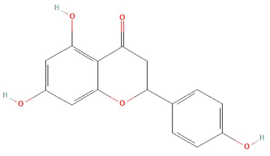
Figure 1. The molecular structure of naringenin.
2. Skeletal Effects of Naringenin on Osteoblasts
The currently available evidence shows that naringenin exerts stimulatory effects on osteoblasts through MAPK, PI3K/Akt, and CXCR4/SDF-1 pathways. Osteoblasts are responsible for osteogenesis by synthesising and mineralising organic bone matrix (osteoid) during skeleton construction and bone remodelling [20][41]. The MAPK cascades regulate Runx2 phosphorylation and transcription, which promote osteoblast differentiation. MAPK pathways and their components, JNK, ERK, and p38, which enforce osteoblastogenesis and establish the non-canonical BMP-2 signal transduction pathways [21][22][23][24][42,43,44,45]. Naringenin stimulates osteogenic gene activation, indicating that it has a stimulatory effect on osteogenic differentiation [25][26][21,22]. Activation of the PI3K/Akt signalling pathway also promotes osteoblast proliferation, differentiation, and bone formation activity [27][46]. Naringenin has been reported to stimulate BMP-2-dependent osteoblastogenesis through the activation of the PI3K/Akt signalling pathway [28][27]. Activation of the CXCR4/SDF-1 signalling pathway is critical in early osteoblastogenesis and its suppression leads to lower bone formation and mineralisation [29][30][47,48].
Osteoclasts are bone resorption cells originating from hematopoietic lineage cells [31][49]. Bone resorption is vital in bone remodelling, but excessive resorption can result in pathological bone loss. Osteoclast differentiation and activation are governed by various hormones and cytokines. The cytokines RANKL and M-CSF, in particular, are required for osteoclastic differentiation [32][50]. To promote osteoclast differentiation, preservation and bone resorption, the M-CSF binds to the colony-stimulating factor 1 receptor, whilst RANKL binds to the RANK receptor [33][34][51,52]. TRAF factors such as TRAF 6 are recruited by RANK-RANKL binding [35][53], leading to the activation of NF-kB, Akt, and MAPKs (ERK/p38/JNK) pathways. Furthermore, the RANKL signalling stimulates c-Fos and then NFATc1, a major switch that plays a role in controlling osteoclast terminal differentiation [36][37][54,55]. Naringenin was reported to reduce M-CSF and RANKL-induced expression of critical markers of osteoclast differentiation markers such as cathepsin K, c-Fos, and NFATc1 [38][39][24,25].
The biological properties of naringenin suggest a broad range of clinical applications. Naringenin decreased CAB-CEJ distance in buccal maxilla and mandible as well as in lingua maxilla and mandible, indicating that naringenin supplementation protects against alveolar bone loss in rats with induced periodontal disease [40][33]. Supplementation of naringenin also improved bone mineral microstructure, mineral, and biomechanical strength [28][41][42][43][23,27,31,32]. However, the bone loss models that have been used to test the effects of naringenin have been limited to OVX and retinol-induced models. Thus, results from other models, such as testosterone deficiency and glucocorticoid models, are indispensable before it is tested on patients with other causes of osteoporosis. Following joint replacement, the abrasive particles initiated by the prosthesis are primarily responsible for osteolysis [44][56]. Naringenin supplementation prevented Ti-particle-induced osteolysis, implying that it may be preferable for treating periprosthetic osteolysis [39][25].
Pharmacokinetics and safety issues of naringenin should be considered before it is used clinically. From the pharmacokinetic aspects, naringenin has very low in vivo bioavailability due to its hydrophobic nature, which limits its practical use. It has a short half-life and is easily converted to its crystalline form, and therefore it is poorly absorbed by the digestive system [45][46][47][48][57,58,59,60]. Previous researchers developed a variety of methods to improve naringenin absorption and low bioavailability, including particle size reduction, complexation with cyclodextrins [49][61], salt formation, solid dispersions [50][62], surfactant usage, nanoparticles, nanocarriers [51][63], and self-emulsifying drug delivery system, as well as prodrug formation [52][64]. Nanotechnology proved to be an efficient way to improve the bioavailability of naringenin by multiple delivery routes to enhance its effectiveness in the treatment of cancer, inflammation, diabetes, liver, brain, and ocular diseases mostly through numerous in vitro and in vivo methods [53][65]. Meanwhile, Rodríguez-Fragoso et al., (2011) [54][66] found out that naringenin inhibits some drug-metabolizing cytochrome P450 enzymes, including CYP3A4 and CYP1A2, potentially leading to drug-drug interactions in the intestine and liver, where phytochemical concentrations are higher. Modification in cytochrome P450 and other enzymatic activity may influence the outcome of drugs that go through extensive first-pass metabolism. An acute toxicity study using Wistar rats reported the lethal dose (LD50) value of naringenin to be 340 mg/kg body weight [55][67]. Using body surface ratio conversation [56][68], the human equivalent dose is 64 mg/kg.
The term “naringenin” was searched for on https://clinicaltrials.gov/ (accessed on 31 August 2022) and the search revealed thirteen registered clinical trials on naringenin. The trials investigate the effects of naringenin on healthy subjects (NCT02627547, NCT04867655, NCT05073523, NCT02380144), hepatitis virus/HCV infection/chronic HCV/Hepatitis C (NCT01091077), energy expenditure/safety issues/glucose metabolism (NCT04697355), safety issues/pharmacokinetics (NCT03582553), subjective cognitive decline (NCT04744922), cardiovascular disease risk factors (NCT00539916), intestinal disease (NCT03032861), metabolic syndrome/vascular compliance/predisposition to cardiovascular disease (NCT04731987), pharmacokinetics of new curcumin formulations/safety of new curcumin formulations (NCT01982734) and cardiovascular risk factor/type-2 diabetes mellitus/insulin sensitivity/metabolic syndrome (NCT03527277). Seven of these clinical trials have been completed, four are still recruiting, one with an unknown status, and lastly one trial is still active but not recruiting. However, no attempt has been made to conduct a human clinical trial to evaluate the impact of naringenin on skeletal diseases. Since pure naringenin has only been studied in limited clinical trials, more research on free drug and naringenin-loaded nanosystems in humans is warranted. Further exploration into the interactions of these nanoformulations with the human body is required before they can be translated into pharmaceuticals and nutraceutical supplements [57][69].