Rapamycin Signaling at Muscle Fiber Fate in Sarcopenia: Comparison
Please note this is a comparison between Version 2 by Amina Yu and Version 1 by Giuseppe Sirago.

Sarcopenia, the age-related decline of muscle mass and strength/function is a major risk factor for disability and loss of independence in late life. Studies have shown that behavioral interventions (e.g., physical activity, adapted nutrition) reduce the rate of muscle wasting during aging. However, an incomplete understanding of the mechanisms driving age-related muscle loss has hampered the development of effective drugs to prevent or treat sarcopenia. Altered muscle protein metabolism is considered to be one of the main factors underlying the development and progression of sarcopenia. While basal rates of muscle protein synthesis (MPS) and degradation (MPD) seem to be unaffected by age, the anabolic response to a variety of stimuli (e.g., exercise, nutrient ingestion) is blunted during aging. The mammalian target of rapamycin (mTOR) is a key regulator of muscle anabolic and catabolic pathways and, hence, a promising target for interventions against sarcopenia.

  • aging
  • skeletal muscle
  • autophagy
  • mitophagy
  • protein synthesis
  • protein degradation

1. Supramolecular Organization of the Mammalian Target of Rapamycin

Growing evidence points to the mTOR pathway as a major regulator of skeletal myofiber viability. This serine-threonine kinase is at the crossroad of cell proliferation and survival and, through the supramolecular mTOR complexes mTORC1 (mammalian target of rapamycin complex 1) and mTORC2, controls muscle fiber metabolism, growth, and proliferation [7][1]. Cryo-electron microscopy resolution experiments have revealed the supramolecular organization of the two mTOR complexes. In particular, mTORC1 appears to be organized into three main core proteins (i.e., mTOR, Raptor, and mammalian homolog of protein lethal with sec 13 protein 8), and two accessory regulatory proteins (i.e., GβL and Deptor) [7,8][1][2]. mTORC2, instead, seems to be organized into four main core proteins (i.e., mTOR, Rictor, the stress-activated map kinase-interacting protein 1 Sin1 (mSIN1), and mLST8), with three accessory regulatory proteins (i.e., PRR5/Protor-1, DEPTOR, and GβL) [7,9,10][1][3][4]. The molecular characterization of these supramolecular machineries is still incomplete and novel functions have recently been described. For instance, a direct role of Raptor in stimulating mitochondrial protein synthesis has been reported during muscle hypertrophy in mice [11][5]. It is noteworthy that the mTOR structure adapts to the myocyte metabolic milieu via chemical modifications and/or interactions with other protein complexes to modulate cell metabolism [7][1]. In the next sections, the integration of anabolic and catabolic signals by mTOR in the setting of muscle metabolism is described.

2. Muscle Protein Synthesis: The Mammalian Target of Rapamycin Complex 1 Axis

Muscle growth is regulated by several stimuli, such as mechanical loading and calcium uptake during physical exercise, insulin, insulin growth factor 1 (IGF-1), and amino acid signaling, that ultimately activate the phosphatidyl inositol 3-kinase (PI3K)–serine/threonine kinase 1 (AKT)–mTORC1 axis [3,12][6][7]. This axis stimulates protein synthesis and inhibits proteolysis, thereby switching muscle protein metabolism towards MPS and preserving cellular homeostasis in a balance with autophagy. Such modulation has been observed along with the stabilization of the neuromuscular junction (NMJ) and allows efficient excitation–contraction coupling [13,14,15][8][9][10]. Reduced mTORC1 signaling in murine models has been reported to trigger fiber denervation with the appearance of myofibers positive for neural cell adhesion molecules and segmented NMJ morphology [15][10]. This indicates that skeletal muscle fibers may be able to exert direct control over innervation to model motor neuron plasticity in response to molecular and metabolic changes. Non-coding RNAs (ncRNAs) are emerging as important regulators of muscle metabolism, and miR-29c seems to be able to trigger MPS and block MPD independent of the mTOR axis [16][11]. Conversely, mir-145-5p may inhibit mTOR phosphorylation, resulting in its deactivation [17][12].

mTORC1 Axis: Different Stimuli for Muscle Protein Synthesis

The two main upstream activators of mTOR (i.e., amino acids/glucose and insulin/IGFs) operate via specific signaling pathways depending on the type of stimulus [18,19][13][14]. Upon binding with their sarcolemmal receptors, insulin and IGF-1 trigger differential signaling pathways. In particular, the insulin receptor preferentially activates mTORC1 and AKT signaling to regulate metabolism, while the IGF-1 receptor preferentially activates Rho GTPases to regulate cellular growth [20][15]. mTORC2, instead, controls cell proliferation and survival via phosphorylation and activation of AKT [21][16].
Physical exercise stimulates PI3K signaling via integrins, located in the sarcolemma, which receive and integrate mechanical stimuli [22][17]. These are then converted into phosphorylation of focal adhesion kinases, mainly anchored to integrins at the sarcoplasmic side of the sarcolemma, ultimately resulting in the activation of the PI3K–phosphoinositide-dependent kinase 1–AKT–mTOR cascade [22][17]. Raptor seems to be the main factor phosphorylated by PI3K signaling during exercise [23,24][18][19]. The differential activation of the mTOR pathway is accomplished via a molecular feedback loop that blocks the activation of AKT upon mTORC1 and, therefore, represents a checkpoint for muscle growth or proteostasis [7][1]. The modulation of Raptor by exercise indicates that mTORC1 may control MPS under an intense rate of contraction, but not at a basal rate, or at least it could not be the only factor involved, which would explain its apparent loss of function during aging [24][19].
Nutrients, such as glucose and amino acids, are also able to activate protein translation and promote MPS via mTOR [25,26][20][21]. In particular, amino acids can activate mTORC1 through four small guanosine triphosphatases (GTPases), called Rag GTPases A–D, that bind Raptor and also activate Rheb, another GTP-binding protein, resulting in the activation of mTOR through a Rheb-GTP-binding-dependent mechanism [27][22]. The exact mechanism through which Rag activates mTOR is not well understood. However, Rag interaction with Raptor seems to be strongly inhibited in catabolic conditions, such as alcohol abuse and sarcopenia [28][23].

3. Muscle Protein Degradation: The Other Face of the mTORC1 Axis during Aging
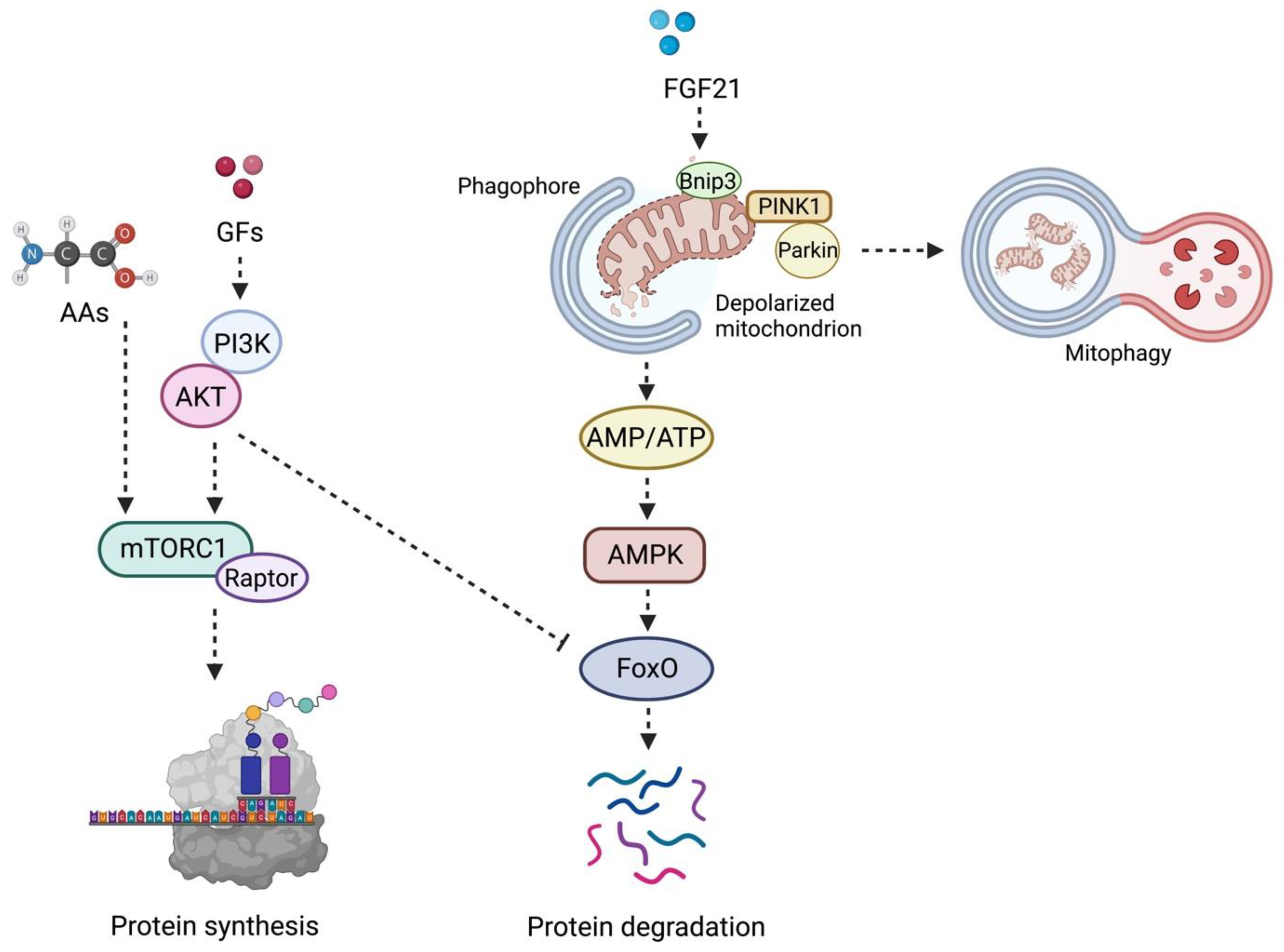
The PI3K–AKT–mTORC1 axis can trigger MPD via AKT signaling, especially during aging and in the setting of oxidative stress [29][24] (Figure 1). AKT is a serine-threonine protein kinase that regulates cell proteostasis, apoptosis, and turnover [29][24]. Upon activation, AKT phosphorylates Forkhead box protein O (FoxO) transcriptional regulators, thereby limiting their access to the nucleus and resulting in downregulation of atrogenes expression.
Figure 1. Schematic Representation of the Coordinated Regulation of the Mammalian Target of Rapamycin and Mitophagy in Muscle Protein Synthesis and Degradation. In the presence of growth factors (e.g., insulin, insulin-like growth factor 1, growth hormone), the phosphoinositide 3-kinases-protein kinase B (PI3K) is activated and triggers muscle protein synthesis via the mammalian target of rapamycin complex 1 (mTORC1). The latter complex is also positively modulated by amino acid availability. Conversely, downregulation of PI3K signaling induces translocation of Forkhead box O (FoxO) into the nucleus, where it regulates the transcription of the ubiquitin-ligases muscle ring finger 1 (MuRF1) and muscle atrophy F-box (MAFbx) genes. The activation of this signaling pathway induces degradation of sarcomere components and ignites a muscle pro-atrophy response. The same degradative molecular program is also triggered by fibroblast growth factor 21 (FGF21) in the setting of mitochondrial dysfunction and oxidative stress. In this case, the release of FGF21 stimulates the expression of the mitophagy-related protein B-cell lymphoma 2 interacting protein 3, paralleled by recruitment of the phosphatase and tensin homolog-induced kinase 1 (PINK1) through the translocases of the inner and the outer membranes and its activation at the site of depolarized mitochondria. This event promotes the sequestration of the E3 ubiquitin ligase Parkin at the outer mitochondrial membrane and guides the clearance of dysfunctional organelles. Finally, depolarized mitochondria are coated and prepared for disposal by the ubiquitin-binding adaptor protein p62/sequestosome-1 and the recruitment of the microtubule-associated proteins 1A/1B light chain 3B (LC3). This enables the transfer of mitochondria to lysosomes. FoxO-dependent atrophy is also pursued when severely damaged and bioenergetically incompetent mitochondria are not efficiently removed and, thus, the AMP/ATP ratio increases, which engages 5′ AMP-activated protein kinase (AMPK). Abbreviations: AA, amino acid; AMP, adenosine monophosphate; ATP, adenosine triphosphate; GF, growth factor; TIM23, translocase of the inner membrane 23; TOM, translocase of the outer membrane. Created with BioRender.com, accessed on 26 August 2022.
Atrogenes are E3 ubiquitin ligases that catalyze the rate-limiting step of the ubiquitin–proteosome system and allow recognition of substrates to be degraded. Muscle RING-finger 1 (MuRF1) and muscle atrophy F-box (Atrogin-1/MAFbx) are the two muscle-specific ubiquitin ligases. Among other emerging E3 ubiquitin ligases, Trim32 is specifically involved in the degradation and renewal of skeletal muscle sarcomere components [30][25]. Trim32 mutations have been found in limb–girdle muscular dystrophy 2H, and mice knock-out for Trim32 develop premature sarcopenia [31][26]. The maintenance of adequate levels of Trim32 has been associated with the preservation of muscle mass, reinnervation capacity, and NMJ plasticity during human aging [32][27]. A balance between AKT activation and repression is instrumental in achieving muscle protein homeostasis and preventing the accrual of defective unfolded proteins. Polymorphisms in FoxO genes have been linked to longevity and are included among the genetic mechanisms contributing to aging [33][28]. The existence of genetic variants and/or pools of genetic variants can also explain, at least partly, the variability existing among older adults in muscle responses to internal or external stimuli and, ultimately, in the susceptibility to develop sarcopenia [34][29].
Finally, sestrins, a class of biomolecules produced in response to stressful conditions, have also been described as relevant regulators of anabolic and degradative pathways orchestrated by mTORC1 [35][30]. In flies, sestrins are crucial for detecting leucine-containing food [36][31]. Sestrin-null flies lose the ability to detect leucine-free food and are unable to preferentially feed the progeny with leucine-containing food [36][31]. This ability is conveyed to flies by the capacity of sestrins to bind leucine. The latter is the main regulator of mTORC1 activity, and this nutrient-sensing role of mTORC1 is pivotal for mediating not only detection, but also adaptation to low-leucine diets in Drosophila [36][31]. Sestrin1, the skeletal muscle isoform of sestrin, shows a high affinity for leucine. Sestrin1 can bind to a complex named GTPase-activating protein activity toward Rags 2 (GATOR2), which sequesters Sestrin1. Leucine-induced activation of mTORC1 is directly controlled by the dissociation of Sestrin1 from GATOR2, due to the high affinity of Sestrin1 for leucine [37][32].
Together with ncRNAs, these stress-induced molecules represent emerging factors that may be able to modulate the mTOR pathway and that could, therefore, be exploited for drug development.

References

  1. Saxton, R.A.; Sabatini, D.M. MTOR Signaling in Growth, Metabolism, and Disease. Cell 2017, 168, 960–976.
  2. Aylett, C.H.S.; Sauer, E.; Imseng, S.; Boehringer, D.; Hall, M.N.; Ban, N.; Maier, T. Architecture of Human mTOR Complex 1. Science 2016, 351, 48–52.
  3. Yu, Z.; Chen, J.; Takagi, E.; Wang, F.; Saha, B.; Liu, X.; Joubert, L.M.; Gleason, C.E.; Jin, M.; Li, C.; et al. Interactions between mTORC2 Core Subunits Rictor and MSin1 Dictate Selective and Context-Dependent Phosphorylation of Substrate Kinases SGK1 and Akt. J. Biol. Chem. 2022, 298, 102288.
  4. Scaiola, A.; Mangia, F.; Imseng, S.; Boehringer, D.; Berneiser, K.; Shimobayashi, M.; Stuttfeld, E.; Hall, M.N.; Ban, N.; Maier, T. The 3.2-Å Resolution Structure of Human mTORC2. Sci. Adv. 2020, 6, eabc1251.
  5. Baraldo, M.; Nogara, L.; Dumitras, G.A.; Dondjang, A.H.T.; Geremia, A.; Scalabrin, M.; Türk, C.; Telkamp, F.; Zentilin, L.; Giacca, M.; et al. Raptor Is Critical for Increasing the Mitochondrial Proteome and Skeletal Muscle Force during Hypertrophy. FASEB J. 2021, 35, e22031.
  6. Sartori, R.; Romanello, V.; Sandri, M. Mechanisms of Muscle Atrophy and Hypertrophy: Implications in Health and Disease. Nat. Commun. 2021, 12, 330.
  7. Baehr, L.M.; Hughes, D.C.; Waddell, D.S.; Bodine, S.C. SnapShot: Skeletal Muscle Atrophy. Cell 2022, 185, 1618–1618.e1.
  8. Vainshtein, A.; Sandri, M. Signaling Pathways That Control Muscle Mass. Int. J. Mol. Sci. 2020, 21, 5759.
  9. Ham, D.J.; Börsch, A.; Lin, S.; Thürkauf, M.; Weihrauch, M.; Reinhard, J.R.; Delezie, J.; Battilana, F.; Wang, X.; Kaiser, M.S.; et al. The Neuromuscular Junction Is a Focal Point of MTORC1 Signaling in Sarcopenia. Nat. Commun. 2020, 11, 4510.
  10. Baraldo, M.; Geremia, A.; Pirazzini, M.; Nogara, L.; Solagna, F.; Türk, C.; Nolte, H.; Romanello, V.; Megighian, A.; Boncompagni, S.; et al. Skeletal Muscle mTORC1 Regulates Neuromuscular Junction Stability. J. Cachexia Sarcopenia Muscle 2020, 11, 208–225.
  11. Ketilly, P.; Alves, N.; Cruz, A.; Silva, W.J.; Labeit, S.; Moriscot, A.S. MiR-29c Increases Protein Synthesis in Skeletal Muscle Independently of AKT/mTOR. Int. J. Mol. Sci. 2022, 23, 7198.
  12. Jin, J.; Li, F.; Fan, C.; Wu, Y.; He, C. Elevated Mir-145-5p Is Associated with Skeletal Muscle Dysfunction and Triggers Apoptotic Cell Death in C2C12 Myotubes. J. Muscle Res. Cell Motil. 2022, 43, 135–145.
  13. Picca, A.; Calvani, R.; Sirago, G.; Coelho-Junior, H.J.; Marzetti, E. Molecular Routes to Sarcopenia and Biomarker Development: Per Aspera Ad Astra. Curr. Opin. Pharmacol. 2021, 57, 140–147.
  14. Liu, G.Y.; Sabatini, D.M. mTOR at the Nexus of Nutrition, Growth, Ageing and Disease. Nat. Rev. Mol. Cell Biol. 2020, 21, 183–203.
  15. Nagao, H.; Cai, W.; Albrechtsen, N.J.W.; Steger, M.; Batista, T.M.; Pan, H.; Dreyfuss, J.M.; Mann, M.; Kahn, C.R. Distinct Signaling by Insulin and IGF-1 Receptors and Their Extra- And Intracellular Domains. Proc. Natl. Acad. Sci. USA 2021, 118, e2019474118.
  16. Sarbassov, D.D.; Guertin, D.A.; Ali, S.M.; Sabatini, D.M. Phosphorylation and Regulation of Akt/PKB by the Rictor-mTOR Complex. Science 2005, 307, 1098–1101.
  17. Philp, A.; Hamilton, D.L.; Baar, K. Highlighted Topic Signals Mediating Skeletal Muscle Remodeling by Activity Signals Mediating Skeletal Muscle Remodeling by Resistance Exercise: PI3-Kinase Independent Activation of mTORC1. J. Appl. Physiol. 2011, 110, 561–568.
  18. Frey, J.W.; Jacobs, B.L.; Goodman, C.A.; Hornberger, T.A. A Role for Raptor Phosphorylation in the Mechanical Activation of mTOR Signaling. Cell. Signal. 2014, 26, 313–322.
  19. You, J.S.; Mcnally, R.M.; Jacobs, B.L.; Privett, R.E.; Gundermann, D.M.; Lin, K.H.; Steinert, N.D.; Goodman, C.A.; Hornberger, T.A. The Role of Raptor in the Mechanical Load-Induced Regulation of mTOR Signaling, Protein Synthesis, and Skeletal Muscle Hypertrophy. FASEB J. 2019, 33, 4021–4034.
  20. Vary, T.C.; Jefferson, L.S.; Kimball, S.R. Amino Acid-Induced Stimulation of Translation Initiation in Rat Skeletal Muscle. Am. J. Physiol. 1999, 277, 1077–1086.
  21. Jeyapalan, A.S.; Orellana, R.A.; Suryawan, A.; O’Connor, P.M.J.; Nguyen, H.V.; Escobar, J.; Frank, J.W.; Davis, T.A. Glucose Stimulates Protein Synthesis in Skeletal Muscle of Neonatal Pigs through an AMPK- and mTOR-Independent Process. Am. J. Physiol. Endocrinol. Metab. 2007, 293, 595–603.
  22. Sancak, Y.; Peterson, T.R.; Shaul, Y.D.; Lindquist, R.A.; Thoreen, C.C.; Bar-Peled, L.; Sabatini, D.M. The Rag GTPases Bind Raptor and Mediate Amino Acid Signaling to mTORC1. Science 2008, 320, 1496–1501.
  23. Korzick, D.H.; Sharda, D.R.; Pruznak, A.M.; Lang, C.H. Aging Accentuates Alcohol-Induced Decrease in Protein Synthesis in Gastrocnemius. Am. J. Physiol. Regul. Integr. Comp. Physiol. 2013, 304, R887–R898.
  24. Tang, H.; Inoki, K.; Brooks, S.V.; Okazawa, H.; Lee, M.; Wang, J.; Kim, M.; Kennedy, C.L.; Macpherson, P.C.D.; Ji, X.; et al. mTORC1 Underlies Age-Related Muscle Fiber Damage and Loss by Inducing Oxidative Stress and Catabolism. Aging Cell 2019, 18, e12943.
  25. Cohen, S.; Zhai, B.; Gygi, S.P.; Goldberg, A.L. Ubiquitylation by Trim32 Causes Coupled Loss of Desmin, Z-Bands, and Thin Filaments in Muscle Atrophy. J. Cell Biol. 2012, 198, 575–589.
  26. Mokhonova, E.I.; Avliyakulov, N.K.; Kramerova, I.; Kudryashova, E.; Haykinson, M.J.; Spencer, M.J. The E3 Ubiquitin Ligase TRIM32 Regulates Myoblast Proliferation by Controlling Turnover of NDRG2. Hum. Mol. Genet. 2015, 24, 2873–2883.
  27. Skoglund, E.; Grönholdt-Klein, M.; Rullman, E.; Thornell, L.E.; Strömberg, A.; Hedman, A.; Cederholm, T.; Ulfhake, B.; Gustafsson, T. Longitudinal Muscle and Myocellular Changes in Community-Dwelling Men over Two Decades of Successful Aging–The ULSAM Cohort Revisited. J. Gerontol. A Biol. Sci. Med. Sci. 2020, 75, 654–663.
  28. Kenyon, C.J. The Genetics of Ageing. Nature 2010, 464, 504–512.
  29. Sirago, G.; Picca, A.; Giacomello, E.; Marzetti, E.; Toniolo, L. The Contribution of Genetics to Muscle Disuse, Retraining, and Aging. Genes 2022, 13, 1378.
  30. Segalés, J.; Perdiguero, E.; Serrano, A.L.; Sousa-Victor, P.; Ortet, L.; Jardí, M.; Budanov, A.V.; Garcia-Prat, L.; Sandri, M.; Thomson, D.M.; et al. Sestrin Prevents Atrophy of Disused and Aging Muscles by Integrating Anabolic and Catabolic Signals. Nat. Commun. 2020, 11, 189.
  31. Gu, X.; Jouandin, P.; Lalgudi, P.V.; Binari, R.; Valenstein, M.L.; Reid, M.A.; Allen, A.E.; Kamitaki, N.; Locasale, J.W.; Perrimon, N.; et al. Sestrin Mediates Detection of and Adaptation to Low-Leucine Diets in Drosophila. Nature 2022, 608, 209–216.
  32. Xu, D.; Shimkus, K.L.; Lacko, H.A.; Kutzler, L.; Jefferson, L.S.; Kimball, S.R. Evidence for a Role for Sestrin1 in Mediating Leucine-Induced Activation of mTORC1 in Skeletal Muscle. Am. J. Physiol. Endocrinol. Metab. 2019, 316, E817–E828.
More
Video Production Service