Your browser does not fully support modern features. Please upgrade for a smoother experience.
Please note this is a comparison between Version 1 by Sonja Radjenovic and Version 2 by Peter Tang.
Transcranial ultrasound holds much potential as a safe, non-invasive modality for navigated neuromodulation, with low-intensity focused ultrasound (FUS) and transcranial pulse stimulation (TPS) representing the two main modalities.
- transcranial ultrasound
- neuromodulation
- TPS
- FUS
- clinical neuromodulation
1. Introduction
Over the past decade, there have been considerable advances in developing new sonication methods for non-invasive brain stimulation [1][2][3][1,2,3]. Although deep brain stimulation (DBS) is known as an optimistic treatment method for various clinical indications including epilepsy, essential tremor, dystonia, and Parkinson’s disease [4], it is a surgical procedure, and the risk of side-effects cannot be completely ruled out [5]. The growing interest in ultrasound-based modalities over electrical stimulation techniques, such as transcranial magnetic stimulation [6] or transcranial direct current stimulation [7], is due to a number of advantages. These include, for instance, the high spatial accuracy [8] or possibility of non-invasive subcortical stimulation [9], which have been limitations for electrical stimulation techniques, especially in clinical settings [10][11][10,11]. While these characteristics apply in general to ultrasonic neuromodulation, notable varieties in available transducer systems produce different stimulation pulses. Low-intensity focused ultrasound (FUS) systems generate sonication trains of sinus tones with a fixed fundamental frequency that is usually (though not necessarily) applied in a pulsed fashion [12]. Next to FUS, transcranial pulse stimulation (TPS) represents another ultrasound-based stimulation technique with markedly different characteristics. TPS generates ultrashort pressure pulses consisting of multiple frequencies with higher amplitude [13]. The pulse duration is in the range of a few microseconds, and typically administered with a repetition frequency between 1 and 5 Hz, reducing the risk of tissue heating associated with continuous application of ultrasound [14]. Both FUS and TPS can be described with a number of interacting parameters that can be controlled to change the physical properties of the stimulation pulse. Among these are, for instance, the fundamental frequency (typically between 250–1000 kHz), pulse repetition frequency (determining the pulse rate), duty cycle (the percentage of the time the ultrasound pulse is “on” over the smallest stimulation period) and the intensity.
Transcranial ultrasound neuromodulation has been extensively investigated in pre-clinical settings, both in animal and human studies, yet clinical applications are still scarce. The first uses were in patients with disorders of consciousness, both with a non-navigated TPS precursor [15] and navigated FUS [16]. Although neither trial had sham controls, results were promising and showed no signs of neurological damage. The first clinical study with highly focused navigated pulses has been performed with the TPS technology [13]. In this multicenter clinical study with Alzheimer patients, TPS was shown to be safe and specifically improve cognition, memory and depression scores [13][17][18][13,17,18]. Later, navigated FUS studies investigated patients with disorders of consciousness [19][20][19,20], Alzheimer’s disease [21] and epilepsy [22]. While the possible clinical applications for precise neural stimulation are immense, it is vital to deepen our understanding of the exact mechanisms and consequences of brain sonication in neural tissue to offer safe and effective treatment options.
Clinical ultrasound has the potential to produce two major types of effects with relevance for clinical safety, namely heating and cavitation [23]. These modalities strongly differ from others. Tissue heating occurs due to the absorption of the ultrasonic waves, and heating increases with ultrasound frequency and the applied acoustic intensity [14]. Cavitation relates to the expansion and collapse of local tissue exposed to a tensile pressure. If local gas bodies exist, harming effects on local tissue cells may increase.
2. Main Bioeffects
2.1. Cavitation Effects
The Mechanical Index (MI) was developed to adequately indicate the likelihood of cavitation [24]. The formula for the MI is based on the assumption that a nucleation site of only the resonant site is available for inertial cavitation [23]: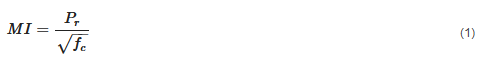 with Pr being the derated peak rarefactional pressure [MPa] and fc the center frequency of the ultrasound pulse [MHz]. Notably, the MI is not applicable for TPS pressure pulses since the timescale of the expansion phase of a bubble forced by a TPS pulse is much longer than the TPS pulse length [25]. Concerning conditions for occurrence of cavitation in biological tissues, a wide range of cavitation thresholds are reported in the literature, demonstrating that this phenomenon largely depends on exposure and experimental conditions. The findings from several studies looking at cavitation effects are summarized below. One study investigated the cavitation threshold in sheep brains exposed to 660 kHz ultrasonic pulses (2 cycles) [25]. No cavitation could be detected for peak tensile pressures below 11.6 MPa, but systematically at 22.4 MPa. Another study found a reversible opening of the blood brain barrier (BBB) in rats after 50 focused shockwaves with tensile pressures of 9.8 MPa [26]. With the prior adjunction of microbubbles, BBB disruption and FUS induced cavitation using 20 ms bursts at 1.5 MHz at peak tensile pressures around 0.5 MPa were reported [27]. In the investigation of cavitation thresholds induced by ultrasound pulses (1–2 cycles) with a 1.1 MHz transducer in different media [28], a cavitation threshold (50% probability) from 14 to 30 MPa tensile pressure, depending on the sonicated medium, was produced. Other studies reported cavitation onset in a tissue mimicking phantom (agar) at 1 MHz for peak tensile pressures between 3 and 10 MPa [29][30]. The threshold for cavitation onset depended on the number of cycles (minimum 10 cycles), the duration of sonication (up to 3 s) and previous exposures. While researchers studying cavitation thresholds for 1–2 cycle histotripsy pulses at frequencies from 300 kHz to 3 MHz in water, phantoms and ex vivo bovine tissues [31] reported cavitation onset (50% probability) for peak tensile pressures from 24 to 30.6 MPa. In another study using a 1 MHz transducer delivering 5 histotripsy pulses repeated at a frequency of 100 to 1000 Hz, the same researchers reported initiation of dense bubble clouds in ex vivo porcine tissues from 1.5 (in lungs) up to 27 MPa (in other soft tissues) peak tensile pressures. They noted the importance of the tissue stiffness and mentioned the need for single bubbles to expand to a sufficient size during the initial cycles of the pulse in order to initiate a dense bubble cloud, as well as the reduction of the cavitation threshold at higher PRFs [32]. the center frequency of the ultrasound pulse [MHz]. Notably, the MI is not applicable for TPS pressure pulses since the timescale of the expansion phase of a bubble forced by a TPS pulse is much longer than the TPS pulse length [25]. Concerning conditions for occurrence of cavitation in biological tissues, a wide range of cavitation thresholds are reported in the literature, demonstrating that this phenomenon largely depends on exposure and experimental conditions. The findings from several studies looking at cavitation effects are summarized below. One study investigated the cavitation threshold in sheep brains exposed to 660 kHz ultrasonic pulses (2 cycles) [25]. No cavitation could be detected for peak tensile pressures below 11.6 MPa, but systematically at 22.4 MPa. Another study found a reversible opening of the blood brain barrier (BBB) in rats after 50 focused shockwaves with tensile pressures of 9.8 MPa [26]. With the prior adjunction of microbubbles, BBB disruption and FUS induced cavitation using 20 ms bursts at 1.5 MHz at peak tensile pressures around 0.5 MPa were reported [27]. In the investigation of cavitation thresholds induced by ultrasound pulses (1–2 cycles) with a 1.1 MHz transducer in different media [28], a cavitation threshold (50% probability) from 14 to 30 MPa tensile pressure, depending on the sonicated medium, was produced. Other studies reported cavitation onset in a tissue mimicking phantom (agar) at 1 MHz for peak tensile pressures between 3 and 10 MPa [29,30]. The threshold for cavitation onset depended on the number of cycles (minimum 10 cycles), the duration of sonication (up to 3 s) and previous exposures. While researchers studying cavitation thresholds for 1–2 cycle histotripsy pulses at frequencies from 300 kHz to 3 MHz in water, phantoms and ex vivo bovine tissues [31] reported cavitation onset (50% probability) for peak tensile pressures from 24 to 30.6 MPa. In another study using a 1 MHz transducer delivering 5 histotripsy pulses repeated at a frequency of 100 to 1000 Hz, the same researchers reported initiation of dense bubble clouds in ex vivo porcine tissues from 1.5 (in lungs) up to 27 MPa (in other soft tissues) peak tensile pressures. They noted the importance of the tissue stiffness and mentioned the need for single bubbles to expand to a sufficient size during the initial cycles of the pulse in order to initiate a dense bubble cloud, as well as the reduction of the cavitation threshold at higher PRFs [32].
This variability in conditions and pressure ranges for generation of cavitation indicates that the MI alone is not a sufficient predictor of bioeffects [33][34][33,34]. Beyond the MI, other conditions have a major influence, such as the number of consecutive pulses applied, burst duration, or use of ultrasound contrast agents (UCA). The FDA guidelines limit the MI to 1.9 for diagnostic ultrasound devices [35]. No adverse non-thermal bioeffects have been observed for MI under the FDA limit in tissues without gas bodies, and the lowest threshold for cavitation in vivo and related adverse effects more likely lies at or above MI values of 4 [35].
The presence of injected or endogenous gas bodies in the sonicated medium is a critical condition for the onset of acoustic cavitation. Several authors reported a dramatic reduction in cavitation thresholds in organs naturally containing air bubbles, such as lungs and intestine, and in other tissues after systemic injection of UCA [34]. In brain injuries in rats following shockwave exposure from 7 up to 14 MPa tensile pressures after injection of UCA, a higher threshold was observed when no UCA was administered [36]. Another study reported only minor cavitation injuries in tissues exposed to clinically relevant lithotripsy exposures (>40 Mpa) [37]. However, this threshold decreased to 2 MPa in tissues containing endogenous gas bodies or when a UCA was injected.
with Pr being the derated peak rarefactional pressure [MPa] and fc the center frequency of the ultrasound pulse [MHz]. Notably, the MI is not applicable for TPS pressure pulses since the timescale of the expansion phase of a bubble forced by a TPS pulse is much longer than the TPS pulse length [25]. Concerning conditions for occurrence of cavitation in biological tissues, a wide range of cavitation thresholds are reported in the literature, demonstrating that this phenomenon largely depends on exposure and experimental conditions. The findings from several studies looking at cavitation effects are summarized below. One study investigated the cavitation threshold in sheep brains exposed to 660 kHz ultrasonic pulses (2 cycles) [25]. No cavitation could be detected for peak tensile pressures below 11.6 MPa, but systematically at 22.4 MPa. Another study found a reversible opening of the blood brain barrier (BBB) in rats after 50 focused shockwaves with tensile pressures of 9.8 MPa [26]. With the prior adjunction of microbubbles, BBB disruption and FUS induced cavitation using 20 ms bursts at 1.5 MHz at peak tensile pressures around 0.5 MPa were reported [27]. In the investigation of cavitation thresholds induced by ultrasound pulses (1–2 cycles) with a 1.1 MHz transducer in different media [28], a cavitation threshold (50% probability) from 14 to 30 MPa tensile pressure, depending on the sonicated medium, was produced. Other studies reported cavitation onset in a tissue mimicking phantom (agar) at 1 MHz for peak tensile pressures between 3 and 10 MPa [29][30]. The threshold for cavitation onset depended on the number of cycles (minimum 10 cycles), the duration of sonication (up to 3 s) and previous exposures. While researchers studying cavitation thresholds for 1–2 cycle histotripsy pulses at frequencies from 300 kHz to 3 MHz in water, phantoms and ex vivo bovine tissues [31] reported cavitation onset (50% probability) for peak tensile pressures from 24 to 30.6 MPa. In another study using a 1 MHz transducer delivering 5 histotripsy pulses repeated at a frequency of 100 to 1000 Hz, the same researchers reported initiation of dense bubble clouds in ex vivo porcine tissues from 1.5 (in lungs) up to 27 MPa (in other soft tissues) peak tensile pressures. They noted the importance of the tissue stiffness and mentioned the need for single bubbles to expand to a sufficient size during the initial cycles of the pulse in order to initiate a dense bubble cloud, as well as the reduction of the cavitation threshold at higher PRFs [32]. the center frequency of the ultrasound pulse [MHz]. Notably, the MI is not applicable for TPS pressure pulses since the timescale of the expansion phase of a bubble forced by a TPS pulse is much longer than the TPS pulse length [25]. Concerning conditions for occurrence of cavitation in biological tissues, a wide range of cavitation thresholds are reported in the literature, demonstrating that this phenomenon largely depends on exposure and experimental conditions. The findings from several studies looking at cavitation effects are summarized below. One study investigated the cavitation threshold in sheep brains exposed to 660 kHz ultrasonic pulses (2 cycles) [25]. No cavitation could be detected for peak tensile pressures below 11.6 MPa, but systematically at 22.4 MPa. Another study found a reversible opening of the blood brain barrier (BBB) in rats after 50 focused shockwaves with tensile pressures of 9.8 MPa [26]. With the prior adjunction of microbubbles, BBB disruption and FUS induced cavitation using 20 ms bursts at 1.5 MHz at peak tensile pressures around 0.5 MPa were reported [27]. In the investigation of cavitation thresholds induced by ultrasound pulses (1–2 cycles) with a 1.1 MHz transducer in different media [28], a cavitation threshold (50% probability) from 14 to 30 MPa tensile pressure, depending on the sonicated medium, was produced. Other studies reported cavitation onset in a tissue mimicking phantom (agar) at 1 MHz for peak tensile pressures between 3 and 10 MPa [29,30]. The threshold for cavitation onset depended on the number of cycles (minimum 10 cycles), the duration of sonication (up to 3 s) and previous exposures. While researchers studying cavitation thresholds for 1–2 cycle histotripsy pulses at frequencies from 300 kHz to 3 MHz in water, phantoms and ex vivo bovine tissues [31] reported cavitation onset (50% probability) for peak tensile pressures from 24 to 30.6 MPa. In another study using a 1 MHz transducer delivering 5 histotripsy pulses repeated at a frequency of 100 to 1000 Hz, the same researchers reported initiation of dense bubble clouds in ex vivo porcine tissues from 1.5 (in lungs) up to 27 MPa (in other soft tissues) peak tensile pressures. They noted the importance of the tissue stiffness and mentioned the need for single bubbles to expand to a sufficient size during the initial cycles of the pulse in order to initiate a dense bubble cloud, as well as the reduction of the cavitation threshold at higher PRFs [32].
This variability in conditions and pressure ranges for generation of cavitation indicates that the MI alone is not a sufficient predictor of bioeffects [33][34][33,34]. Beyond the MI, other conditions have a major influence, such as the number of consecutive pulses applied, burst duration, or use of ultrasound contrast agents (UCA). The FDA guidelines limit the MI to 1.9 for diagnostic ultrasound devices [35]. No adverse non-thermal bioeffects have been observed for MI under the FDA limit in tissues without gas bodies, and the lowest threshold for cavitation in vivo and related adverse effects more likely lies at or above MI values of 4 [35].
The presence of injected or endogenous gas bodies in the sonicated medium is a critical condition for the onset of acoustic cavitation. Several authors reported a dramatic reduction in cavitation thresholds in organs naturally containing air bubbles, such as lungs and intestine, and in other tissues after systemic injection of UCA [34]. In brain injuries in rats following shockwave exposure from 7 up to 14 MPa tensile pressures after injection of UCA, a higher threshold was observed when no UCA was administered [36]. Another study reported only minor cavitation injuries in tissues exposed to clinically relevant lithotripsy exposures (>40 Mpa) [37]. However, this threshold decreased to 2 MPa in tissues containing endogenous gas bodies or when a UCA was injected.
2.2. Heating Effects
The energy deposit in tissues can be high enough to generate substantial local heating [14], which is desired for ablative indications such as MRI-guided focused ultrasound [38]. The local temperature rise depends on the tissue characteristics, such as absorption or perfusion. For instance, skull bone, due to its specific tissue properties, absorbs more acoustic energy and is subsequently more susceptible to heating [39]. The difference in temperature increase between skull and brain tissue was recently illustrated in a simulation using non-human primate data, where the authors showed an increase of 0.5 °C at the target site compared to 2.9 °C where the transducer was placed at the skull [40]. The Thermal Index (TI) was established to provide a reasonable estimate of temperature rise when ultrasound propagates through tissues, which is the ratio of the attenuated acoustic power on the acoustic power needed to raise the temperature by 1 °C at a specified point [41]. It is a unit-less value and has a recommended maximum of 6 in adults and should be adjusted according to the planned exposure time [42]. Another important physical metric related to thermal effects is the spatial-peak temporal average intensity (ISPTA), which gives the fraction of the sonication intensity per second, i.e., the time average over a continued pulse train: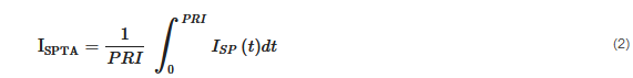 where PRI is the pulse repetition interval and ISP is the spatial average intensity [W/cm2]. This is related to the spatial-peak pulse average intensity (ISPTA), which gives the average over a single pulse:
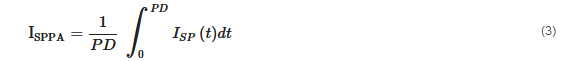 where PD is the pulse duration. Note that, depending on the mode of application, which is described by the fraction of sonication duration per second or duty cycle (DC), the ISPTA will be different (pulsed application) or equal (continuous wave) to the ISPPA [43]. Importantly, FDA guidelines for diagnostic imaging applications set the ISPTA limit to 720 mW/cm2, which is often used as a reference, but this limit has been exceeded in past transcranial ultrasound stimulation studies without clinically relevant adverse events.