Perivascular Adipose Tissue-Derived Extracellular Vesicle Exosomes: Comparison
Please note this is a comparison between Version 2 by Dean Liu and Version 1 by Melvin R Hayden.

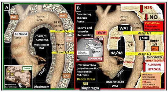
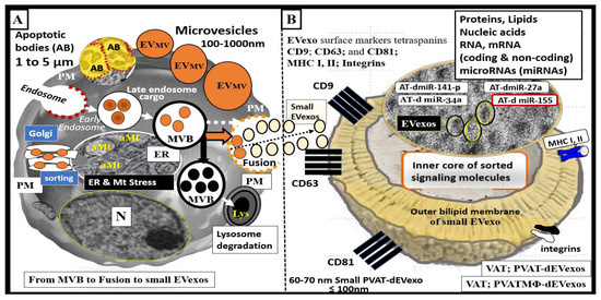
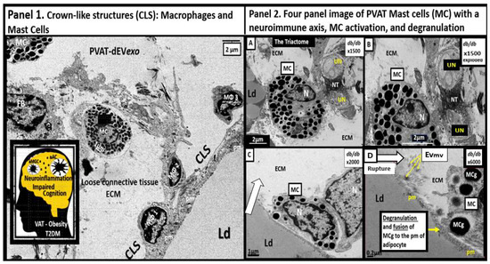
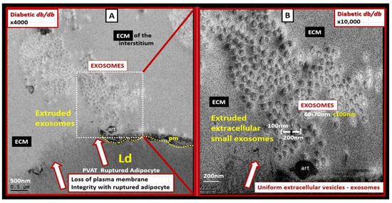
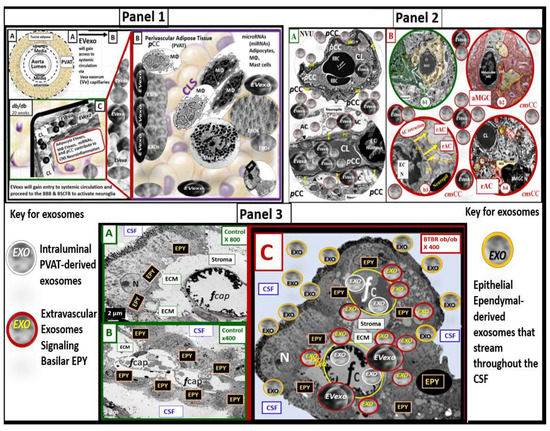
Perivascular adipose tissue (PVAT)-derived extracellular vesicles (EVs) with small exosome(s) (PVAT-dEVexos) from the descending aorta are capable of entering capillaries and systemic circulation. These PVAT-dEVexos are delivered to the central nervous system (CNS) in preclinical, obese, insulin and leptin resistant, diabetic, db/db mouse models and humans with T2DM. Once within the CNS, these exosomes are capable of traversing the blood–brain barrier and the blood-cerebrospinal fluid barrier resulting in activation of the neuroglia microglia cell(s) (aMGCs) and the formation of reactive astrocytes (rACs). The chronic peripheral inflammation in the PVAT via crown-like structures consists of activated macrophages and mast cells, which harbor peripheral adipokines, cytokines, and chemokines (pCC) in addition to the EV exosomes. 

  • adipokines
  • chemokines
  • cytokines
  • EVexosomes

1. Introduction

Obesity has now gained global epidemic proportions and parallels the current increasing trend of the global type 2 diabetes mellitus (T2DM) epidemic [1]. For example, in the United States approximately 60% of the population has been classified as being overweight as defined by a body mass index of ≥25 kg/m2, or obese as defined by a body mass index of ≥30 kg/m2 [1]. The incidence of T2DM has also increased in parallel with the obesity epidemic and thus, T2DM could be considered a major co-morbid condition associated with obesity [2,3,4][2][3][4]. Recent epidemiologic findings have shown that ~85% of individuals with T2DM are also obese and it is currently estimated that by 2025 more than 300 million people will have T2DM that is related to obesity [5]. Importantly, obesity and T2DM are associated with metabolic syndrome (MetS), which increases the risk of cerebrocardiovascular disease (CCVD). Further, CCVD includes the vasculature system, heart, and brain, which are involved in the clinical utility of the MetS and its increased morbidity and mortality by 2-fold or greater [6,7,8,9][6][7][8][9].

1.1. Adipose Tissue and Remodeling in Obesity, MetS, and T2DM

Adipose tissue (AT) may be considered a multi-depot and endocrine organ that is divided into brown adipose tissue (BAT) and white adipose tissue (WAT) in mammals and humans [10,11,12,13][10][11][12][13]. BAT is responsible for non-shivering thermogenesis and is known to stain positive for uncoupling proteins (UCP-1, 2) and WAT is known to be responsible for the storage of excess fat (free fatty acids that are taken up by WAT adipocytes and stored as triglycerides and do not stain positive for UCP-1, 2 [10,11,12,13,14][10][11][12][13][14]. WAT may be further defined as being either subcutaneous AT (SAT) or visceral AT (VAT) [10,11,12,13][10][11][12][13]. VAT may be further subdivided into omental, mesenteric, retroperitoneal, perinephric, epicardial, pericardial, and perivascular AT (PVAT) [11,12,13][11][12][13]. The VAT is known to develop an inflammatory phenotype in conditions of excess nutrient intake and is more closely associated with the development of obesity, T2DM, insulin resistance (IR), and CCVD as compared to SAT [6,7,15][6][7][15]. Importantly, VAT mass correlates positively with the development of IR, while SAT tissue mass does not [16,17,18][16][17][18]. Additionally, mesenteric VAT of obese T2DM individuals has a significant increase in leptin and a downregulation of the adiponectin gene expression with decreased adiponectin as compared to SAT [19].
VAT develops chronic low-grade inflammation (metainflammation) in obesity and T2DM, which includes the PVAT of the descending aorta. PVAT is the VAT that envelops the outermost region of the aorta and is contained within the tunica adventitia layer. There is a definite ultrastructural demarcation of the PVAT within the tunica adventitia such that some have recently defined this region as the PVAT of the tunica adiposa (the periadventitial VAT or the PVAT) [20,21,22][20][21][22].
AT is known to be a highly plastic organ and capable of transdifferentiation from BAT to WAT and vice versa [23]. The PVAT depot in the descending thoracic aorta in health (control mice and humans) consists of a near-complete multilocular BAT depot replete with multiple mitochondria typical of BAT. In contrast, the abdominal aorta PVAT consists of a unilocular WAT depot with a relative paucity of mitochondria as compared to the BAT depot [24]. The descending thoracic aorta undergoes near-complete transdifferentiation to WAT with marked hypertrophic unilocular adipocytes in the obese, diabetic db/db mouse. Thus, it was very interesting that BAT of descending thoracic aorta in control models underwent transdifferentiation from BAT to that of a markedly enhanced volume of unilocular WAT (Figure 1) [20].
Figure 1. The perivascular adipose tissue (PVAT) of descending aorta in the obese, diabetic db/db model undergoes transdifferentiation from multilocular brown adipose tissue (BAT) to unilocular white adipose tissue (WAT). (A) demonstrates the normal PVAT depot, which consists of multilocular brown adipose tissue (BAT) in control models (insert). (B) depicts the transdifferentiation from BAT to WAT with hypertrophic unilocular adipocytes that are prone to rupture and secrete multiple cytokines/chemokines and PVAT-derived small extracellular vesicles exosomes (PVAT-dEVexo) in the obese, diabetic db/db mice (insert). Interestingly, the transdifferentiation of BAT in controls to WAT in the db/db models is associated with aortic vascular stiffing. This modified image is published with permission by CC 4.0 [20]. ADRF = adipose- derived relaxing factor; AGE/RAGE = advanced glycation end products and their receptor for AGE; ALDO = aldosterone; DESC = descending aorta; Dys = dysfunction; EC = endothelial cell(s); ECM = extracellular matrix; FFA = free fatty acids; H2S = hydrogen sulfide; MR = mineralocorticoid receptor; NO = nitric oxide; OS = oxidative stress; pCC = peripheral cytokines/chemokines; RSI = reactive species interactome.
The group and various colleagues have been extremely fortunate to have had the opportunity to examine multiple models of obesity, insulin resistance (IR), leptin resistance (LR), and models with no measurable leptin such as the BTBR ob/ob, with MetS and diabetes over the past decade. The data obtained from these studies have allowed for the creation of Venn diagrams to compare these multiple models and how they overlap (Figure 2) [14,20,25,26,27,28,29,30,31,32,33,34,35,36,37,38,39][14][20][25][26][27][28][29][30][31][32][33][34][35][36][37][38][39].
Figure 2. Venn diagrams depicting multiple models in sets/circles (1 through 3) of brain remodeling. The model most studied was the obese, insulin-, and leptin-resistant (IR and LR, respectively) db/db mouse model at 20 weeks of age (circle 2) [14,20,25,26,30,31,32,33,34,35,36,37,38][14][20][25][26][30][31][32][33][34][35][36][37][38]. Furthermore, the Zucker fa/fa rat (circle 2) [25[25][26][27],26,27], diet-induced obesity (DIO) Western (circle 1) [25[25][26][28][31],26,28,31], and most recently the novel BTBR ob/ob mouse (circle 3) [39] in addition to the streptozotocin-induced diabetic model (circle 4-upper right colored in purple) [29] were studied. Note the overlapping circles depicting IR and LR between circles 1 and 2 (light blue ellipse) and only IR between circles 1 and 3 that share IR (yellow ellipse). Importantly, note that where all three circles (1–3) of the Venn diagram intersect the arrow points to the common features of obesity, IR, dysglycemia, prediabetes, and manifest diabetes (multicolored triangle lower-right). Only the db/db, Zucker obese fa/fa, and the Western models share in common LR. Deficient cellular leptin signaling is common in all models studied (circles 1–3). This modified image is provided with permission by CC 4.0 [39]. IFG = impaired fasting glucose; IGT = impaired glucose tolerance; IR = insulin resistance; LR = leptin resistance.
Notably, these above studies allowed for the current transmission electron microscopic (TEM) observations of small PVAT-derived EVexosomes (PVAT-dEVexos) in the unilocular engorged rupture-prone WAT of the descending thoracic aorta and macrophages that create crown-like structures in the PVAT of obese, diabetic db/db models.

1.2. EVexosomes in VAT and PVAT Are Capable of Interorgan Signaling to the Brain and Result in Neuroglial Activation

The history of extracellular vesicle exosomes (EVexos) is rich and this field of research has greatly expanded since they were first discovered in reticulocytes by Harding et al., and Pan et al. [40,41,42][40][41][42]. Notably, it was Rose Johnstone who coined the term exosomes [43]. A major breakthrough occurred when it was found that parenteral EVexos contained both messenger ribonucleic acid (mRNA) and microRNA (miRNA) and that they could be translated into proteins by target cells [44,45][44][45]. The interest and fascination with exosomes have expanded exponentially in the past few years because it has been found that these exosomes not only signal via an autocrine, paracrine (intercellular communication) but also an endocrine mechanism to signal distant organs and cells (inter-organ communication) from the parental cells generating exosomes [46]. Furthermore, it has been found that exosomes contain multiple biologically versatile cargoes including various proteins, fats (fatty acids, cholesterol, sphingomyelins, and ceramides), and nucleic acids including coding and long-non-coding RNA (incRNAs), mRNA, and miRNAs that are capable of signaling adjacent and distant cells and organs [46,47][46][47].
Stressed cells (especially those with endoplasmic reticulum stress) as in obesity and T2DM are capable of producing greater numbers of exosomes as occur in the obese PVAT adipocytes with hypertrophy and crown-like structures (CLS) of macrophages with or without rupture that may be capable of producing more exosomes than quiescent homeostatic non-stressed adipocytes in healthy lean models [48]. This concept is especially important given that WAT (SAT and VAT) may be the largest secretory, endocrine organ in the human body [49].
Notably, many publications regarding AT derived-exosomes are related to their protective roles such as adipocyte-derived mesenchymal stem cells with anti-inflammatory and regenerative potential. However, wresearchers are only recently beginning to learn of the potential negative and damaging roles that obese adipocytes (VAT and PVAT) are playing in multiple diseases such as occurs in obesity, MetS, IR, LR, and T2DM [50,51,52][50][51][52]. This overview will primarily focus on small EVexos that are generated in PVAT adipocytes (PVAT-dEVexos) and macrophages (PVATMΦ-dEVexos) as observed with TEM in the obese, IR, LR, female, diabetic, db/db preclinical rodent models. Extracellular vesicles including exosomes, microvesicles, and apoptotic bodies are identified in the VAT-PVAT of obese, prediabetic, and manifest diabetic models such as occurs in the db/db preclinical models (Figure 3).
Figure 3. Extracellular vesicles (EV): Exosomes, microvesicles, and apoptotic bodies as found in the obese and diabetic preclinical models. (A) illustrates a representative cell with early and late endosomes and multivesicular body formation and fusion with the plasma membrane (pm) and secretion of exosomes along with the alternative pathway for lysosomal degradation of MVB contents. (B) illustrates a cross-section view of a small exosome. Exosomes are derived from late endosome-multivesicular bodies (MVB), which fuse with the plasma membrane and are secreted into the extracellular space for paracrine intercellular and/or endocrine inter-organ communication. They are dependent on endosomal sorting complexes required for transport (ESCRT), cluster of differentiation tetraspanins (CD-9, CD-63, CD-81), ceramides, and various stimuli for proper secretion. In contrast, microvesicles are a heterogeneous population of membrane vesicles produced by membrane budding, and apoptotic bodies (AB) are liberated via membrane blebbing during the late stages of apoptotic cellular death. Exosomes are considered to be small when they are less than 100 nm as depicted in panel B. Note the cropped image of uniform and multiple small exosomes from ruptured adipocytes in the obese, diabetic db/db model that are approximately 60–70 nm in diameter. Exosomes have an outer bilipid membrane (colored yellow) and surround an inner core that contains various proteins, lipids, nucleic acids, coding messenger RNAs (mRNA) coding and long-non-coding RNA (incRNAs), microRNAs (miRNAs), which are small single-stranded non-coding molecules (19–23 nucleotides) that function in RNA silencing and/or post-transcriptional regulation of gene expression in recipient cells. miRNAs accomplish this via cleavage, destabilization, and less efficient translation of mRNAs into proteins [53,54][53][54]. EV exosomes such as perivascular adipose tissue-derived EV exosomes (PVAT-dEVexos) may include various miRNAs including miR-155 (promoting proinflammatory macrophage (MΦ) M1 polarization), miR-34a (inhibiting MΦ M2 polarization), miR-27a (promoting insulin resistance (IR) in skeletal muscle), miR-141-3p (promoting IR in hepatocytes) [55]. Importantly, small EVexos are able to be transferred horizontally to adjacent cells (paracrine signaling) or to distant cells and organs (endocrine–inter-organ signaling). aMt = aberrant mitochondria; AT = adipose tissue; ER = endoplasmic reticulum; M1 = classically activated macrophage polarization-proinflammatory; MHC = major histocompatibility complex; Mt = mitochondria; PVAT-dEVexo = perivascular adipose tissue-derived small extracellular vesicle exosomes; PVATMΦ-dEVexo = perivascular adipose tissue macrophage-derived small extracellular vesicle exosomes.

2. PVAT Adipokines, Peripheral Cytokines/Chemokines (pCC), Adipocyte PVAT-dEVexos and Macrophages PVATMΦ-dEVexos

The PVAT synthesizes and secretes a variety of proinflammatory and anti-inflammatory factors, including the peripheral adipokines leptin (proinflammatory-upregulated), adiponectin (anti-inflammatory-downregulated), resistin (proinflammatory), and visfatin (proinflammatory), and omentin (anti-inflammatory-downregulated) in obesity. Furthermore, the PVAT synthesizes and secretes proinflammatory cytokines such as TNF-α, IL-6, and chemokines including monocyte chemoattractant protein-1 (MCP-1) also termed CXCL2, in obesity [56,57][56][57].
The normal healthy SAT and VAT depots are known to consist of multiple cells in a bed of loose connective tissue. These cells consist of the parenchymal unilocular (WAT) or multilocular (BAT) adipocytes and the stromal-vascular fraction, which includes endothelial cells, vascular smooth muscles cells and pericytes, unmyelinated neurons, resident innate immune cells consisting of macrophage(s) (MΦ), mast cell(s) (MC), fibrocytes, pre-adipocytes, and adipocyte mesenchymal stem cells [10,11,12,13][10][11][12][13]. However, in obesity, there are multiple remodeling changes that may be observed such as the obese models and obese, diabetic db/db models. In these models, the VAT becomes inflamed and more so in the VAT as compared to the SAT. Importantly, in the transdifferentiated PVAT (Figure 1) the unilocular VAT becomes inflamed along with the other multiple VAT depots. These depots form what are classically known as crown-like structures (CLS) with MΦs forming the crowns over the adjacent engorged, ruptured, dying unilocular adipocytes that are frequently adherent to the plasma membrane of these adipocytes. The most frequent innate inflammatory cell is the MΦ and the second most frequent is the MC, which acts as first responder cells [49,58][49][58]. Notably, the adaptive immune cell lymphocytes were only rarely noted in the db/db models by TEM studies (Figure 4) [13,20,58][13][20][58].
Figure 4. Crown-like structures (CLS) with activated macrophages (MΦ), a neuroimmune axis (triactome), and activated Mast Cells (MC) with degranulation. Panel 1 depicts three activated and attached MΦs to the plasma membrane (pm) of a hypertrophic adipocyte within the Perivascular adipose tissue (PVAT) of the descending aorta. Furthermore, note a MC and a fibroblast (FB). Insert depicts neuroinflammation within the brain and impaired cognition due to obesity, type 2 diabetes mellitus (T2DM), and PVAT metainflammation. Panel 2 with (AD). (A,B) depict unmyelinated neurons innervating a MC that is also in contact with an adipocyte creating the triactome (neuro–immune–adipose interaction). (C) depicts the homing of a MC to the pm of the unilocular adipocyte that is undergoing rupture (open arrow). (D) depicts the actual fusion of the MC and its active degranulation of its mast cell granule (MCg) to fuse with the pm of the adjacent unilocular adipocyte. Importantly, note the liberation of extracellular vesicle microvesicles (EVmv) (yellow arrows) measuring ~100–120 nm and larger than small EVexosomes (<100 nm). These modified images are provided with permission by CC 4.0 [20]. ECM = extracellular matrix; Ld = lipid droplet; N = nucleus; NT = neurotransmitters; PVAT-dEVexos = perivascular adipose tissue-derived extracellular small exosomes; UN = unmyelinated neuron.
Notably, the PVAT in the db/db model is known to be richly innervated with unmyelinated neurons that are capable of interacting with the resident immune cells (MΦs and MCs) to form a neuro-immune-adipose axis or triactome of the descending thoracic aorta [20,21,22][20][21][22]. The aortic PVAT contained numerous MCs that were innervated by unmyelinated neurons and interestingly, there was observed degranulation of the MCs in these regions that could contribute to the neuroimmunomodulation and remodeling of the PVAT (Figure 4) [59]. MC degranulation contributes to a proinflammatory environment via MC secretion of numerous preformed cytokines and chemokines within the MC granules and contributes the formation of CLS due to activation of resident MΦs as previously depicted (Figure 4).
The continued hypertrophic expansion and hyperplasia in the PVAT (in combination with multiple other VAT depots) combine to contribute to damaging ectopic lipid deposition in multiple tissues [60] and certainly may outstrip their vascular supply of the vasa vasorum capillaries in the PVAT and result in hypoxic, dying, and dead adipocytes that trigger CLS (Figure 4). As the constantly engorging PVAT adipocytes expand with triglyceride storage, their lipid droplet expands with eventual plasma membrane thinning, loss of integrity, and rupture. These adipocyte ruptures allow their contents (multiple toxic lipids including toxic free fatty acids, oxidized lipids, cholesterol-oxidized cholesterol, sphingomyelins, ceramides, and PVAT-dEVexos) to be extruded,.
PVAT-derived adipokines and pCC are known to signal adjacent cells via a paracrine mechanism but can also signal distant organs such as the CNS via an endocrine, inter-organ mechanism. Likewise, adipocyte PVAT-dEVexos and PVATMΦ-dEVexos are also capable of adjacent paracrine cellular and long distant endocrine signaling such as the CNS [31,61,62][31][61][62]. Importantly, PVAT-dEVexos and PVATMΦ-dEVexos are extruded in the descending thoracic aorta of the PVAT and are capable of entering the systemic circulation from the PVAT via the vasa vasorum capillary beds within these depots to signal the brain (Figure 5 and Figure 6).
Figure 5. Ruptured adipocyte in the perivascular adipose tissue (PVAT) of the descending aorta in the obese, diabetic db/db model at 20 weeks of age. (A) depicts a ruptured adipocyte in the PVAT (open red arrows) and note the dashed boxed-in region depicting uniform small extracellular vesicles within the interstitial extracellular matrix (ECM). Magnification ×4000; scale bar = 500 nm. (B) depicts a higher magnification of the dashed boxed-in region in panel A in order to increase the clarity and measurement of the small extracellular vesicle exosomes. Upon careful measurements, these ECM vesicles measured approximately 60–70 nm and are therefore considered to be adipocyte PVAT-derived extracellular vesicles small exosome(s) (PVAT-dEVexos). Magnification ×10,000; scale bar = 200 nm. Notably, these findings of PVAT adipocyte rupture and extracellular vesicle extrusions were not observed in control or db/db models treated for 10-weeks with the antidiabetic sodium-glucose transporter 2 (SGLT2) inhibitor empagliflozin. These modified images (highly magnified) are provided by CC 4.0 [20]. art = artifact; Ld = lipid droplet; pm = plasma membrane.
Figure 6. Perivascular adipose tissue macrophage-derived extracellular vesicle small exosomes (PVATMΦ-dEVexos) in the descending thoracic of the obese, diabetic db/db models. (AC) depict the increasing magnification (×800–×4000) of the PVAT macrophage that is adherent to the adipocyte plasma membrane (pm). (DF) depict the progressive exploded images of panel C in Microsoft paint with intact scale bars. Note how the protrusions from the PVAT macrophage depict definite multiple small extracellular vesicles (sEVs) (outlined with dashed lines and labeled with asterisks). (E) is brightened and contrasted and (F) is darkened and contrasted to better illustrate these sEVs that are definitely less than 100 nm (~ 60–70 nm and similar to adipocyte derived EVexos) in diameter allowing them to be considered PVATMΦ-derived small exosomes (PVATMΦ-dEVexo). Similar to Figure 5, these images of PVATMΦ-dEVexos were not observed in control models or db/db treated for 10-weeks with the antidiabetic sodium-glucose transporter 2 (SGLT2) inhibitor empagliflozin [20]. These modified images (highly magnified) are provided by CC 4. [20]. Magnifications and scale bars are identified in each panel.

Mechanisms That Help Explain Why Peripherally Derived VAT and PVAT-dEVexos Signal the CNS

Based on these TEM observational findings in the PVAT of the descending thoracic aorta of the obese, IR, LR, female diabetic db/db models (Figure 5 and Figure 6) and the knowledge that EVexosomes may not only result in intercellular paracrine communication but also endocrine inter-organ and long-distance communication a working model was inspired [51,52][51][52]. Indeed, Figure 5 and Figure 6 were the inspiration for the presented concept of neuroglia activation by peripherally derived PVAT EVexosomes. PVAT-dEVexos of the VAT and PVAT depots could signal the CNS via an endocrine inter-organ signaling mechanism to activate neuroglia [51,52][51][52]. Additionally, the exosomes released from these two cells (ruptured adipocytes and the CLS activated macrophages) would be capable of being absorbed by the local vasa vasorum capillaries within the PVAT of the aorta and the regional capillaries of VAT. Subsequently, these adipocyte PVAT-dEVexos and PVATMΦ-dEVexos could enter the systemic circulation to enter and pass the blood–brain barrier (BBB) and the choroid plexus blood-cerebrospinal fluid barrier (BCSFB) interfaces. Further, once these small EVexos are delivered to the brain they could signal the component cells of the neurovascular unit endothelial cells, pericytes, astrocytes, and the adjacent ramified microglia to result in neuroglial activation with neuroinflammation, CNS remodeling and impaired cognition. Importantly, it is known from previous TEM studies that the neurovascular unit and neuroglia become highly remodeled in the 20-week-old obese, diabetic preclinical db/db models [32,33,34,36][32][33][34][36]. The neurovascular unit (NVU), BBB, and brain endothelial cell(s) (BECs) become activated and remodel their tight and adherens junctions (TJ/AJ) to become attenuated and/or lost with increased permeability. The ramified microglia cell(s) (rMGCs) undergo polarization to become activated proinflammatory M1-like microglia cell(s) (aMGCs). Astrocytes become reactive (rACs) and retract from the NVU with NVU uncoupling and regional ischemia, retract from neuronal dendritic synapses and instigate reactive astrogliosis. Oligodendrocytes remodel with nuclear chromatin condensation, and dysmyelination develops with myelin splitting, separation, and ballooning with subsequent axonal collapse. These multiple neuroglia activation remodeling changes are associated with impaired cognition in the obese, diabetic db/db models [32,33,34,36][32][33][34][36]. The following illustrations depict these proposed events (endocrine, inter-organ long distance signaling) of small EVexos being delivered from the PVAT to the brain via the systemic circulation that enter the CNS via the BBB and BCSFB interfaces (Figure 7).
Figure 7. Illustration demonstrating how the perivascular-derived small extracellular vesicle exosomes (PVAT-dEVexos) and PVAT macrophage-derived extracellular vesicles exosomes (PVATMΦ-dEVexos) signals the brain via the endocrine, inter-organ, and long-distance signaling by the systemic circulation. Panel 1 depicts PVAT-dEVexos that are synthesized and secreted by the activated, stressed, and ruptured hypertrophic adipocytes and the accumulated activated crown-like structure (CLS) macrophages to enter the systemic circulation to enter the brain (images (AC)). Panel 2 depicts the systemically derived PVAT-dEVexos and PVATMΦ-dEVexos entering the neurovascular unit blood–brain barrier (BBB) (image (A)) and activating the microglia and the resulting reactive AC (image (B)). Panel 3 depicts the PVAT-dEVexos readily entering the blood-CSF barrier (BCSFB) via its fenestrated capillaries (images (A,B)) to activate the choroid plexus basilar ependymal cells (EPY), which in turn result in the synthesis and secretion of EPY-derived EVexos that traverse and stream across the CSF to enter the epithelial ependymal lining cells of the lateral ventricles of the CSF that then enter the CNS interstitium in the subventricular zone to undergo bulk flow throughout the CNS interstitium and result in microglia activation and the subsequent formation of reactive astrocyte formation (image (C)). Note the color-coded key describing the different exosomes. Please see the text for further descriptions of these processes. Of course, the exosomes depicted are not to scale in order to emphasize their importance. The modified cropped transmission electron microscopic images were provided with permission by CC 4.0 [20,32,33,34,36][20][32][33][34][36]. AC = astrocyte; aMGC = activated MGC; aMt = aberrant mitochondria; CL = capillary lumen; cnsCC = central nervous system cytokines/chemokines; CSF = cerebrospinal fluid; EC = endothelial cell(s); ECM = extracellular matrix; EPY = ependymal cells of the choroid plexus; EVexos = extracellular vesicle exosomes; fCap = fenestrated capillary; microRNA = micro ribosomal nucleic acid; MΦ = macrophage; NVU = neurovascular unit; pCC = peripheral cytokines/chemokines; rAC = reactive astrocyte; WBC = white blood cell.
These PVAT-dEVexos and PVATMΦ-dEVexos will enter the PVAT capillaries along with the peripheral PVAT adipokines, pCC, and enter the systemic circulation in an endocrine mechanism to eventually traverse the BBB and BCSFB of the CNS and will synergistically result in the activation of the CNS neuroglia. Importantly, Morales-Prieta et al., have demonstrated that small EVs pass the BBB and induce neuroglia activation [63]. Once MGC and AC neuroglia are activated and in a reactive state, they will result in a chronic and ongoing vicious cycle wherein reactive neuroglia promote more reactive neuroglia and contribute to chronic neuroinflammation via increased cnsCC. This chronic neuroinflammation and CNS remodeling could certainly result in clinically impaired cognition (Figure 7) [62,63][62][63].
Peripheral inflammation including the descending aorta PVAT and its PVAT-dEVexos and PVATMΦ-dEVexos, along with adipokines, and pCC provide endocrine, inter-organ long distance communication between the periphery and the CNS at the NVU BBB [62] and also the BCSFB [64]. In an elegant study by Balusu et al., they were able to demonstrate that soluble EVexos were capable of activating choroid plexus epithelial ependymal cells to induce ependymal sEVexos to be secreted into the CSF and deliver their miRNA cargo to the brain via the cerebrospinal fluid (Figure 7 [64]. Furthermore, Li JJ et al., were able to demonstrate that the microglia were activated primarily around the lateral and third ventricles following LPS infusions to simulate peripheral inflammation and activation of neuroglia cells (Figure 7 Panel 3) [65]. As the peripheral PVAT-dEVexos and PVATMΦ-EVexos enter the choroid plexus capillaries via the CNS choroidal arterial supply they will readily cross the fenestrated capillaries that supply the CSF epithelial ependymal cells (EPY) to result in epithelial ependymal cell activation to produce small EVexos and EPY derived cytokines and chemokines and liberate them and their cargo into the cerebrospinal fluid. In turn, these EPY-dEVexos will stream throughout the CSF to enter the EPY CSF lining ventricular cells to enter the subventricular zone interstitium. Upon this entry into the CNS interstitium, the EPY-derived EVexos (EPY-dEVexos) will be capable of diffusing via interstitial fluid bulk flow throughout the CNS interstitium to activate neuroglia with aMGCs and subsequent rACs that increase CNS CC (cnsCC) to result in neuroglia activation, CNS remodeling, neurodegeneration, and impaired cognition. Additionally, these peripherally derived exosomes will also readily enter the circumventricular organs with their fenestrated capillaries.

References

  1. Flegal, K.M.; Carroll, M.D.; Kit, B.K.; Ogden, C.L. Prevalence of obesity and trends in the distribution of body mass index among US adults, 1999–2010. JAMA 2012, 307, 491–497.
  2. Abdullah, A.; Peeters, A.; de Courten, M.; Stoelwinder, J. The magnitude of association between overweight and obesity and the risk of diabetes: A meta-analysis of prospective cohort studies. Diabetes Res. Clin. Pract. 2010, 89, 309–319.
  3. Flegal, K.M.; Carroll, M.D.; Kuczmarski, R.J.; Johnson, C.L. Overweight and obesity in the United States: Prevalence and trends, 1960–1994. Int. J. Obes. Relat. Metab. Disord. 1998, 22, 39–47.
  4. Centers for Disease Control and Prevention (CDC). Prevalence of overweight and obesity among adults with diagnosed diabetes–United States, 1988–1994 and 1999–2002. MMWR Morb Mortal Wkly Rep. 2004, 53,1066–1068. Prevalence of overweight and obesity among adults with diagnosed diabetes--United States, 1988–1994 and 1999–2002. MMWR Morb. Mortal Wkly. Rep. 2004, 53, 1066–1068.
  5. NCD Risk Factor Collaboration (NCD-RisC). Worldwide trends in diabetes since 1980: A pooled analysis of 751 population-based studies with 4.4 million participants. Lancet 2016, 387, 1513–1530.
  6. National Cholesterol Education Program (NCEP). Expert Panel on Detection, Evaluation, and Treatment of High Blood Cholesterol in Adults: Executive summary of the third report of the National Cholesterol Education Program (NCEP) Expert Panel on Detection, Evaluation, and Treatment of High Blood Cholesterol in Adults (Adult Treatment Panel III). JAMA 2002, 285, 2486–2497.
  7. Grundy, S.M.; Brewer, H.B., Jr.; Cleeman, J.I.; Smith, S.C., Jr.; Lenfant, C.; American Heart Association; National Heart, Lung, and Blood Institute. Definition of metabolic syndrome: Report of the National Heart, Lung, and Blood Institute/American Heart Association conference on scientific issues related to definition. Circulation 2004, 109, 433–438.
  8. Flint, A.J.; Hu, F.B.; Glynn, R.J.; Caspard, H.; Manson, J.E.; Willett, W.C.; Rimm, E.B. Excess weight and the risk of incident coronary heart disease among men and women. Obesity 2010, 18, 377–383.
  9. Lee, C.M.; Huxley, R.R.; Wildman, R.P.; Woodward, M. Indices of abdominal obesity are better discriminators of cardiovascular risk factors than bmi: A meta-analysis. J. Clin. Epidemiol. 2008, 61, 646–653.
  10. Cinti, S. The adipose organ: Morphological perspectives of adipose tissues. Proc. Nutr. Soc. 2001, 60, 319–328.
  11. Cinti, S. The adipose organ. Prostaglandins Leukot. Essent. Fat. Acids 2005, 73, 915.
  12. Cinti, S. The adipose organ at a glance. Dis. Model Mech. 2012, 5, 588–594.
  13. Chait, A.; den Hartigh, L.J. Adipose Tissue Distribution, Inflammation and Its Metabolic Consequences, Including Diabetes and Cardiovascular Disease. Front. Cardiovasc. Med. 2020, 7, 22.
  14. Hayden, M.R. The Mighty Mitochondria Are Unifying Organelles and Metabolic Hubs in Multiple Organs of Obesity, Insulin Resistance, Metabolic Syndrome, and Type 2 Diabetes: An Observational Ultrastructure Study. Int. J. Mol. Sci. 2022, 23, 4820.
  15. Bjørndal, B.; Burri, L.; Staalesen, V.; Skorve, J.; Berge, R.K. Different Adipose Depots: Their Role in the Development of Metabolic Syndrome and Mitochondrial Response to Hypolipidemic Agents. J. Obes. 2011, 2011, 490650.
  16. Wajchenberg, B.L. Subcutaneous and visceral adipose tissue: Their relation to the metabolic syndrome. Endocr. Rev. 2000, 21, 697–738.
  17. Chowdhury, B.; Sjöström, L.; Alpsten, M.; Kostanty, J.; Kvist, H.; Löfgren, R. A multicompartment body composition technique based on computerized tomography. Int. J. Obes. Relat. Metab. Disord. 1994, 18, 219–234.
  18. Hoffstedt, J.; Arner, P.; Hellers, G.; Lönnqvist, F. Variation in adrenergic regulation of lipolysis between omental and subcutaneous adipocytes from obese and non-obese men. J. Lipid Res. 1997, 38, 795–804.
  19. Yang, Y.K.; Chen, M.; Clements, R.H.; Abrams, G.A.; Aprahamian, C.J.; Harmon, C.M. Human mesenteric adipose tissue plays unique role versus subcutaneous and omental fat in obesity related diabetes. Cell Physiol. Biochem. 2008, 22, 531–538.
  20. Hayden, M.R. Empagliflozin Ameliorates Tunica Adiposa Expansion and Vascular Stiffening of the Descending Aorta in Female db/db Mice. Adipobiology 2020, 10, 1.
  21. Chaldakov, G.N.; Beltowsky, J.; Ghenev, P.I.; Fiore, M.; Panayotov, P.; Rančič, G.; Aloe, L. Adipoparacrinology-vascular periadventitial adipose tissue (tunica adiposa) as an example. Cell Biol. Int. 2012, 36, 327–330.
  22. Chaldakov, G.N.; Fiore, M.; Ghenev, P.I.; Beltowski, J.; Ranćić, G.; Tunçel, N.; Aloe, L. Triactome: Neuro-immune-adipose interactions. Implication in vascular biology. Front. Immunol. 2014, 5, 130.
  23. Cinti, S. Adipocyte differentiation and transdifferentiation: Plasticity of the adipose organ. J. Endocrinol Invest. 2002, 25, 823–835.
  24. Padilla, J.; Jenkins, N.T.; Vieira-Potter, V.J.; Laughlin, M.H. Divergent phenotype of rat thoracic and abdominal perivascular adipose tissues. Am. J. Physiol. Regul. Integr. Comp. Physiol. 2013, 304, R543–R552.
  25. Hayden, M.R.; Joginpally, T.; Salam, M.; Sowers, J.R. Childhood and adolescent obesity in cardiorenal metabolic syndrome and Type 2 diabetes: A clinical vignette and ultrastructure study. Diabetes Manag. 2011, 1, 601–614.
  26. Hayden, M.R.; Sowers, J.R. Childhood-Adolescent Obesity in the Cardiorenal Syndrome: Lessons from Animal Models. Cardiorenal Med. 2011, 1, 75–86.
  27. Aroor, A.R.; Sowers, J.R.; Bender, S.B.; Hayden, M.R.; Nistala, R.; DeMarco, V.G.; Hayden, M.R.; Johnson, M.S.; Salam, M.; Whaley-Connell, A.; et al. Dipeptidylpeptidase Inhibition Is Associated with Improvement in Blood Pressure and Diastolic Function in Insulin-Resistant Male Zucker Obese Rats. Endocrinology 2013, 154, 2501–2513.
  28. Hayden, M.R.; Banks, W.A.; Shah, G.N.; Gu, Z.; Sowers, J.R. Cardiorenal metabolic syndrome and diabetic cognopathy. Cardiorenal Med. 2013, 3, 265–282.
  29. Salameh, T.S.; Shah, G.N.; Price, T.O.; Hayden, M.R.; Banks, W.A. Blood-Brain Barrier Disruption and Neurovascular Unit Dysfunction in Diabetic Mice: Protection with the Mitochondrial Carbonic Anhydrase Inhibitor Topiramate. J. Pharm. Exp. Ther. 2016, 359, 452–459.
  30. Habibi, J.; Aroor, A.R.; Sowers, J.R.; Jia, G.; Hayden, M.R.; DeMarco, V.G.; Barron, B.; Mayoux, E.; Rector, R.S.; Whaley-Connell, A.; et al. Sodium glucose transporter 2 (SGLT2) inhibition with empagliflozin improves cardiac diastolic function in a female rodent model of diabetes. Cardiovasc. Diabetol. 2017, 16, 9.
  31. Aroor, A.R.; Habibi, J.; Kandikattu, H.K.; Hayden, M.R.; Garro-Kacher, M.; Bender, S.B.; Hayden, M.R.; Whaley-Connell, A.; Bender, S.B.; Klein, T.; et al. Dipeptidyl peptidase-4 (DPP-4) inhibition with linagliptin reduces western diet-induced myocardial TRAF3IP2 expression, inflammation and fibrosis in female mice. Cardiovasc. Diabetol. 2017, 16, 61.
  32. Hayden, M.R.; Grant, D.G.; Aroor, A.R.; Demarco, V.G. Ultrastructural Remodeling of The Neurovascular Unit in The Female Diabetic db/db Model—Part I: Astrocyte. Neuroglia. Neuroglia 2018, 1, 220–244.
  33. Hayden, M.R.; Grant, D.G.; Aroor, A.R.; Demarco, V.G. Ultrastructural Remodeling of The Neurovascular Unit in in the Female Diabetic db/db Model–Part II: Microglia and Mitochondria. Neuroglia 2018, 1, 311–326.
  34. Hayden, M.R.; Grant, D.G.; Aroor, A.R.; Demarco, V.G. Ultrastructural Remodeling of the Neurovascular Unit in the Female Diabetic db/db Model—Part III: Oligodendrocyte and Myelin. Neuroglia 2018, 1, 351–364.
  35. Aroor, A.R.; Das, N.A.; Carpenter, A.J.; Habibi, J.; Jia, G.; Ramirez-Perez, F.I.; Martinez-Lemus, L.; Manrique-Acevedo, C.M.; Hayden, M.R.; Duta, C.; et al. Glycemic control by the SGLT2 inhibitor empagliflozin decreases aortic stiffness, renal resistivity index and kidney injury. Cardiovasc. Diabetol. 2018, 17, 108.
  36. Hayden, M.R. Hypothesis: Astrocyte Foot Processes Detachment from the Neurovascular Unit in Female Diabetic Mice May Impair Modulation of Information Processing-Six Degrees of Separation. Brain Sci. 2019, 9, 83.
  37. Hayden, M.R. Type 2 Diabetes Mellitus Increases the Risk of Late-Onset Alzheimer’s Disease: Ultrastructural Remodeling of the Neurovascular Unit and Diabetic Gliopathy. Brain Sci. 2019, 9, 262.
  38. Hayden, M.R.; Grant, D.G.; Aroor, A.R.; DeMarco, V.G. Empagliflozin Ameliorates Type 2 Diabetes-Induced Ultrastructural Remodeling of the Neurovascular Unit and Neuroglia in the Female db/db Mouse. Brain Sci. 2019, 9, 57.
  39. Hayden, M.R.; Banks, W.A. Deficient Leptin Cellular Signaling Plays a Key Role in Brain Ultrastructural Remodeling in Obesity and Type 2 Diabetes Mellitus. Int. J. Mol. Sci. 2021, 22, 5427.
  40. Harding, C.; Heuser, J.; Stahl, P. Endocytosis and intracellular processing of transferrin and colloidal gold-transferrin in rat reticulocytes: Demonstration of a pathway for receptor shedding. Eur. J. Cell Biol. 1984, 35, 256–263.
  41. Pan, B.T.; Johnstone, R.M. Fate of the transferrin receptor during maturation of sheep reticulocytes in vitro: Selective externalization of the receptor. Cell 1983, 33, 967–978.
  42. Harding, C.V.; Heuser, J.E.; Stahl, P.D. Exosomes: Looking back three decades and into the future. J. Cell Biol. 2013, 200, 367–371.
  43. Johnstone, R.M. Revisiting the road to the discovery of exosomes. Blood Cells Mol. Dis. 2005, 34, 214–219.
  44. Ratajczak, J.; Wysoczynski, M.; Hayek, F.; Janowska-Wieczorek, A.; Ratajczak, M.Z. Membrane-derived microvesicles: Important and underappreciated mediators of cell-to-cell communication. Leukemia 2006, 20, 1487–1495.
  45. Valadi, H.; Ekström, K.; Bossios, A.; Sjöstrand, M.; Lee, J.J.; Lötvall, J.O. Exosome-mediated transfer of mRNAs and microRNAs is a novel mechanism of genetic exchange between cells. Nat. Cell Biol. Nat. Cell Biol. 2007, 9, 654–659.
  46. Huang, Z.; Xu, A. Adipose Extracellular Vesicles in Intercellular and Inter-Organ Crosstalk in Metabolic Health and Diseases. Front. Immunol. 2021, 12, 608680.
  47. Raposo, G.; Stoorvogel, W. Extracellular vesicles: Exosomes, microvesicles, and friends. J. Cell Biol. 2013, 200, 373–383.
  48. Kanemoto, S.; Nitani, R.; Murakami, T.; Kaneko, M.; Asada, R.; Matsuhisa, K.; Saito, A.; Imaizumi, K. Multivesicular body formation enhancement and exosome release during endoplasmic reticulum stress. Biochem. Biophys. Res. Commun. 2016, 480, 166–172.
  49. Coelho, M.; Oliveira, T.; Fernandes, R. Biochemistry of adipose tissue: An endocrine organ. Arch. Med. Sci. 2013, 9, 191–200.
  50. Gao, X.; Salomon, C.; Freeman, D.J. Extracellular Vesicles from Adipose Tissue-A Potential Role in Obesity and Type 2 Diabetes? Front. Endocrinol. 2017, 8, 202.
  51. Zhang, Y.; Liu, Y.; Liu, H.; Tang, W.H. Exosomes: Biogenesis, biologic function and clinical potential. Cell Biosci. 2019, 9, 19.
  52. Liu, Y.; Wang, C.; Wei, M.; Yang, G.; Yuan, L. Multifaceted Roles of Adipose Tissue-Derived Exosomes in Physiological and Pathological Conditions. Front. Physiol. 2021, 12, 669429.
  53. Bartel, D.P. MicroRNAs: Target recognition and regulatory functions. Cell 2009, 136, 215–233.
  54. Fabian, M.R.; Sonenberg, N.; Filipowicz, W. Regulation of mRNA translation and stability by microRNAs. Annu. Rev. Biochem. 2010, 79, 351–379.
  55. Zhou, Y.; Tan, C. miRNAs in Adipocyte-Derived Extracellular Vesicles: Multiple Roles in Development of Obesity-Associated Disease. Front. Mol. Biosci. 2020, 7, 171.
  56. Fantuzzi, G. Adipose tissue, adipokines, and inflammation. J. Allergy Clin. Immunol. 2005, 115, 911–919.
  57. Tilg, H.; Moschen, A.R. Nat Adipocytokines: Mediators linking adipose tissue, inflammation and immunity. Rev. Immunol. 2006, 6, 772–783.
  58. Altintas, M.M.; Azad, A.; Nayer, B.; Contreras, G.; Zaias, J.; Faul, C.; Jochen Reiser, J.; Ali Nayer, A. Mast cells, macrophages, and crown-like structures distinguish subcutaneous from visceral fat in mice. J. Lipid Res. 2011, 52, 480–488.
  59. Zelechowska, P.; Agier, J.; Kozłowska, E.; Brzezińska-Błaszczyk, E. Mast cells participate in chronic low-grade inflammation within adipose tissue. Obes. Rev. 2018, 19, 686–697.
  60. Longo, M.; Zatterale, F.; Naderi, J.; Parrillo, L.; Formisano, P.; Raciti, G.A.; Beguinot, F.; Miele, C. Adipose Tissue Dysfunction as Determinant of Obesity-Associated Metabolic Complications. Int. J. Mol. Sci. 2019, 20, 2358.
  61. Gurung, S.; Perocheau, D.; Touramanidou, L.; Baruteau, J. The exosome journey: From biogenesis to uptake and intracellular signalling. Cell Commun Signal 2021, 19, 47.
  62. Hayden, M.R. Hypothesis: Neuroglia Activation Due to Increased Peripheral and CNS Proinflammatory Cytokines/Chemokines with Neuroinflammation May Result in Long COVID. Neuroglia 2021, 2, 7–35.
  63. Morales-Prieto, D.M.; Murrieta-Coxca, J.M.; Stojiljkovic, M.; Diezel, C.; Streicher, P.E.; Henao-Restrepo, J.A.; Röstel, F.; Lindner, J.; Witte , O.W.; Weis, S.; et al. Small Extracellular Vesicles from Peripheral Blood of Aged Mice Pass the Blood-Brain Barrier and Induce Glial Cell Activation. Cells 2022, 11, 625.
  64. Balusu, S.; Van Wonterghem, E.; De Rycke, R.; Raemdonck, K.; Stremersch, S.; Gevaert, K.; Brkic, M.; Demeestere, D.; Vanhooren, V.; Hendrix, A.; et al. Identification of a novel mechanism of blood-brain communication during peripheral inflammation via choroid plexus-derived extracellular vesicles. EMBO Mol. Med. 2016, 8, 1162–1183.
  65. Li, J.J.; Wang, B.; Kodali, M.C.; Chen, C.; Kim, E.; Patters, B.J.; Kumar, S.; Xinjun Wang, X.; Yue, J.; Liao, F.F. In vivo evidence for the contribution of peripheral circulating inflammatory exosomes to neuroinflammation. J. Neuroinflammation 2018, 15, 8.
More
Video Production Service