The Role of Metallophores in Staphylococcus aureus: Comparison
Please note this is a comparison between Version 2 by Beatrix Zheng and Version 3 by Beatrix Zheng.

Staphylococcus aureus (S. aureus) is considered one of the most widespread infectious bacteria. It is found in the environment as well as being part of the human skin and nasal microbiota. Normally, S. aureus is harmless on healthy skin, but once it enters the blood or internal tissues, diverse infections occur including pneumonia, infection of surgical site and nosocomial bacteremia. Systemic S. aureus infection depends on the bacteria breaking through the epithelial protective layer. The incidence rate of this serious medical condition is between 20 and 50 cases/100,000 per year, with fatality rate ranging from 10% to 30%. Moreover, S. aureus forms biofilms that are associated with medical device infections such as prosthetic joints and endocarditis. The prevalence of antibiotic resistance S. aureus isolates, methicillin-resistant S. aureus (MRSA), is posing a serious problem for combating infectious diseases caused by this pathogen.

  • metallophores
  • virulence
  • Staphylococcus aureus

1. Siderophores

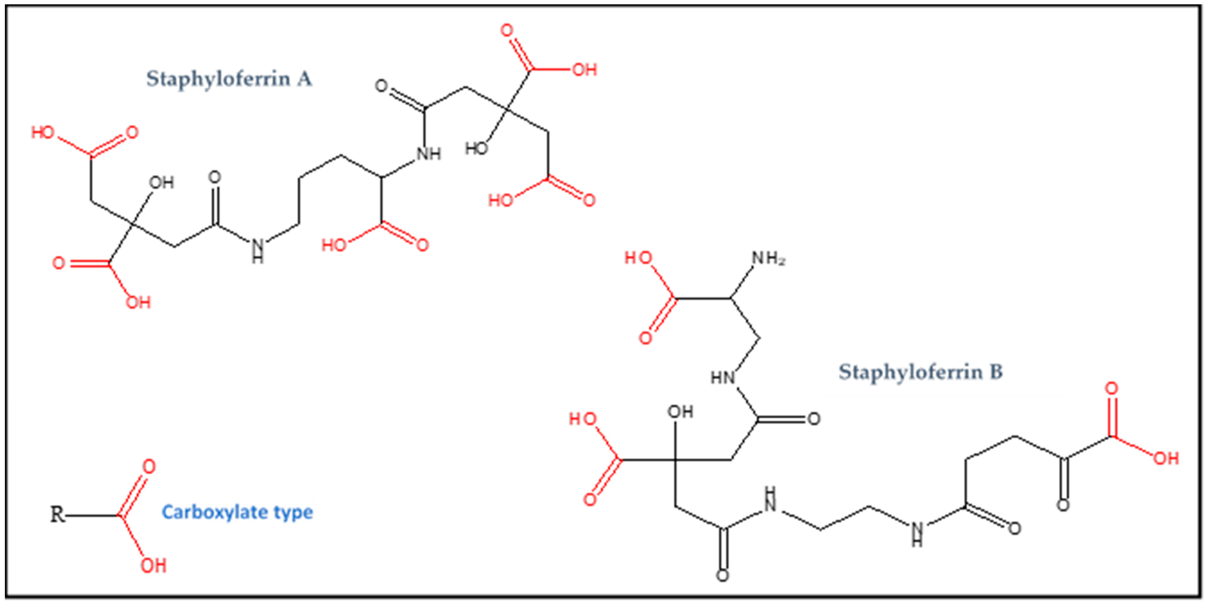
Iron, which has multiple oxidative states, is needed in many vital life processes such as electron transfer and DNA replication [1]. Bacteria require iron as an enzyme cofactor in the catalysis of redox reactions included in their basic cellular processes [2]. In the human body the level of free iron ions is extremely low since most iron is confined to storage, metabolic molecules and transport. Ferrous Fe2+ ions are exceedingly toxic due to their association with the Fenton reaction that produces harmful hydroxyl radicals [3]. On the other hand, ferric Fe3+ ions are insoluble at physiological pH and thus not readily bioavailable [4]. In order to endure this iron shortage iron, bacteria developed several mechanisms for obtaining iron from the host since it is important for their colonization during infection. These mechanisms include acquiring heme-bound iron, absorption by membrane-bound intake systems and siderophore secretion [5]. Siderophores are iron high-affinity metallophores and are essential pathogenicity factors in bacteria including S. aureus. The latter produces and secretes two staphyloferrins (siderophores) into the extracellular environment to scavenge iron. In addition, S. aureus has specific uptake systems for these staphyloferrins and for siderophores produced by other microorganisms as well [6].
There are four distinguished types of siderophores, catecholate, phenolate, hydroxamate and carboxylate, classified according to their iron chelation moieties [7][8]. The synthesis of siderophores is achieved either by non-ribosomal peptide synthesis (NRPS) or by NRPS-independent siderophore (NIS) synthesis (polyketide synthase (PKS) domains) that function together with NRPS units [9]. Also, a small quantity of siderophores is produced independent of these two pathways [10]. The NRPS pathway is the most common while that of NIS is less characterized.
NRPS siderophores have peptidic scaffolds, often incorporating nonproteinogenic amino acids and their derivatives, which are assembled stepwise with covalently bound intermediates [11]. In the NIS pathway, the covalent attachment of intermediates to the enzymes was not noted [9]. In the first synthesis route, siderophores are manufactured by the assembly of individual enzymes where dicarboxylic acids are condensed with diamines, amino alcohols, and alcohols in alternating subunits. Further subunit modifications (decarboxylation, oxidation or isomerization) are performed by distinct enzymes encoded by clusters of genes located near those related to synthetases encoding. Composite pathways using both assembly types were also reported [12]. S. aureus uses the NIS pathway in the synthesis of its staphyloferrins. Siderophores secretion is an active process (energy driven) and is flowed out through transport pumps [7].
The intake of iron chelated by siderophores varies between Gram-negative and Gram-positive bacteria due to the presence of an outer membrane in Gram-negative bacteria through which they should be transported [13][14]. In Gram-negative bacteria, the loaded siderophores are recognized specifically by receptors (β-barrel) found in the outer membrane. The change in the receptors conformation once the ligand is bound allows the translocation of loaded siderophores into the periplasm. [15]. Then, the transport into the cytoplasm is mediated by an ABC transporter located in the inner membrane [16]. The iron is reduced in the periplasm in some cases, and only Fe2+ ion is brought into the cytosol [17]. Concerning Gram-positive bacteria, the import of siderophores is directly achieved by an ABC transporter extending across the cell membrane because there is no outer-membrane receptors [18]. After iron release, siderophores may be either recycled [19] or hydrolyzed [20].

2. Additional Metal Acquiring Systems

In addition to the iron uptake system, S. aureus have other various systems of transportation for transitional metal ions, such as Cnt, Adc, NixA and Nik [21][22][23]. The latter is an ABC transporter and is essential for bacterial acquisition of nickel. This system (Nik) is effective in delivering nickel by the means of small chelating molecules (e.g., L-histidine), determining the activity of urease, and having a crucial role in the mouse urinary tract colonization [24][25]. Another nickel-acquiring system in S. aureus is NixA, which is a secondary transporter of NiCoT (nickel-cobalt transporter) membrane protein family. Along with Nik, NixA is also critical for the activity of urease and colonization in kidney [26]. Adc system is responsible for zinc uptake in Gram-positive bacteria including S. aureus and it is composed of AdcA that is a metal acquiring unit and AdcBC which is an ABC transporter [27]. Cnt, on the other hand, can transport several metals such as nickel, zinc, copper, cobalt, zinc, and manganese [27] but at zinc scarce conditions, it serves as a zinc uptake system [27].

2.1. Metallophore Staphylopine

In the first place, S. aureus utilizes Adc for importing zinc. Cnt system will be aroused when Adc alone becomes unable to meet zinc cellular requirement. A distinctive characteristic of Cnt is utilizing staphylopine, which is a nicotianamine-like metallophore [27]. This last contains imidazole ring and three carboxylic groups. It is an opine metallophore and can chelate several metal ions (nickel, zinc, cobalt, iron and copper), so it is considered a broad-spectrum metallophore [28]. The import of these wide range of metal ions via staphylopine depends on the metal nature and concentration along with the S. aureus growth status [25]. The structure of staphylopine is shown in Figure 12.
Figure 12. The structure of metallophore staphylopine.

2.2. Staphylopine Synthesis

Nine genes in the operon cnt (cntKLMABCDFE) encodes the multiple functions needed for the synthesis and the exportof staphylopine in addition to the import of the complexe (Staphylopine-metal ion). The three genes, cntKLM, encode the needed enzymes for staphylopine biosynthesis CntK, CntL and CntM respectively. The five genes cntABCDF encode the transporter ABC implicated in metal loaded staphylopine import [29][30]. Concerning cntE, it is involved in staphylopine export by encoding transport protein located in the bacterium membrane. All Cnt genes are most expressed in metal scant medium [28]. The importer protein CntA plays a main role in initiation of metal loaded staphylopine recognition and importation [21][24][25]. The mechanism of recognition and transportation of metal loaded staphylopine at the molecular level was verified by the interdomain change that occurs in CntA conformation upon binding to metal loaded staphylopine loaded [31]. The two CntB and CntC proteins located in membrane form a channel that may have a role in staphylopine loaded transportation [21][25][28]. As for ATP-binding CntD and CntF membrane proteins, they supply the energy needed for transportation [21][24]. The uptake of iron, nickel, zinc and cobalt decreases in S. aureus cntL and cntA-F mutant strains [25]. Zinc represses the transcription from the promoter cntA [21]. S. aureus also has Zur which represses the operon that encodes the two proteins related to ABC transporter [32]. If iron is available, Fur represses cnt genes [30]. These data designate that the expression of cnt gene is limited in zinc and iron rich environment and that Cnt system is controlled by both Fur and Zur [33] so, the synthesis of staphylopine is under negative control by Fur/Zur binding. On the other hand, staphylopine export and staphylopine metal recovery is less repressed by cooperative Fur/Zur repression [33].
Three steps are involved in the biosynthesis of staphylopine [28]. Firstly, D-histidine is produced via CntK which is a histidine racemase. Then, the enzyme CntL, that resembles nicotianamine-synthase, uses D-histidine as a substrate and catalyzes the production of xNA (the name comes from its nicotianamine correlation). The latter in an intermediate that is produced through the addition of aminobutyrate (an S-adenosyl methionine moiety). The last step involves the enzyme CntM that condensates pyruvate with xNA producing staphylopine [25]. CntM has the biochemical characteristics of opine synthase enzyme members [25]. A study done in vitro has found that metals employ several effects on the CntM catalyzed reaction [34]. They noticed that at low concentration of copper and zinc, the reaction was moderately activated but totally inhibited at high concentration. Manganese, on the other hand, was an activator only while nickel and cobalt were inhibitors only so it was proposed that the metal affinity toward xNA and an enzyme inhibitory binding site controlled the activation or inhibition according to the concentration of metals. This regulation of the enzyme involved in staphylopine synthesis is dependent on metal may happen in vivo as well and can could help in the adjustment of the production of metallophore [34].
Table 1 summarizes the properties of each S. aureus metallophore mentioned in in this research and Table 2 summarizes their regulation and transportation.
Table 1. Different types of metallophores produced by S. aureus.
S. aureus metallophores regulation and transportation.
Table 2.

2.3. Staphylopine as Zincophore

Nutritional immunity drastically limits the bioavailability of zinc during bacterial infection [35]. In spite of this essential nutrient restriction, S. aureus remains capable of causing severe disease because it is able to compete for zinc with the host [36]. As previously mentioned, S. aureus has two distinct ABC permease types involved in zinc acquisition, AdcABC and CntABCDF. AdcABC is homologous to ABC permeases associated with direct zinc recruitment, while CntABCDF belongs to the NikA/Opp family of ABC permeases. CntABCDF functions in conjunction with staphylopine to specifically promote zinc acquisition. This indicates that staphylopine functions as a staphylococcal zincophore although it can bind various metals in vitro. In a study performed by Grim et al. [27], they found that in zinc depleted medium, strains lacking the Cnt-staphylopine system and Adc permease had major growth defects and failed specifically in zinc accumulation. These results demonstrated that both systems serve as the major zinc importers of this bacterium. Concerning other metal ions such as Co, Ni, and Cu, they found that they are not physiological substrates of the Cnt-staphylopine system, and it is modestly responsive to Mn and Fe [30] which only exert transcriptional influence in the absence of zinc. These findings suggest that the abundance of zinc is the main regulatory factor that controls the system expression [27].

2.4. Important Features of Cnt-Staphylopine System

The Cnt system, as previously mentioned, is essential for the optimal metals import metal-limiting conditions and contributes to S. aureus virulence. The failure to efflux staphylopine results in its intracellular accumulation thus impairing the fitness of S. aureus [37]. A recent study has shown that CntE loss resulted in a stronger virulence defect than other components of the Cnt-staphylopine system, even in zinc restricted tissues. The toxicity associated with intracellular staphylopineaccumulation contributed to the virulence defect of strains lacking CntE, even when S. aureus is zinc starved during infection. Moreover, they noticed that the intracellular accumulation of staphylopine did not increase metal importer expression or altered cellular metal concentrations, suggesting that contrary to prevailing models, the toxicity associated with staphylopine is not strictly due to intracellular chelation of metals [38]. CntK catalyzes the first step of staphylopine synthesis by converting L -histidine to D -histidine in order to provide an essential building block of staphylopine. It was found, by structural modeling, that CntK is specific for histidine, whereas other proteinogenic amino acids, with the exception of arginine, do not show any binding with it. These findings helped in developing powerful antibiotics targeting the staphylopine-mediated metal acquisition process in bacteria via designing irreversible inhibitors [39]. Another study confirmed that during the synthesis of staphylopine, CntL stereoselectively carries out the catalysis of D-histidine and not L-histidine. These findings provided critical structural and mechanistic insights into CntL for a better understanding of of nicotianamine-like metallophores biosynthesis and the discovery of inhibitors of this process [40]. Concerning CntA, responsible for the recognition and transport of diverse solutes, a study was performed to investigate the structural conformation upon staphylopine binding. CntA has a fork-like structure formed by three domains (Ia and Ib and II). It uses a bi-domain architectural form of domain II assisted by inter-domain hinge cluster residues. Important clustered communities regulat the conformational changes in CntA. In addition to open (without staphylopine) and close states (with staphylopine) [31], the fluctuating regions sampled two additional intermediate states that were considered closed or open previously. CntA prefers fluctuating the non-conserved regions rather than conserved where domain II turned out to be rigid and maintains a stable fold. Such findings are important to the researcher in field of drug-designing [41].
As for the regulation of cnt operon, a novel regulator (Rsp) was identified that activates the system, in addition to the metal-dependent Fur and Zur repressors. This regulator is an AraC-type regulator. Rsp activation in S. aureus may act to maintain basal cellular levels of staphylopine to scavenge free metals when needed [42]. It is worth mentioning that the AraC family regulators are an abundant group of transcriptional regulators in bacteria, acting mostly as gene expression activators, that controls diverse cellular functions such as virulence and stress response [43]. A study has reported the establishment of a fast and efficient method for directly converting adenine to guanine in bacterial genomes. A systematic screening that targets the possibly editable adenine sites of S. aureus cntBC locates key residues for metal importation, demonstrating that the application of the system might greatly facilitate the bacterial genomic engineering [44].

References

  1. Gkouvatsos, K.; Papanikolaou, G.; Pantopoulos, K. Regulation of iron transport and the role of transferrin. Biochim. Biophys. Acta 2012, 1820, 188–202.
  2. Liu, L.; Wang, W.; Wu, S.; Gao, H. Recent Advances in the Siderophore Biology of Shewanella. Front. Microbiol. 2022, 13, 823758.
  3. Winterbourn, C.C. Toxicity of iron and hydrogen peroxide: The Fenton reaction. Toxicol. Lett. 1995, 82, 969–974.
  4. Emerson, D.; Roden, E.; Twining, B.S. The microbial ferrous wheel: Iron cycling in terrestrial, freshwater, and marine environments. Front. Microbiol. 2012, 3, 383.
  5. Kramer, J.; Özkaya, O.; Kümmerli, R. Bacterial siderophores in community and host interactions. Nat. Rev. Microbiol. 2020, 18, 152–163.
  6. Sebulsky, M.T.; Hohnstein, D.; Hunter, M.D.; Heinrichs, D.E. Identification and characterization of a membrane permease ivolved in iron-hydroxamate transport in Staphylococcus aureus. J. Bacteriol. 2000, 182, 4394–4400.
  7. Miethke, M.; Marahiel, M.A. Siderophore-based iron acquisition and pathogen control. Microbiol. Mol. Biol. Rev. 2007, 71, 413–451.
  8. Cassat, J.E.; Skaar, E.P. Iron in infection and immunity. Cell Host Microbe 2013, 13, 509–519.
  9. Challis, G.L. A widely distributed bacterial pathway for siderophore biosynthesis independent of nonribosomal peptide synthetases. Chem. Bio. Chem. 2005, 6, 601–661.
  10. Carroll, C.S.; Moore, M.M. Ironing out siderophore biosynthesis: A review of non-ribosomal peptide synthetase (NRPS)-independent siderophore synthetases. Crit. Rev. Biochem. Mol. Biol. 2018, 53, 356–381.
  11. Crosa, J.H.; Walsh, C.T. Genetics and assembly line enzymology of siderophore biosynthesis in bacteria. Microbiol. Mol. Biol. Rev. 2002, 66, 223–249.
  12. Lee, J.Y.; Janes, B.K.; Passalacqua, K.D.; Pfleger, B.F.; Bergman, N.H.; Liu, H.; Håkansson, K.; Somu, R.V.; Aldrich, C.C.; Cendrowski, S.; et al. Biosynthetic analysis of the petrobactin siderophore pathway from Bacillus anthracis. J. Bacteriol. 2007, 189, 1698–1710.
  13. Andrews, S.C.; Robinson, A.K.; Rodriguez-Quinones, F. Bacterial iron homeostasis. FEMS Microbiol Rev. 2003, 27, 215–237.
  14. Sheldon, J.R.; Heinrichs, D.E. Recent developments in understanding the iron acquisition strategies of gram positive pathogens. FEMS Microbiol. Rev. 2015, 39, 592–630.
  15. Faraldo-Gómez, J.D.; Sansom, M.S.P. Acquisition of siderophores in gram-negative bacteria. Nat. Rev. Mol. Cell Biol. 2003, 4, 105–116.
  16. Schalk, I.J.; Guillon, L. Fate of ferrisiderophores after import across bacterial outer membranes: Different iron release strategies are observed in the cytoplasm or periplasm depending on the siderophore pathways. Amino Acids 2013, 44, 1267–1277.
  17. Ganne, G.; Brillet, K.; Basta, B.; Roche, B.; Hoegy, F.; Gasser, V.; Schalk, I.J. Iron release from the siderophore pyoverdine in Pseudomonas aeruginosa involves three new actors: FpvC, FpvG, and FpvH. ACS Chem. Biol. 2017, 12, 1056–1065.
  18. Krewulak, K.D.; Vogel, H.J. Structural biology of bacterial iron uptake. Biochim. Biophys. Acta 2008, 1778, 1781–1804.
  19. Imperi, F.; Tiburzi, F.; Visca, P. Molecular basis of pyoverdine siderophore recycling in Pseudomonas aeruginosa. Proc. Natl. Acad. Sci. USA 2009, 106, 20440–20445.
  20. Lin, H.; Fischbach, M.A.; Liu, D.R.; Walsh, C.T. In vitro characterization of salmochelin and enterobactin trilactone hydrolases IroD, IroE, and Fes. J. Am. Chem. Soc. 2005, 127, 11075–11084.
  21. Remy, L.; Carrière, M.; Derré-Bobillot, A.; Martini, C.; Sanguinetti, M.; Borezée-Durant, E. The Staphylococcus aureus Opp1 ABC transporter imports nickel and cobalt in zinc-depleted conditions and contributes to virulence. Mol. Microbiol. 2013, 87, 730–743.
  22. Lebrette, H.; Brochier-Armanet, C.; Zambelli, B.; de Reuse, H.; Borezée-Durant, E.; Ciurli, S.; Cavazza, C. Promiscuous nickel import in human pathogens: Structure, thermodynamics, and evolution of extracytoplasmic nickel-binding proteins. Structure 2014, 22, 1421–1432.
  23. Eitinger, T.; Suhr, J.; Moore, L.; Smith, J.A. Secondary transporters for nickel and cobalt ions: Theme and variations. Biometals 2005, 18, 399–405.
  24. Lebrette, H.; Borezée-Durant, E.; Martin, L.; Richaud, P.; Erba, E.B.; Cavazza, C. Novel insights into nickel import in Staphylococcus aureus: The positive role of free histidine and structural characterization of a new thiazolidine- type nickel chelator. Metallomics 2015, 7, 613–621.
  25. Ghssein, G.; Brutesco, C.; Ouerdane, L.; Fojcik, C.; Izaute, A.; Wang, S.; Hajjar, C.; Lobinski, R.; Lemaire, D.; Richaud, P.; et al. Biosynthesis of a broad-spectrum nicotianamine-like metallophore in Staphylococcus aureus. Science 2016, 352, 1105–1109.
  26. Hiron, A.; Posteraro, B.; Carrière, M.; Remy, L.; Delporte, C.; La Sorda, M.; Sanguinetti, M.; Juillard, V.; Borezée-Durant, E. A nickel ABC-transporter of Staphylococcus aureus is involved in urinary tract infection. Mol. Microbiol. 2010, 77, 1246–1260.
  27. Grim, K.P.; San Francisco, B.; Radin, J.N.; Brazel, E.B.; Kelliher, J.L.; Párraga Solórzano, P.K.; Kim, P.C.; McDevitt, C.A.; Kehl-Fie, T.E. The Metallophore Staphylopine Enables Staphylococcus aureus To Compete with the Host for Zinc and Overcome Nutritional Immunity. mBio 2017, 8, e01281-17.
  28. Ghssein, G.; Matar, S.F. Chelating Mechanisms of Transition Metals by Bacterial Metallophores “Pseudopaline and Staphylopine”: A Quantum Chemical Assessment. Computation 2018, 6, 56.
  29. Hiron, A.; Borezee-Durant, E.; Piard, J.C.; Juillard, V. Only one of four oligopeptide transport systems mediates nitrogen nutrition in Staphylococcus aureus. J. Bacteriol. 2007, 189, 5119–5129.
  30. Ding, Y.; Fu, Y.; Lee, J.C.; Hooper, D.C. Staphylococcus aureus NorD, a putative efflux pump coregulated with the Opp1 oligopeptide permease, contributes selectively to fitness in vivo. J. Bacteriol. 2012, 194, 6586–6593.
  31. Song, L.; Zhang, Y.; Chen, W.; Gu, T.; Zhang, S.Y.; Ji, Q. Mechanistic insights into staphylopine-mediated metal acquisition. Proc. Natl. Acad. Sci. USA 2018, 115, 3942–3947.
  32. Lindsay, J.A.; Foster, S.J. zur: A Zn(21)-responsive regulatory element of Staphylococcus aureus. Microbiology 2001, 147, 1259–1266.
  33. Fojcik, C.; Arnoux, P.; Ouerdane, L.; Aigle, M.; Alfonsi, L.; Borezée-Durant, E. Independent and Cooperative Regulation of Staphylopine Biosynthesis and Trafficking by Fur and Zur. Mol. Microbiol. 2018, 108, 159–177.
  34. Hajjar, C.; Fanelli, R.; Laffont, C.; Brutesco, C.; Cullia, G.; Tribout, M.; Nurizzo, D.; Borezée-Durant, E.; Voulhoux, R.; Pignol, D.; et al. Control by Metals of Staphylopine Dehydrogenase Activity during Metallophore Biosynthesis. Am. Chem. Soc. 2019, 141, 5555–5562.
  35. Corbin, B.D.; Seeley, E.H.; Raab, A.; Feldmann, J.; Miller, M.R.; Torres, V.J.; Anderson, K.L.; Dattilo, B.M.; Dunman, P.M.; Gerads, R.; et al. Metal chelation and inhibition of bacterial growth tissue abscesses. Science 2008, 319, 962–965.
  36. Kehl-Fie, T.E.; Zhang, Y.; Moore, J.L.; Farrand, A.J.; Hood, M.I.; Rathi, S.; Chazin, W.J.; Caprioli, R.M.; Skaar, E.P. MntABC and MntH contribute to systemic Staphylococcus aureus infection by competing with calprotectin for nutrient manganese. Infect. Immun. 2013, 81, 3395–3405.
  37. Chen, C.; Hooper, D.C. Intracellular accumulation of staphylopine impairs the fitness of Staphylococcus aureus cntE mutant. FEBS Lett. 2019, 593, 1213–1222.
  38. Grim, K.P.; Radin, J.N.; Solórzano, P.K.P.; Morey, J.R.; Frye, K.A.; Ganio, K.; Neville, S.L.; McDevitt, C.A.; Kehl-Fie, T.E. Intracellular Accumulation of Staphylopine Can Sensitize Staphylococcus aureus to Host-Imposed Zinc Starvation by Chelation-Independent Toxicity. J. Bacteriol. 2020, 202, 00014–00020.
  39. Luo, S.; Ju, Y.; Zhou, J.; Gu, Q.; Xu, J.; Zhou, H. Crystal structure of CntK, the cofactor-independent histidine racemase in staphylopine-mediated metal acquisition of Staphylococcus aureus. Int. J. Biol. Macromol. 2019, 135, 725–733.
  40. Luo, Z.; Luo, S.; Ju, Y.; Ding, P.; Xu, J.; Gu, Q.; Zhou, H. Structural insights into the ligand recognition and catalysis of the key aminobutanoyltransferase CntL in staphylopine biosynthesis. FASEB J. 2021, 5, 21575.
  41. Abideen, Z.U.; Ahmad, A.; Usman, M.; Majaz, S.; Ali, W.; Noreen, S.; Mahmood, T.; Nouroz, F. Dynamics and conformational propensities of staphylococcal CntA. J. Biomol. Struct. Dyn. 2021, 39, 4923–4935.
  42. Vinué, L.; Hooper, D.C. Rsp activates expression of the Cnt system in Staphylococcus aureus. BMC Microbiol. 2020, 20, 327.
  43. Kotecka, K.; Kawalek, A.; Kobylecki, K.; Bartosik, A.A. The AraC-Type Transcriptional Regulator GliR (PA3027) Activates Genes of Glycerolipid Metabolism in Pseudomonas aeruginosa. Int. J. Mol. Sci. 2021, 22, 5066.
  44. Zhang, Y.; Zhang, H.; Wang, Z.; Wu, Z.; Wang, Y.; Tang, N.; Xu, X.; Zhao, S.; Chen, W.; Ji, Q. Programmable adenine deamination in bacteria using a Cas9-adenine-deaminase fusion. Chem. Sci. 2020, 6, 1657–1664.
More
Video Production Service