Factors Contributing to Children Avoiding Severe COVID-19: Comparison
Please note this is a comparison between Version 2 by Conner Chen and Version 1 by Karen Angeliki Krogfelt.

Coronavirus Disease 2019 (COVID-19) manifests differently in children than adults, as children usually have a milder course of disease, mild symptoms if any, and lower fatality rates are recorded among children. Severe acute respiratory syndrome coronavirus 2 (SARS-CoV-2) transmission also seems to be different between children and adults. Many factors are proposed to explain the milder outcome in children, e.g., a more appropriate immune response (especially active innate response), trained immunity, a lack of immunosenescence, and the reduced prevalence of comorbidities. A better understanding of the differences in susceptibility and outcome in children compared with adults could lead to greater knowledge of risk factors for complicated COVID-19 cases and potential treatment targets.

  • SARS-CoV-2
  • COVID-19
  • coronavirus
  • children
  • adolescents
  • young people
  • ACE2
  • immune system
  • transmission
  • viral infection

1. Strong Innate Response

Children are frequently introduced to viral infections and vaccinations, which keeps their innate immune system active. An increased activation of neutrophils and reduced circulation of various innate immune cells (monocytes, dendritic cells, and natural killer cells) was observed in children with mild severe acute respiratory syndrome coronavirus 2 (SARS-CoV-2) infection in a study, suggesting that innate immune cells might be recruited to infected areas [15][1]. In SARS-CoV-2-infected adults only a reduction in circulating non-classical monocytes was observed [15][1]. This could indicate that children’s innate immune response is generally much stronger and efficient at acting against SARS-CoV-2. Specifically, the mucosal immune response appears more vigorous in children compared to adults upon SARS-CoV-2 infection [16][2]. It has also been reported that the production of IL-17A and IFN-γ during an early immune response can result in a more rapid clearance of viral infection [17][3].
Children’s adaptive immune systems seem to differ from adults’, which may partly explain why children’s immune systems are less likely to overreact to infection with SARS-CoV-2. Adults show a stronger T-cell response to the spike protein of SARS-CoV-2 and a higher neutralizing antibody count compared to children. However, the boosted adaptive response in adults does not result in better outcomes [17][3]. A less vigorous adaptive immune response in children and a strong innate response may prevent a cytokine storm, potentially eliminating an unwanted overreaction to the virus.

2. Activated Immunity Providing Cross-Protection

The cells in the innate immune system can be modified by exposure to antigens [18][4]. The milder disease in children can be linked to trained immunity offering protection against SARS-CoV-2 by cross-reactive neutralizing antibodies. These cross-reactive antibodies are thought to be more common in children than adults [4,19][5][6]. It is possible to activate cross-protection through Bacille Calmette Guerin (BCG) vaccination [19][6], and has also been demonstrated that measles-containing vaccines (MCVs) can induce adaptive immunomodulation [20][7]. Although both vaccines reduce overall mortality and protect against viral infections in general [21[8][9][10],22,23], more research is needed in the area, and currently, the effects of BCG vaccination on Coronavirus Disease 2019 (COVID-19) is under investigation [24][11]. Since a large proportion of children have received a number of different vaccines more recently than adults, it could indicate that vaccines play a part in the observed age-related difference in COVID-19 manifestation. However, one study did not find age-related differences in pre-existing antibodies to other coronaviruses, which suggests that cross-immunity does not play an important role in clinical responses to SARS-CoV-2 [17][3].

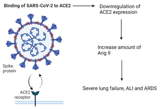
3. The Role of Angiotensin Converting Enzyme-2

The entry point for SARS-CoV-2 into cells is via the receptor, angiotensin converting enzyme-2 (ACE2). ACE2 is part of the renin–angiotensin–aldosterone system (RAAS), which is important for keeping the homeostasis of cardiovascular and respiratory systems [25][12]. Under normal circumstances, ACE2 is a negative regulator of the RAAS system, it has anti-inflammatory properties, and is involved in converting angiotensin II (Ang II) to angiotensin 1–7 [26][13]. The virus is able to enter the cells by attaching its Spike (S)-protein to ACE2, which is located on the apical membranes of respiratory epithelium, endothelial cells of blood vessels and heart epithelium, and on enterocytes in the small intestines [18,27,28,29][4][14][15][16]. After the entry of SARS-CoV-2 into the body, the expression of ACE2 is downregulated, which causes an increase in the amount of Ang II [18,26][4][13]. Elevated Ang II levels can cause severe lung failure, such as that seen with acute lung injury (ALI) and Acute Respiratory Distress syndrome (ARDS) [25,30][12][17] (Figure 1).
Figure 1. The effect of SARS-CoV-2 binding to the ACE2 receptor.
A study with mice showed that the loss of expression of ACE2 resulted in enhanced vascular permeability, increased lung edema, neutrophil accumulation, and worsened lung function. In addition, mice treated with catalytically active recombinant ACE2 protein showed improvements in the symptoms of ALI and ARDS [30,31][17][18]. These findings suggest that ACE2 has a protective role against ARDS and ALI via the negative regulation of Ang II. A study reported the lower expression of ACE2 in nasal and bronchial tissue of children when compared to adults [32][19]. Studies have supported the findings that the expression of ACE2 in nasal and lung epithelium increases with age and have suggested that this could be an explanation for the difference in COVID-19 severity observed between children and adults [33,34][20][21]. However, in the elderly, ACE2 is suggested to decrease again, primarily based on findings in animal models [26[13][22],35], and this age group is most susceptible to severe SARS-CoV-2 infection. Therefore, even though the virus gains access to cells via ACE2, having less of the receptor is not an efficient sole defense against disease, supporting the important role of an active innate immune system in children.

4. Immunosenescence

Immunosenescence is a term used for the deterioration of the immune system seen with age. It involves changes to both innate and adaptive immune functions, increases the susceptibility to infectious diseases, and decreases the response to vaccinations [36,37][23][24]. Since older people seem to be more affected by SARS-CoV-2 infection, it is likely that immunosenescence may contribute to the poor clearance of the virus. Furthermore, the lack of immunosenescence in children could explain why they are less likely to present with a severe infection.
The risk of developing ARDS during a COVID-19 infection is age dependent and can be the cause of death for people infected with SARS-CoV-2 [38][25]. Adults produce less inflammatory cytokines and antibodies during infection than children due to waning of the immune system [39][26]. Ageing of the immune system can result in the dysregulation of the production of inflammatory cytokines, which causes damage to the body. Elevated levels of neutrophils and cytokines indicate an exaggerated innate immune response. In adult patients, severe COVID-19 is associated with significantly lower counts of lymphocytes, especially T cells [40,41][27][28]. Children have an inherently higher percentage of lymphocytes. Some have suggested that this has a defensive role against SARS-CoV-2. Two studies showed that only 3.5–5.2% of pediatric patients had lymphocytopenia, and the majority had normal leukocytes [42,43][29][30].

5. Comorbidities

A systematic review and meta-analysis from November 2020 considering over 9000 pediatric patients with COVID-19 reported that children with pre-existing conditions had higher risks of severe infection when compared to children with no underlying disease [44][31]. Pre-existing cardiac conditions were reported in a large proportion of children becoming critically ill, but also, childhood obesity has been suggested to correlate positively with the severity of COVID-19 [44,45][31][32]. However, while comorbidities are associated with severe disease in children, these conditions are not commonly observed. Both adults and children with pre-existing comorbidities have worse outcomes when infected with SARS-CoV-2 than those without any comorbidities, but adults are more likely to suffer from comorbidities [46][33].

6. Melatonin Levels

Melatonin is an indoleamine hormone produced at night, mainly by the pineal gland, and secreted into the blood stream. It plays a big role in the biological processes of the circadian system, regulating night–day as well as sleep–wake cycles, and has anti-inflammatory and anti-oxidative properties [47,48][34][35]. SARS-CoV-2 is considered to originate from bats; however, it has minimal to no impact on the health of the host. Bats are nocturnal animals and have high levels of melatonin compared to humans, which may contribute to their increased resistance toward illness from the virus [49,50][36][37]. Melatonin levels in humans are negatively correlated with age [51[38][39],52], potentially contributing to the worse symptoms experienced in the elderly and explaining why children are better protected when infected with SARS-CoV-2. Melatonin has been shown to increase the proliferation of T and B cells, natural killer cells, granulocytes, and monocytes in bone marrow and tissues [53][40] and can also protect against ARDS [54][41]. A recently published review highlights several steps where melatonin can interfere with damages caused by COVID-19. Infection by SARS-CoV-2 can result in the high production of neutrophil myeloperoxidase (MOP) and reactive oxygen species (ROS), both involved in combating pathogens, but can cause tissue damage if the response is too strong. Melatonin can inhibit MOP activity, as well as scavenger ROS, thereby potentially reducing COVID-19 severity [55][42]. Melatonin as a possible supplement against COVID-19 is particularly interesting due to the stability, accessibility, safety, and cost of the drug.

7. Difference in Microbiota

Another less-examined hypothesis as to why children may fare better against COVID-19 involves the microbiota. The microbiota is important for human well-being; it is involved in regulating the immune system, inflammation, and gut homeostasis and plays an important part in protection against pathogens. The gut microbiome seems to be altered in patients with COVID-19, with an under-representation of Faecalibacterium prausnitzii, Eubacterium rectale, and bifidobacteria, which all have immunomodulatory prospects. This change in microbial composition, associated with elevated levels of inflammatory cytokines and blood markers of tissue damage, suggests the involvement of the gut microbiome in COVID-19 disease severity [56][43]. The gut microbiota changes with age, and children generally have higher numbers of Bifidobacterium than adults [57][44], potentially providing a better defense against infection. Children might also have a higher nasopharyngeal colonization of viruses and bacteria, which by competition might limit the growth of SARS-CoV-2 and lead to the reduced colonization of the pathogen. However, it is important to remember that the microbiota is affected by many factors and that the reported changes might be a cause of the disease and not vice versa.

References

  1. Neeland, M.R.; Bannister, S.; Clifford, V.; Dohle, K.; Mulholland, K.; Sutton, P.; Curtis, N.; Steer, A.C.; Burgner, D.P.; Crawford, N.W.; et al. Innate cell profiles during the acute and convalescent phase of SARS-CoV-2 infection in children. Nat. Commun. 2021, 12, 1084.
  2. Loske, J.; Röhmel, J.; Lukassen, S.; Stricker, S.; Magalhães, V.G.; Liebig, J.; Chua, R.L.; Thürmann, L.; Messingschlager, M.; Seegebarth, A.; et al. Pre-activated antiviral innate immunity in the upper airways controls early SARS-CoV-2 infection in children. Nat. Biotechnol. 2021, 39, 1–6.
  3. Pierce, C.A.; Preston-Hurlburt, P.; Dai, Y.; Aschner, C.B.; Cheshenko, N.; Galen, B.; Garforth, S.J.; Herrera, N.G.; Jangra, R.K.; Morano, N.C.; et al. Immune responses to SARS-CoV-2 infection in hospitalized pediatric and adult patients. Sci. Transl. Med. 2020, 12, eabd5487.
  4. Dhochak, N.; Singhal, T.; Kabra, S.K.; Lodha, R. Pathophysiology of COVID-19: Why Children Fare Better than Adults? Indian J. Pediatr. 2020, 87, 537–546.
  5. Snape, M.D.; Viner, R.M. COVID-19 in children and young people. Science 2020, 370, 286–288.
  6. Moorlag, S.; Arts, R.; Van Crevel, R.; Netea, M. Non-specific effects of BCG vaccine on viral infections. Clin. Microbiol. Infect. 2019, 25, 1473–1478.
  7. Messina, N.; Zimmermann, P.; Curtis, N. The impact of vaccines on heterologous adaptive immunity. Clin. Microbiol. Infect. 2019, 25, 1484–1493.
  8. Higgins, J.P.T.; Soares-Weiser, K.; López, J.; Kakourou, A.; Chaplin, K.; Christensen, H.; Martin, N.K.; Sterne, J.; Reingold, A.L. Association of BCG, DTP, and measles containing vaccines with childhood mortality: Systematic review. BMJ 2016, 355, i5170.
  9. Kleinnijenhuis, J.; Quintin, J.; Preijers, F.; Joosten, L.A.B.; Ifrim, D.C.; Saeed, S.; Jacobs, C.; van Loenhout, J.; de Jong, D.; Stunnenberg, H.G.; et al. Bacille Calmette-Guérin induces NOD2-dependent nonspecific protection from reinfection via epigenetic reprogramming of monocytes. Proc. Natl. Acad. Sci. USA 2012, 109, 17537–17542.
  10. Mysore, V.; Cullere, X.; Settles, M.L.; Ji, X.; Kattan, M.W.; Desjardins, M.; Durbin-Johnson, B.; Gilboa, T.; Baden, L.R.; Walt, D.R.; et al. Protective heterologous T cell immunity in COVID-19 induced by the trivalent Measles-Mumps-Rubella and Tetanus-Diptheria-Pertussis vaccine antigens. Med 2021, 2, 1050–1071.e7.
  11. Moorlag, S.J.C.F.M.; van Deuren, R.C.; van Werkhoven, C.H.; Jaeger, M.; Debisarun, P.; Taks, E.; Mourits, V.P.; Koeken, V.A.; de Bree, L.C.J.; ten Doesschate, T.; et al. Safety and COVID-19 Symptoms in Individuals Recently Vaccinated with BCG: A Retrospective Cohort Study. Cell Rep. Med. 2020, 1, 100073.
  12. Ingraham, N.E.; Barakat, A.G.; Reilkoff, R.; Bezdicek, T.; Schacker, T.; Chipman, J.G.; Tignanelli, C.; Puskarich, M. Understanding the renin—angiotensin—aldosterone—SARS-CoV axis: A comprehensive review. Eur. Respir. J. 2020, 56, 2000912.
  13. Zimmermann, P.; Curtis, N. Why is COVID-19 less severe in children? A review of the proposed mechanisms underlying the age-related difference in severity of SARS-CoV-2 infections. Arch. Dis. Child. 2021, 106, 429–439.
  14. Song, R.; Preston, G.; Yosypiv, I.V. Ontogeny of angiotensin-converting enzyme 2. Pediatr. Res. 2012, 71, 13–19.
  15. Yan, R.; Zhang, Y.; Li, Y.; Xia, L.; Guo, Y.; Zhou, Q. Structural basis for the recognition of SARS-CoV-2 by full-length human ACE2. Science 2020, 367, 1444–1448.
  16. Liu, M.-Y.; Zheng, B.; Zhang, Y.; Li, J.-P. Role and mechanism of angiotensin-converting enzyme 2 in acute lung injury in coronavirus disease 2019. Chronic Dis. Transl. Med. 2020, 6, 98–105.
  17. Imai, Y.; Kuba, K.; Rao, S.; Huan, Y.; Guo, F.; Guan, B.; Yang, P.; Sarao, R.; Wada, T.; Leong-Poi, H.; et al. Angiotensin-converting enzyme 2 protects from severe acute lung failure. Nature 2005, 436, 112–116.
  18. Sodhi, C.P.; Nguyen, J.; Yamaguchi, Y.; Werts, A.D.; Lu, P.; Ladd, M.R.; Fulton, W.B.; Kovler, M.; Wang, S.; Prindle, T.; et al. A Dynamic Variation of Pulmonary ACE2 Is Required to Modulate Neutrophilic Inflammation in Response to Pseudomonas aeruginosa Lung Infection in Mice. J. Immunol. 2019, 203, 3000–3012.
  19. Sharif-Askari, N.S.; Sharif-Askari, F.S.; Alabed, M.; Temsah, M.-H.; Al Heialy, S.; Hamid, Q.; Halwani, R. Airways Expression of SARS-CoV-2 Receptor, ACE2, and TMPRSS2 Is Lower in Children Than Adults and Increases with Smoking and COPD. Mol. Ther. Methods Clin. Dev. 2020, 18, 1–6.
  20. Muus, C.; Luecken, M.; Eraslan, G.; Waghray, A.; Heimberg, G.; Sikkema, L.; Kobayashi, Y.; Vaishnav, E.D.; Subramanian, A.; Smilie, C.; et al. Integrated analyses of single-cell atlases reveal age, gender, and smoking status associations with cell type-specific expression of mediators of SARS-CoV-2 viral entry and highlights inflammatory programs in putative target cells. BioRxiv 2020.
  21. Wang, A.; Chiou, J.; Poirion, O.B.; Buchanan, J.; Valdez, M.J.; Verheyden, J.M.; Hou, X.; Kudtarkar, P.; Narendra, S.; Newsome, J.M.; et al. Single-cell multiomic profiling of human lungs reveals cell-type-specific and age-dynamic control of SARS-CoV2 host genes. eLife 2020, 9, 1–28.
  22. Xie, X.; Chen, J.; Wang, X.; Zhang, F.; Liu, Y. Age- and gender-related difference of ACE2 expression in rat lung. Life Sci. 2006, 78, 2166–2171.
  23. Caruso, C.; Buffa, S.; Candore, G.; Colonna-Romano, G.; Dunn-Walters, D.; Kipling, D.; Pawelec, G. Mechanisms of immunosenescence. Immun. Ageing 2009, 6, 10.
  24. McElhaney, J.E.; Effros, R.B. Immunosenescence: What does it mean to health outcomes in older adults? Curr. Opin. Immunol. 2009, 21, 418–424.
  25. Gibson, P.G.; Qin, L.; Puah, S.H. COVID -19 acute respiratory distress syndrome (ARDS): Clinical features and differences from typical pre- COVID -19 ARDS. Med. J. Aust. 2020, 213, 54–56.e1.
  26. Ng, K.W.; Faulkner, N.; Cornish, G.H.; Rosa, A.; Harvey, R.; Hussain, S.; Ulferts, R.; Earl, C.; Wrobel, A.G.; Benton, D.J.; et al. Preexisting and de novo humoral immunity to SARS-CoV-2 in humans. Science 2020, 370, 1339–1343.
  27. Qin, C.; Zhou, L.; Hu, Z.; Zhang, S.; Yang, S.; Tao, Y.; Xie, C.; Ma, K.; Shang, K.; Wang, W.; et al. Dysregulation of Immune Response in Patients With Coronavirus 2019 (COVID-19) in Wuhan, China. Clin. Infect. Dis. 2020, 71, 762–768.
  28. Song, J.-W.; Zhang, C.; Fan, X.; Meng, F.-P.; Xu, Z.; Xia, P.; Cao, W.-J.; Yang, T.; Dai, X.-P.; Wang, S.-Y.; et al. Immunological and inflammatory profiles in mild and severe cases of COVID-19. Nat. Commun. 2020, 11, 1–10.
  29. Lu, X.; Zhang, L.; Du, H.; Zhang, J.; Li, Y.Y.; Qu, J.; Zhang, W.; Wang, Y.; Bao, S.; Li, Y.; et al. SARS-CoV-2 Infection in Children. N. Engl. J. Med. 2020, 382, 1663–1665.
  30. Bai, K.; Liu, W.; Liu, C.; Fu, Y.; Hu, J.; Qin, Y.; Zhang, Q.; Chen, H.; Xu, F.; Li, C. Clinical Analysis of 25 COVID-19 Infections in Children. Pediatr. Infect. Dis. J. 2020, 39, e100–e103.
  31. Tsankov, B.K.; Allaire, J.M.; Irvine, M.A.; Lopez, A.A.; Sauvé, L.J.; Vallance, B.A.; Jacobson, K. Severe COVID-19 Infection and Pediatric Comorbidities: A Systematic Review and Meta-Analysis. Int. J. Infect. Dis. 2020, 103, 246–256.
  32. Williams, N.; Radia, T.; Harman, K.; Agrawal, P.; Cook, J.; Gupta, A. COVID-19 Severe acute respiratory syndrome coronavirus 2 (SARS-CoV-2) infection in children and adolescents: A systematic review of critically unwell children and the association with underlying comorbidities. Eur. J. Nucl. Med. Mol. Imaging 2020, 180, 689–697.
  33. Wu, Z.; McGoogan, J.M. Characteristics of and Important Lessons From the Coronavirus Disease 2019 (COVID-19) Outbreak in China: Summary of a Report of 72,314 Cases From the Chinese Center for Disease Control and Prevention. JAMA 2020, 323, 1239–1242.
  34. Posadzki, P.P.; Bajpai, R.; Kyaw, B.M.; Roberts, N.J.; Brzezinski, A.; Christopoulos, G.I.; Divakar, U.; Bajpai, S.; Soljak, M.; Dunleavy, G.; et al. Melatonin and health: An umbrella review of health outcomes and biological mechanisms of action. BMC Med. 2018, 16, 18.
  35. Hardeland, R. Melatonin and inflammation—Story of a double-edged blade. J. Pineal Res. 2018, 65, e12525.
  36. Wang, L.-F.; Shi, Z.; Zhang, S.; Field, H.; Daszak, P.; Eaton, B.T. Review of Bats and SARS. Emerg. Infect. Dis. 2006, 12, 1834–1840.
  37. Shneider, A.; Kudriavtsev, A.; Vakhrusheva, A. Can melatonin reduce the severity of COVID-19 pandemic? Int. Rev. Immunol. 2020, 39, 153–162.
  38. Iguchi, H.; Kato, K.-I.; Ibayashi, H. Age-Dependent Reduction in Serum Melatonin Concentrations in Healthy Human Subjects. J. Clin. Endocrinol. Metab. 1982, 55, 27–29.
  39. Waldhauser, F.; Weiszenbacher, G.; Tatzer, E.; Gisinger, B.; Schemper, M.; Frisch, H. Alterations in Nocturnal Serum Melatonin Levels In Humans With Growth and Aging. J. Clin. Endocrinol. Metab. 1988, 66, 648–652.
  40. Miller, S.C.; Pandi, P.S.R.; Esquifino, A.I.; Cardinali, D.P.; Maestroni, G.J.M. The role of melatonin in immuno-enhancement: Potential application in cancer. Int. J. Exp. Pathol. 2006, 87, 81–87.
  41. Zhang, R.; Wang, X.; Ni, L.; Di, X.; Ma, B.; Niu, S.; Liu, C.; Reiter, R.J. COVID-19: Melatonin as a potential adjuvant treatment. Life Sci. 2020, 250, 117583.
  42. Camp, O.G.; Bai, D.; Gonullu, D.C.; Nayak, N.; Abu-Soud, H.M. Melatonin interferes with COVID-19 at several distinct ROS-related steps. J. Inorg. Biochem. 2021, 223, 111546.
  43. Yeoh, Y.K.; Zuo, T.; Lui, G.C.-Y.; Zhang, F.; Liu, Q.; Li, A.Y.; Chung, A.C.; Cheung, C.P.; Tso, E.Y.; Fung, K.S.; et al. Gut microbiota composition reflects disease severity and dysfunctional immune responses in patients with COVID-19. Gut 2021, 70, 698–706.
  44. Derrien, M.; Alvarez, A.-S.; de Vos, W.M. The Gut Microbiota in the First Decade of Life. Trends Microbiol. 2019, 27, 997–1010.
More
Video Production Service