Cannabis and Cannabinoids for Treatment of Cancer: Comparison
Please note this is a comparison between Version 1 by Igor Kovalchuk and Version 2 by Vivi Li.

CThe endocannancer is a disease which affects approximately 40% of people in their lifetime. Chemotherapy, the primary choice for treatment of cancer, is often ineffective or/and presents itself with many debilitating side effects, including loss of appetite, nausea, insomnia, and anxiety. Cbinoid system (ECS) is an ancient homeostasis mechanism operating from embryonic stages to adulthood. It controls the growth and development of many cells and cell lineages. Dysregulation of the components of cannabis extracts, including cannabinoids and terpenes, may present an alternative for the ECS may result in uncontrolling side effects and may be used for tumor shrinkage together with chemodrugs. Cannabinoids act on so called endocannabinoid system (ECS) that operates in human body to maintain homeostasis. ECS promotes healthyed proliferation, adhesion, invasion, inhibition of apoptosis and increased vascularization, leading to the development of tissues and regulates many processes in our organism and when disbalanced may lead to disease, including cancervarious malignancies. Cancer is the disease of uncontrolled cell division. 

  • endocannabinoid system
  • cancer and carcinogenesis
  • primary care
  • palliative care
  • cannabinol
  • tetrahydrocannabinol

1. Introduction

Endocannabinoid system (ECS) is an ancient (over 600 mln years old), evolutionary stable animal homeostasis system [1]. It consists of three components—ligands, including 2-arachidonoylglycerol (2-AG) and arachidonoyl ethanolamide (AEA or anandamide), receptors, such as cannabinoid receptor 1 (CB1) and cannabinoid receptor 2 (CB2), and the metabolizing enzymes—fatty acid amide hydrolase (FAAH) and monoacylglycerol lipase (MAGL). As a regulator of homeostasis, ECS regulates the activity of brain, endocrine, and immune systems, among others. One such regulatory mechanism is the regulation of energy metabolism. ECS increases the energy intake, facilitates its storage, and decreases the expenditure [2]. Central regulators of energy metabolism, such as hypothalamic orexigenic neuropeptide Y (NPY) and anorexigenic cocaine and amphetamine regulated transcript (CART) peptide and peripheral regulators, such as leptin (LEP), ghrelin (GHRL) adiponectin (ADIPOQ), and cholecystokinin (CCK), are known to be dysregulated in various cancers and to contribute to malignancy (reviewed in [3]).
Genes responsible for energy homeostasis play essential roles in the organism, and dysregulation of energetic metabolism not only results in the development of metabolic syndrome, obesity, and diabetes, but is also linked to many cancers [4]. Detailed description of the role of ECS in controlling energy homeostasis is beyond the scope of this rentryview and is presented well elsewhere [5].
Cancer is a disease of dysregulated and uncontrolled cell division and cell proliferation. Successful malignization requires mutations in multiple genes [6]. Numerous theories of cancer development and progression exist. Currently, most cancers have no cure; even so, significant progress in the development of chemotherapy and immune therapy of cancers has been achieved. Cancer therapy consists of primary care, directed at tumor eradication and palliative care, which aims to reduce side effects and suffering of a patient.
Cannabinoids as endogenous regulators of homeostasis are the molecules that can potentially be used for cancer therapy. They may be particularly useful in palliative care.

2. Role of Endocannabinoids in the Human Body

ECS is active in virtually all cells of houmanr organism. It plays an important role in the reproduction, function, and proper development of gametes [7], fertilization event, embryo implantation, and proper placenta development [8]. It is also active at all stages of embryogenesis, regulating cell division, and tissue and organ development, specifically, regulating differentiation of neural progenitors, synaptogenesis, and axonal migration [9]. During human adult life, it regulates homeostasis of many tissues, playing critical role in proper brain function by regulating neuronal synaptic communications affecting critical organismal functions, including general metabolism, growth and development, reproduction, learning and memory formation, mood, and behavior, among others [10]. In the peripheral tissues, endocannabinoids are involved in endocrine regulation and energy balance [11], as well as regulating the function of innate and adaptive immune system and immune response [12], regulating cell migration and apoptosis. The activity and functionality of ECS depends on many factors, from cell- and tissue-specific differences in the synthesis of endocannabinoids, to the number and the activity of endocannabinoid and auxiliary receptors, to the expression and the activity of enzymes involved in the degradation of circulating endocannabinoids. In the cells, endocannabinoids acting in CB-receptor-dependent and independent manner exhibit anti-oxidative properties, are involved in clearance of damaged molecules and regulate mitochondrial activity. Anti-oxidative properties are associated with the inhibition of production of reactive oxygen species (ROS), metal chelation and prevention/alleviation of ROS-induced cell damage [13]. It should be noted that the anti-oxidative effects of cannabinoids are cell specific—while in most cells of the body, they mitigate oxidative stress, in hepatic cells they may cause it, leading to cell death [14]. Similarly, in cancer cells, such as gliomas and leukemia, cannabinoids promote oxidative stress [13]. Cannabinoids contribute to recycling of damaged molecules and are likely involved in autophagy in health tissues [15]—the activity well documented in cancer cells (discussed below). In normal cells, they increase lysosomal stability and integrity [15] through CB1 receptors found on the surface of lysosomes. CB1 receptors are also present on the surface of mitochondria. They regulate mitochondrial oxidative phosphorylation in a positive and a negative manner, acting through the CB1 receptor, but it is not clear what modulates this activity [13]. When cells are stressed, cannabinoids attenuate mitochondrial damage [16] and decrease calcium-induced cytochrome c release [17].

2.1. Mechanism of Action—Ligand/Receptor

Cannabinoid receptors are ubiquitous and expressed on the cell surface as well on cell organelles, including mitochondria and lysosomes. Classical cannabinoid receptors include CB1 and CB2. CB1 is expressed at a higher level in central and peripheral nervous systems, while CB2 is expressed in many different tissues, including the immune system, internal organs, skin, bone, muscle, and glia in the brain [18]. CB1 and CB2 are GPCR (Gi/o) protein-coupled receptors, and when activated, they modulate various cellular functions through receptor internalization; interaction with other G-protein-coupled receptors; inhibition of adenylyl cyclase activity, changing the activity of calcium and potassium channels; increasing phosphorylation of various mitogen-activated protein kinases (MAPK); and many more functions [12] (Figure 1).
Figure 1. Biosynthesis, degradation, and interaction of endocannabinoids with cannabinoid receptors. Biosynthesis and the inactivation of the two endogenous lipid messengers, such as endocannabinoids N-Arachidonoylethanolamine or anandamide (AEA), 2-arachidonoylglycerol (2-AG), and N-palmitoylethanolamide (PEA) act on cannabinoid receptors. AEA and 2-AG are typically released on demand from membrane lipids. AEA synthesized from N-arachidonoyl-phosphatidylethanolamines (NAPE) via the activity of N-acyl-phosphatidylethanolamine-hydrolyzing phospholipase D (NAPE-PLD) and hydrolyzed by fatty acid amide hydrolase (FAAH) to ethanolamine and arachidonic acid (AA). 2-AG is 2-AG can also be produced from sn-2-arachidonate-containing diacylglycerols by sn-1-acyl-2-arachidonoylglycerol lipase (DAGL), and degraded by lipase (MAGL), releasing glycerol and AA. PEA is hydrolyzed by N-acylethanolamine-hydrolyzing acid amidase (NAAA) into ethanolamine and palmitic acid (PA). Cyclooxygenase-2 (COX-2) can also oxidize anandamide and 2-AG, followed by prostaglandin synthases to produce prostamides (from anandamide) and prostaglandin-ethanolamide, PG-EA (from 2-AG). Both AEA and 2-AG move across the plasma membrane via a purported endocannabinoid membrane transporter (EMT) and target CB1 and CB2, which show an extracellular binding site. 2-AG, AEA, and PEA directly activate orphan G-protein-coupled receptors (GPR55, GPR18, GPR119), the transient receptor potential of vanilloid (TRPV) channel, and peroxisome proliferator-activated nuclear receptors (PPARs). Dashed lines denote low-affinity bindings. Phytocannabinoids Δ9-tetrahydrocannabinol (THC), cannabidiol (CBD), and Δ9-tetrahydrocannabivarin (THCV) showed to activate cannabinoid receptors. CB1, cannabinoid receptor 1; CB2, cannabinoid receptor 2; ER, endoplasmic reticulum. This figure was created using images from Servier Medical Art Commons Attribution 3.0 Unported License (http://smart.servier.com (accessed on 4 May 2022)).
Endo- and phytocannabinoids interact with other receptors throughout the body, including the ionotropic transient receptor potential (TRP) cation channels family, including TRPA1, TRPV2, TRPV3, and TRPV4; nuclear receptors/transcription factors called the peroxisome proliferator-activated receptor (PPAR) α and γ; along with the orphan GPCRs, including GPR18 and GPR55; serotonin 1A receptor (5-HT1A); and the adenosine A2A receptor [19][20][21][19,20,21]. The nature of interaction is not always apparent, but it was shown that phytocannabinoid delta-9-tetrahydrocannabinol (THC) functions as an agonist of GPR55, GPR18, PPARγ receptors, while acting as an antagonist on TRPM8 and 5-HT3A receptors. In contrast, cannabidiol (CBD) has a very weak affinity for CB2 or CB1, although it may work as a negative allosteric regulator of these receptors [22], modulating THC activity. TRPA1, TRPV1, TRPV2, TRPV3, PPARγ, 5-HT1A, A2 and A1 adenosine receptors, and CBD functions as an agonist, while on GPR55, GPR18, and 5-HT3A, it functions as an antagonist. In addition, CBD can have inverse agonist activity on the GPR3, GPR6, and GPR12 receptors [23]. THC and CBD also can affect the levels of anandamide in the brain. Moreover, THC can increase AEA and adenosine levels [24].

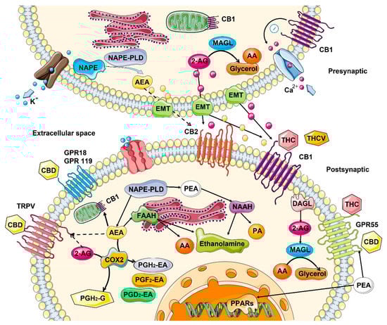
2.2. Role in the Control of Cell Division and Cell Proliferation

It appears that ECS controls the fate of many cells in the organism, regulating the cell division and proliferation, apoptosis, necrosis and autophagy in several organs and organ systems, including the brain, skin, and immune system. In the central nervous system (CNS), the ECS system functions as a neuroprotective system that controls glutamate excitotoxicity, calcium influx, inflammation, and autophagy [25]. In the CNS, the interaction of endocannabinoids with CB1/CB2 and other receptors mediates synaptic plasticity or progenitor cell fate in the central nervous system, promoting self-repair of the brain [26]. It also appears that constitutive release of 2-arachidonoylglycero by late oligodendrocyte progenitors allows oligodendrocyte maturation by activating CB receptors and downstream ERK pathway [27]. In skin, ECS activity maintains the cutaneous homeostasis through the regulation of skin cell proliferation, survival, and differentiation [28]. Locally produced AEA inhibits the cellular growth and the differentiation of cultured NHEK and HaCaT keratinocytes, as well as inducing apoptosis of human HaCaT keratinocytes [28]. CB1 activity is higher in differentiated skin layers [29]. In human cultured hair follicles, AEA but not 2-AG inhibit elongation and proliferation of hair shaft and induce intraepithelial apoptosis in a CB1-dependent manner [30]. Both AEA and 2-AG induce apoptosis of human sebaceous-gland-derived SZ95 sebocytes in a CB2-dependent manner [31]. In the immune system, the central role is played by CB2 receptors that are mainly expressed by cells (T and B lymphocytes) and peripheral tissues of the immune system (spleen and thymus) where it regulates immune suppression, apoptosis, and cell migration [32]. In in vitro studies, it was demonstrated that anandamide inhibits mitogen-induced proliferation of T cells [33], while inhibiting the chemokine SDF-1-induced migration of CD8+ T cells [34]. In contrast, 2-AG, but not anandamide, induced CB2-dependent migration in natural killer cell line KHYG-1 cells [35]. In B cells, 2-AG chemo-attracts naïve B cells and marginal zone B cells and inhibits the function of activated B cells, while 2-AG and anandamide suppress the migration of neutrophils [36]. Additionally, anandamide induces the apoptosis of murine bone-marrow-derived DCs (BMDCs) in a CB1- and CB2-dependent manner [37].

2.3. Changes in the ECS with Age

Cancer can be considered an age-associated disease, due to the accumulation of cellular and DNA damage. From this perspective, it is interesting to understand what happens to ECS with age. In general, information about age-related changes in the ECS is scarce. Most of the data are related to changes in the central nervous system, and even then, the data are very contradictory. In general, it is believed that the activity of ECS declines with age [13]. In rats, in one study, a general decrease in the expression of CB1 and a decrease in density of the receptors in various brain areas with age was observed [38], while in another study—in which only redistribution of the receptors was noted– they were reduced in the postrhinal, but elevated in the entorhinal and temporal cortices in old animals [39] (Table 1). In mice, no changes in the receptor density in most brain regions was found with age, but instead, a significantly reduced receptor/Gi protein coupling was observed [40]. In one study on humans, CB1 expression increased, predominantly in females, most drastically in the basal ganglia, the lateral temporal cortex, and in the hippocampus [41], while another study reported no change [39]. As for endocannabinoids, the picture is not clear either—some studies suggested a decrease, while others found no difference in different brain regions of young and old animals. [13]. However, animals lacking FAAH—the enzyme degrading anandamide showed less pronounced features of aging—decreased expression of pro-inflammatory genes and decreased decline in cardiac function [42] (Table 1).
Table 1.
Age-dependent changes to ECS components in different normal tissues.
Tissues/Organs Endocannabinoids Receptors Metabolizing Enzymes
Skin No reliable data ↓ in CB1 expression [13] FAAH tends to ↓ with age [43][45]
Lung 2-AG ↓ and AEA ↑ in mice [44][46] No reliable data No reliable data
Brain From no change [40] to a ↓ in AEA [45][47]

↓ in 2-AG levels in mice [46][48]
From ↑ in humans [41] to no change [39] to a ↓ [38][47][38,49] in mice/rats in CB1 expression, brain area-specific FAAH activity in rats [48][50]

↑ in MAGL levels in mice [46][48]
Blood Small ↑ in 2-AG and AEA in mice [44][46] No reliable data No reliable data
↑ Indicates increased expression or the amount of circulating product, while ↓ indicates decreased amounts.
Very little reliable data exist on changes in the ECS in skin. Concentration of AEA is 119-fold higher than 2-AG in human skin [49][43], although it is not known how it changes with age. Moderate CB1 activation in skin works as a suppressor of the differentiation, while high activation leads to anti-proliferative and pro-apoptotic events [19]. In mice, CB1 deficiency in skin may lead to premature aging [50][44]. CB1-deficient mice exhibit cognitive impairments and changes in the structure of skin, indicating that CB1 deficiency accelerates aging only in the brain and in the skin, but not other peripheral organs [50][44]. Expression of anandamide degrading enzyme FAAH decreases with age and in response to sunburn in skin [43][45], indicating that ECS may undergo similar changes upon skin aging and in response to UV damage. A decrease in FAAH with age may also mean that there is less anandamide produced with age. There is even less information about ECS activity in other tissues. One report shows a 2-AG decrease in lungs and increase in blood, while AEA increases in lung and blood in mice [44][46].

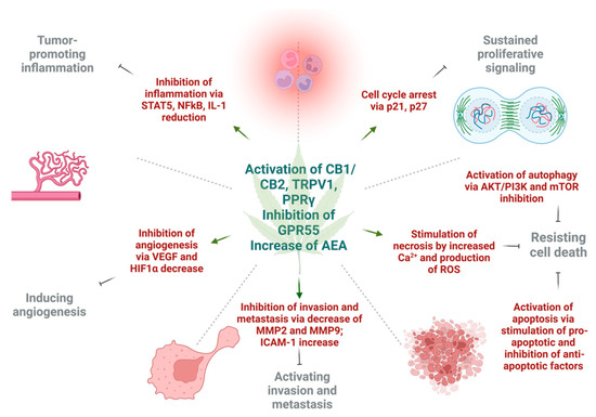
3. Effect of Cannabinoids on Various Hallmarks of Cancer

Various in vitro and in vivo experiments have shown that cannabinoids can target almost every hallmark of cancer (Figure 2) [51][186]. They inhibit proliferation, reduce inflammation, stimulate apoptosis, and inhibit tumor invasiveness, angiogenesis, and metastasis [52][53][54][55][187,188,189,190]. One of the most important effects of cannabinoids, besides their antitumor ability, is that they are less likely to affect non-transformed normal cells surrounding tumors, and they may even have protective effects. For instance, cannabinoids may induce cell death in glioma cells while protecting normal astroglial and oligodendroglial cells from apoptosis via CB1 receptors [52][187]. Studies on animals show the protective effects of cannabinoids against certain types of tumors. For example, a dose-dependent decrease in the incidence of hepatic adenomas and hepatocellular carcinomas in mice that were given THC over 2 years was noted. Additionally, lower incidence rates of benign tumors in mammary glands, uterus, testis, and pancreas were seen in tested rats [56][191].
Figure 2. The effects of cannabinoids on different hallmarks of cancer. The activation of cannabinoid 1 (CB1), cannabinoid 2 (CB2), and transient receptor potential cation channel 1 (TRPV1) increase levels of anandamide (AEA), as well as inhibition of G-protein coupled receptor 55 (GPR55), and exert different effects on tumor cells in respect to cancer hallmarks. Cannabinoids inhibit tumor-promoting inflammation via downregulation of nuclear factor κB (NFκB), signal transducer and activator of transcription 5 (STAT5), and interleukin 1 (IL1). The angiogenesis is inhibited by reduction in vascular endothelial growth factor (VEGF) and hypoxia inducible factor 1α (HIF1α). Next, invasion and metastasis are prevented by decrease of tissue degrading enzymes—matrix metalloproteinase 2 and 9 (MMP2, MMP9), as well as expression of intercellular adhesion molecule 1 (ICAM-1). Sustained proliferative signaling is opposed by the activation of p21 and p27 that leads to cell cycle arrest. Lastly, under the action of cannabinoids, cell death may be achieved by three mechanisms. Autophagy is triggered by inhibition of protein kinase B (AKT), phosphoinositide 3-kinase (PI3K) and mammalian inhibitor of rapamycin (mTOR). Apoptotic cell death is a result of upregulation of pro-apoptotic and downregulation of anti-apoptotic factors under the action of different cannabinoids. Necrosis can result due to high Ca2+ release and formation of ROS in cancer cells. This figure was created with BioRender.com.

3.1. Induction of Autophagy and Apoptosis

Autophagy and apoptosis are two essential mechanisms of regulation of uncontrolled growth. Autophagic activity of cannabinoids observed in several major cancers [57][58][192,193] is partially dependent on the CB1 or CB2 receptor. Mice deficient in CB1 receptor exhibit altered autophagosomal activity [13], while endocannabinoid palmitoylethanolamide increased the phagocytosis of murine microglial cells [59][194]. Additionally, the experimental study using delta-9-THC and a synthetic agonist decreased the cell viability of hepatocellular carcinoma xenografts in nude mice via the CB2 receptors. The anti-cancer effect was explained by activating the endoplasmic reticulum stress response, which leads to macro-autophagy and eventually apoptosis [60][195]. Studies on small-cell lung carcinoma [61] and breast cancer cells [62][67] supported the idea that CB1 and CB2 receptors may be potential targets to achieve apoptosis. The preclinical models of breast cancer showed evidence that CBD may induce apoptosis in estrogen-dependent and estrogen-independent breast cancer cells with little or no effect on normal mammary cells. Surprisingly, this was CB1-, CB2-, and vanilloid receptor-independent [63][176]. The well-established antineoplastic mechanisms of cannabinoids are alterations in ceramide de novo synthesis. In cancer cells, increased ceramide levels, a neutral lipid backbone of complex sphingolipids, can occur under chemotherapy, radiation, and stimulation of CB receptors [64][65][58,196]. As a result, ceramide activates endoplasmic reticulum stress response and causes inhibition of global translation of proteins. At the same time, there is an activation of C/EBP homology protein (CHOP) which can stimulate proapoptotic proteins BAD and BAX [66][197]. Moreover, cannabinoids can cause downregulation of AKT, which may have a variety of intracellular effects. Low AKT leads to activation of autophagy via the mTOR pathway, cell cycle arrest through p21, and activation of caspase 9 and 3, which eventually ends in apoptotic cell death [64][67][68][69][70][71][58,108,155,198,199,200]. Activation of CB1 and CB2 receptors by synthetic cannabinoid agonists could stimulate apoptosis via ceramide synthesis and TNF-receptor activation [64][58]. Another group showed that activation of CB1 receptors in different CRC cell lines causes inhibition of major cancer survival pathways such as RAS/MAPK, ERK1, and PI3K/AKT [68][155]. Additionally, CBD, a partial agonist of CB1/CB2 receptors and antagonist of GPR55, may suppress mTOR/AKT signaling and activate proapoptotic NOXA in CRC cells [72][201]. Moreover, CBD suppressed the production of inhibitors of apoptosis, such as survivin and c-FLIP in colon cancer cells [66][197].

3.2. Reduction of Inflammation and Inhibition of Proliferation

Inflammation is a large component of carcinogenesis. ECS plays a central in the regulation of function of immune system and control of inflammation. Similarly, many phytocannabinoids exert strong anti-inflammatory effects upon local [73][202] or systemic [74][203] application. Cannabinoids inhibited proliferation by suppressing the AKT/PKB prosurvival pathway causing cell cycle arrest in G1/S phase. This was shown in multiple cancers, including melanoma, breast, gastric, lung, and liver carcinomas [75][76][77][78][79][80][93,151,159,160,204,205]. In a breast cancer model, cannabinoids were able to induce cell cycle arrest via inhibition of cyclin dependent kinase 1 (CDK1), induction of p21 and p27, a decrease in cyclin A and E levels, degradation of CDC25A, and finally, inactivation of CDK2 [81][82][206,207]. In the study on head and neck squamous cell carcinoma, cannabinoids were able to stimulate dual specificity phosphatase 1 (DUSP1), which is a negative regulator of MAPK [83][208]. DUSP1 is one of the central mediators in the resolution of inflammation in cells. Moreover, the levels of cyclin dependent kinase inhibitor, p21, as well as growth arrest and DNA damage-inducible protein α (GADD45A) were activated, resulting in cannabinoid’s antiproliferative effects. In human gastric cancer model, CBD upregulated ATM and p21, which caused a decrease in CDK2 and CCNE, resulting in cell arrest in G0/G1 stage [84][209]. In a xenograft model of human glioma, CBD was able to reduce the activity of 5-lipoxygenase, an enzyme that catalyzes synthesis of leukotrienes (LTs) and mediators of inflammation; a decrease in 5-lipoxygenase activity caused inhibition of LTB4 and had antiproliferative effect [85][210]. The eicosanoid system, which contains pro- and anti-inflammatory molecules, plays an important role in cannabinoid-induced tumor cell apoptosis. The addition of R(+)methanandamide to the glioma cells activated de novo ceramide synthesis, which eventually led to COX-2 expression with subsequent production of PGE2 that had proapoptotic effect [86][87][211,212]. It was shown that proapoptotic effects of eicosanoids was PPARγ receptor-dependent [88][89][90][213,214,215]. On the other hand, it was shown that the micromolar concentrations of THC, CB1 agonist—arachidonyl-2-chloroethylamine (ACEA), and CB2 agonist HU308 stimulated the proliferation of cancer cells, which can be explained by transactivation of EGFR [91][92][93][94][169,171,216,217]. The chemoprotective effect of CBD was also shown on colorectal cancer in mice. Adding CBD prevented premalignant and malignant lesions development in the azoxymethane model of colon cancer [95][218]. The effect was explained by DNA protection against oxidative damage, increased levels of endocannabinoids, and decreased cell proliferation [95][218]. The antiproliferative action was CB1 dependent [96][219].

3.3. Inhibition of Angiogenesis, Tumor Invasiveness, and Metastasis

There were multiple reports showing the inhibitory effects of cannabinoids on cancer cell migration, invasion, and metastasis [97][98][99][62,74,220]. CBD was shown to inhibit the invasiveness of lung cancer cell lines by inhibiting ICAM-1 [55][190]. As some experiments indicated, the induction of tissue inhibitor of metalloproteinase-1 (TIMP-1), and ICAM-1 by THC, Met-AEA and CBD had significant anti-invasive effects [55][100][101][105,190,221]. The action of TIMP-1 is achieved via reduction of collagen-degrading enzymes, MMP-2 and MMP-9, that promote cancer cell invasiveness [102][222]. Another way in which cannabinoids are diminishing tumor aggressiveness is inhibition of epithelial-to-mesenchymal transition. A study that involved 2-methyl-2′-F-anandamide (Met-F-AEA) showed a significant reduction in β-catenin, vimentin, N-cadherin, and fibronectin, which are considered mesenchymal markers in tumor invasion. Moreover, Met-F-AEA decreased the levels of EMT markers such as Snail1, Slug, and Twist [103][223]. Other studies showed that CBD may reverse an IL-1β-induced EMT in breast cancer cells [104][224], or TGF-β-induced reorganization of F-actin, which also corresponds to EMT in lung cancer cells [105][60]. Cannabinoids may inhibit the invasion and metastasis of cancer cells through downregulation of vascular endothelial growth factor (VEGF), matrix metalloproteinase 2, matrix metalloproteinase 9, E-cadherin, cyclooxygenase 2 (COX-2), and hypoxia-inducible factor α [106][107][108][225,226,227].