Apoptosis in Acute Myeloid Leukemia: Comparison
Please note this is a comparison between Version 3 by Dean Liu and Version 4 by Dean Liu.

More than 97% of patients with acute myeloid leukemia (AML) demonstrate genetic mutations leading to excessive proliferation combined with the evasion of regulated cell death (RCD). The most prominent and well-defined form of RCD is apoptosis, which serves as a defense mechanism against the emergence of cancer cells. Apoptosis is regulated in part by the BCL-2 family of pro- and anti-apoptotic proteins, whose balance can significantly determine cell survival. Apoptosis evasion plays a key role in tumorigenesis and drug resistance, and thus in the development and progression of AML. Research on the structural and biochemical aspects of apoptosis proteins and their regulators offers promise for new classes of targeted therapies and strategies for therapeutic intervention.

  • acute myeloid leukemia
  • apoptosis
  • neddylation

1. Apoptosis

Apoptosis (from Ancient Greek “falling off”) is a physiological process of cellular suicide required for controlling tissue homeostasis, particularly in rapidly-renewing tissues such as hematopoietic tissue [1]. It is characterized by distinct morphological changes including nuclear condensation, cell shrinkage, membrane blebbing, DNA fragmentation, and the formation of apoptotic bodies [2]. This elimination occurs naturally in the course of organ development or following cellular stress [3]. However, such stress signals can be overcome by cancer cells overexpressing anti-apoptotic proteins, especially those of the BCL-2 (B-cell lymphoma-2) family [3]. The BCL2 gene acts as an oncogene that prevents hemopoietic cell death [4]. It has been proposed that the blockage of apoptotic signaling promotes oncogenesis, and this has been demonstrated in several model systems [5][6].

2. Pathways of Apoptosis

Apoptosis can occur by the intrinsic (or mitochondrial) and extrinsic (or death receptor) pathways [7]. The intrinsic pathway serves to activate apoptosis as an appropriate response to various internal traumas, such as metabolic stress, hypoxia, checkpoint violations, growth factor withdrawal, activation of oncogenes, and irreparable genomic damage. The extrinsic pathway is distinctly triggered by the ligation of proapoptotic transmembrane death receptors from the tumor necrosis factor (TNF) family [8]. A common feature of apoptosis is the involvement of caspases, a family of intracellular cysteine proteases, which are present as inactive zymogens in all animal cells, but can be triggered to assume an active state [1][9]. Caspases can be broadly categorized based on their role in apoptosis (caspase-2, -3, -6, -7, -8, -9, and 10) then further subdivided into two groups: the initiator caspases (caspase-2, -8, -9, and -10) and the effector caspases (caspase-3, -6, and -7) [4].

3. The Mechanism of the Mitochondrial Apoptosis Pathway
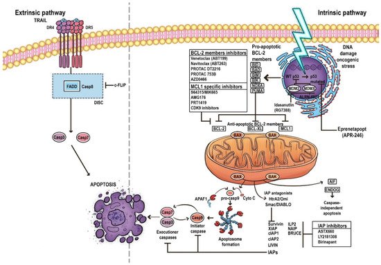
The mitochondrial apoptosis pathway is the dominant form of cell death, leading to the death of over 60 billion cells each day [10]. The process is initiated by stress signals and is triggered by mitochondrial outer membrane permeabilization (MOMP), caused by inter alia BCL-2 family proteins.
MOMP leads to the release of apoptogenic proteins from the intermembrane space. It results in the secretion of various cell death modulators such as cytochrome c, apoptosis-inducing factor (AIF), endonuclease G (ENDOG), direct IAP-binding protein with low pI (DIABLO, also known as second mitochondria-derived activator of caspases, SMAC) or Omi/HtrA2, and BCL-2 family proteins; it also prevents mitochondrial ATP synthesis, inhibits the respiratory chain and increases reactive oxygen species (ROS) production [11]. These events promote the activation of the initiator caspase 9 and executioner caspases (caspases 3, 6, and 7) in order to destroy the cell [12]. A key element in the regulation and induction of intrinsic apoptosis is the BCL-2 protein family, whose pro- and anti-apoptotic members, and the balance between them, play significant roles in determining the fate of cells [10][13] (Figure 1).
Figure 1. The pathways of apoptosis. Arrows represent activation and T bars represent inhibition.

4. BCL-2 Family

The BCL-2 family, which tightly regulates MOMP, consists of three subfamilies: the first is the pro-apoptotic BH3-only members (BIM [BCL-2-like protein 11], BID [BH3 Interacting Domain Death Agonist], PUMA [p53 upregulated modulator of apoptosis], NOXA [Phorbol-12-myristate-13-acetate-induced protein 1], HRK [Activator of apoptosis harakiri], BMF [BCL-2 modifying factor], and BAD [BCL-2 associated agonist of cell death]); the second is the pro-apoptotic effector molecules (BAX [BCL-2 associated X] and BAK [BCL-2 homologous antagonist killer]); and the third is the anti-apoptotic BCL-2 family proteins (BCL-2, BCL-xL [B-Cell Lymphoma-extra-large], BCL-W [BCL-2 like protein 2], MCL-1 [myeloid cell leukemia-1], A1 [BCL-2 related protein A1], and BCL-B [BCL-2 like protein 10]).
The receipt of an apoptotic stimulus upregulates the transcription of the pro-apoptotic BH3-only members. These proteins bind anti-apoptotic members of the BCL-2 family and inhibit their activity. Some direct activators (for example, BID and BIM) can also bind and activate the effectors BAK and BAX, which induces MOMP, resulting in the release of cytochrome c and SMAC from mitochondria and the subsequent activation of initiator caspases [14][15].
Another protein regulating apoptosis is the p53 tumor suppressor, known as the ‘guardian of the genome’. It induces cell-cycle arrest with DNA repair or apoptosis by activating the BCL-2 family. The activation of p53 triggers pro-apoptotic proteins such as PUMA, NOXA, BIM, and BAX to counteract MCL-1, thus overcoming MCL-1 mediated resistance at multiple levels. Moreover, p53 stimulates extrinsic pathway activation by the upregulation of death receptors [16][17][18] (Figure 1).

5. Evasion of Apoptosis in AML

The expression of programmed cell death genes is commonly dysregulated in hematological malignancies, promoting cell accumulation and creating favorable conditions for oncogene activation, genetic instability, and metastasis. Such defects enable the survival of genetically unstable cells, leading to the selection of progressively aggressive clones [19]. Multiple elements of the apoptotic pathway are known to be disturbed in AML, thus facilitating the evasion of apoptosis.
The most common pathway used by cancer cells to evade apoptosis is based on the upregulation of antiapoptotic BCL-2 proteins and the loss of BAX/BAK proteins [20]; indeed, BCL-2 gene overexpression is present in over half of all cancers [11]. As many traditional anti-cancer drugs act on the BCL-2/BAX pathway, any disruption of this target may increase resistance to chemotherapy and radiotherapy by elevating the threshold needed for cell death [20].
TP53 gene mutation prevents apoptotic pathway function [20]. Such mutations are observed in 5–10% of de novo AML and 30–40% of therapy-related cases and are considered important indicators of poor outcome [21][22]. In a study conducted on 500 AML patients, Hou et al. found TP53 mutation to be associated with an inferior response rate (complete remission [CR] rate 28.6% vs. 80.2%, p < 0.0001) and shorter overall survival (OS) (median, 5 vs. 35 months, p < 0.001) compared to unmutated patients [23].
Unlike solid tumors, in most cases of non-complex karyotype de novo AML, the TP53 locus is found to be the wild-type. Most TP53 changes result in missense mutations, i.e., resulting in changes in the amino acid sequence of the DNA-binding domain (encoded by exons 5–8). These are more common in complex karyotypes, relapsed, and elderly AML patients, as well as therapy-related AML [22][24][25]. Where the mutation is absent, as noted in the majority of AML cases, it is possible that apoptosis is blocked by alternate mechanisms [26]. Intriguingly, patients with a TP53 mutation have a lower-than-expected frequency of mutations in other myeloid-related genes, involving splicing, epigenetics, and signal transduction [22][27]. However, irrespective of TP53 mutational status, many AML cases are characterized by p53 dysfunction, presumably through the alteration of p53-regulatory proteins, resulting in the disruption of apoptosis [16][28].
The wild-type TP53 may be significantly inhibited by high levels of mouse double minute 2 homolog (MDM2) or inhibitor of apoptosis proteins (IAP); this presents an appealing targeted strategy for AML treatment [29].
IAPs consist of several proteins which inhibit apoptosis pathway at different levels, namely X-linked inhibitor of apoptosis (XIAP), cellular (cIAP1, cIAP2), neuronal (NIAP), testis-specific (Ts-IAP), Bir-ubiquitin conjugating enzyme (BRUCE), livin, and survivin [30]. IAPs take part in modulating autophagy, necroptosis, and immune regulation, and can inhibit both intrinsic and extrinsic apoptosis; they also stimulate cell survival and increase tumor growth and metastasis when present at elevated levels [31][32]. Moreover, IAP overexpression reduces CR rate in AML patients and increases chemoresistance in several types of cancer. In addition, low IAP expression has also been associated with longer OS in AML patients [30][33][34]. Lack of IAP expression is also associated with higher sensitivity to standard chemotherapy [16][35][36]. Several molecules antagonize IAP activity, with the most prominent ones being SMAC/DIABLO, Omi/HtrA2 (HTRA serine peptidase 2), and XAF-1 (XIAP-associated factor 1) [37][38].

References

  1. Testa, U.; Riccioni, R. Deregulation of apoptosis in acute myeloid leukemia. Haematologica 2007, 92, 81–94.
  2. Li, B.; Dou, S.-X.; Yuan, J.-W.; Liu, Y.-R.; Li, W.; Ye, F.; Wang, P.-Y.; Li, H. Intracellular transport is accelerated in early apoptotic cells. Proc. Natl. Acad. Sci. USA 2018, 115, 12118–12123.
  3. Fernald, K.; Kurokawa, M. Evading apoptosis in cancer. Trends Cell Biol. 2013, 23, 620–633.
  4. Kontomanolis, E.N.; Koutras, A.; Syllaios, A. Role of Oncogenes and Tumor-suppressor Genes in Carcinogenesis: A Review. Anticancer Res. 2020, 40, 6009–6015.
  5. Hanahan, D.; Weinberg, R.A. Hallmarks of cancer: The next generation. Cell 2011, 144, 646–674.
  6. Nechiporuk, T.; Kurtz, S.E.; Nikolova, O.; Liu, T.; Jones, C.L.; D’Alessandro, A.; Culp-Hill, R.; D’Almeida, A.; Joshi, S.K.; Rosenberg, M.; et al. The TP53 Apoptotic Network Is a Primary Mediator of Resistance to BCL2 Inhibition in AML Cells. Cancer Discov. 2019, 9, 910–925.
  7. Letai, A. Apoptosis and Cancer. Annu. Rev. Cancer Biol. 2017, 1, 275–294.
  8. Koren, E.; Fuchs, Y. Modes of Regulated Cell Death in Cancer. Cancer Discov. 2021, 11, 245–265.
  9. MacKenzie, S.H.; Clark, A.C. Death by Caspase Dimerization. In Protein Dimerization and Oligomerization in Biology; Springer: Berlin/Heidelberg, Germany, 2012; Volume 747, pp. 55–73.
  10. Singh, R.; Letai, A.; Sarosiek, K. Regulation of apoptosis in health and disease: The balancing act of BCL-2 family proteins. Nat. Rev. Mol. Cell Biol. 2019, 20, 175–193.
  11. Galluzzi, L.; Vitale, I.; Abrams, J.M.; Alnemri, E.S.; Baehrecke, E.H.; Blagosklonny, M.V.; Dawson, T.M.; Dawson, V.L.; El-Deiry, W.S.; Fulda, S.; et al. Molecular definitions of cell death subroutines: Recommendations of the Nomenclature Committee on Cell Death 2012. Cell Death Differ. 2012, 19, 107–120.
  12. Boice, A.; Bouchier-Hayes, L. Targeting apoptotic caspases in cancer. Biochim. Biophys. Acta 2020, 1867, 118688.
  13. Hatok, J.; Racay, P. Bcl-2 family proteins: Master regulators of cell survival. Biomol. Concepts 2016, 7, 259–270.
  14. Nagata, S. Apoptosis and Clearance of Apoptotic Cells. Annu. Rev. Immunol. 2018, 36, 489–517.
  15. Zhang, L.-N.; Li, J.-Y.; Xu, W. A review of the role of Puma, Noxa and Bim in the tumorigenesis, therapy and drug resistance of chronic lymphocytic leukemia. Cancer Gene Ther. 2012, 20, 1–7.
  16. Valentin, R.; Grabow, S.; Davids, M.S. The rise of apoptosis: Targeting apoptosis in hematologic malignancies. Blood 2018, 132, 1248–1264.
  17. Cassier, P.A.; Castets, M.; Belhabri, A.; Vey, N. Targeting apoptosis in acute myeloid leukaemia. Br. J. Cancer 2017, 117, 1089–1098.
  18. Toufektchan, E.; Toledo, F. The Guardian of the Genome Revisited: P53 Downregulates Genes Required for Telomere Maintenance, DNA Repair, and Centromere Structure. Cancers 2018, 10, 135.
  19. Kitada, S.; Pedersen, I.M.; Schimmer, A.D.; Reed, J.C. Dysregulation of apoptosis genes in hematopoietic malignancies. Oncogene 2002, 21, 3459–3474.
  20. Pfeffer, C.M.; Singh, A.T.K. Apoptosis: A Target for Anticancer Therapy. Int. J. Mol. Sci. 2018, 19, 448.
  21. Hunter, A.M.; Sallman, D.A. Current status and new treatment approaches in TP53 mutated AML. Best Pract. Res. Clin. Haematol. 2019, 32, 134–144.
  22. Wang, C.; Sallman, D.A. What Are the Prospects for Treating TP53 Mutated Myelodysplastic Syndromes and Acute Myeloid Leukemia? Cancer J. 2022, 28, 51–61.
  23. Hou, H.-A.; Chou, W.-C.; Kuo, Y.-Y.; Liu, C.-Y.; Lin, L.-I.; Tseng, M.-H.; Chiang, Y.-C.; Liu, M.-C.; Liu, C.W.; Tang, J.-L.; et al. TP53 mutations in De Novo acute myeloid leukemia patients: Longitudinal follow-ups show the mutation is stable during disease evolution. Blood Cancer J. 2015, 5, e331.
  24. Barbosa, K.; Li, S.; Adams, P.D.; Deshpande, A.J. The role of TP53 in acute myeloid leukemia: Challenges and opportunities. Genes Chromosom. Cancer 2019, 58, 875–888.
  25. Shallis, R.M.; Bewersdorf, J.P.; Stahl, M.F.; Halene, S.; Zeidan, A.M. Are We Moving the Needle for Patients with TP53-Mutated Acute Myeloid Leukemia? Cancers 2022, 14, 2434.
  26. Campos, L.; Rouault, J.P.; Sabido, O.; Oriol, P.; Roubi, N.; Vasselon, C.; Archimbaud, E.; Magaud, J.P.; Guyotat, D. High expression of bcl-2 protein in acute myeloid leukemia cells is associated with poor response to chemotherapy. Blood 1993, 81, 3091–3096.
  27. Niparuck, P.; Police, P.; Noikongdee, P.; Siriputtanapong, K.; Limsuwanachot, N.; Rerkamnuaychoke, B.; Chuncharunee, S.; Siriboonpiputtana, T. TP53 mutation in newly diagnosed acute myeloid leukemia and myelodysplastic syndrome. Diagn. Pathol. 2021, 16, 100.
  28. Quintás-Cardama, A.; Hu, C.; Qutub, A.; Qiu, Y.H.; Zhang, X.; Post, S.M.; Zhang, N.; Coombes, K.; Kornblau, S.M. P53 pathway dysfunction is highly prevalent in acute myeloid leukemia independent of TP53 mutational status. Leukemia 2016, 31, 1296–1305.
  29. Nag, S.; Qin, J.; Srivenugopal, K.S.; Wang, M.; Zhang, R. The MDM2-p53 pathway revisited. J. Biomed. Res. 2013, 27, 254–271.
  30. Jan, R.; Chaudhry, G.-E. Understanding Apoptosis and Apoptotic Pathways Targeted Cancer Therapeutics. Adv. Pharm. Bull. 2019, 9, 205–218.
  31. Hrdinka, M.; Yabal, M. Inhibitor of apoptosis proteins in human health and disease. Genes Immun. 2019, 20, 641–650.
  32. Cheung, C.H.A.; Chang, Y.-C.; Lin, T.-Y.; Cheng, S.M.; Leung, E. Anti-apoptotic proteins in the autophagic world: An update on functions of XIAP, Survivin and BRUCE. J. Biomed. Sci. 2020, 27, 31.
  33. Oberoi-Khanuja, T.K.; Murali, A.; Rajalingam, K. IAPs on the move: Role of inhibitors of apoptosis proteins in cell migration. Cell Death Dis. 2013, 4, e784.
  34. Pluta, A.; Wierzbowska, A.; Cebula-Obrzut, B.; Pluta, P.; Stępka, K.; Szmigielska-Kapłon, A.; Grzybowska-Izydorczyk, O.; Czemerska, M.; Smolewski, P.; Wrzesien-Kus, A.; et al. Prognostic value of inhibitor of apoptosis protein family expression in patients with acute myeloid leukemia. Leuk. Lymphoma 2015, 56, 2529–2535.
  35. Rathore, R.; McCallum, J.E.; Varghese, E.; Florea, A.-M.; Büsselberg, D. Overcoming chemotherapy drug resistance by targeting inhibitors of apoptosis proteins (IAPs). Apoptosis 2017, 22, 898–919.
  36. Zhang, Y.; Huang, F.; Luo, Q.; Wu, X.; Liu, Z.; Chen, H.; Huang, Y. Inhibition of XIAP increases carboplatin sensitivity in ovarian cancer. Onco Targets Ther. 2018, 11, 8751–8759.
  37. Martinez-Ruiz, G.; Maldonado, V.; Ceballos-Cancino, G.; Grajeda, J.P.R.; Melendez-Zajgla, J. Role of Smac/DIABLO in cancer progression. J. Exp. Clin. Cancer Res. 2008, 27, 48.
  38. Mastrangelo, E.; Vachette, P.; Cossu, F.; Malvezzi, F.; Bolognesi, M.; Milani, M. The Activator of Apoptosis Smac-DIABLO Acts as a Tetramer in Solution. Biophys. J. 2015, 108, 714–723.
More
Video Production Service