Neuropeptides Inhibitors for the Development of Cancer Therapy: Comparison
Please note this is a comparison between Version 2 by Jessie Wu and Version 1 by Andrea Ragusa.

Neuropeptides are mainly secreted from the human central and peripheral nervous systems. Neuropeptides bind to its cognate rhodopsin-like G-protein coupled receptor (GPCR) and perform various physiological functions. Conventional cancer treatments in clinical practice still present many drawbacks due to the lack of selectivity toward the target cell, drug-resistance, and side-effects, thus pushing for the development of new therapeutic agents and therapies. Recent research suggests that neuropeptides influence cancer cell proliferation, invasion, metastasis, and angiogenesis and, therefore, they could be exploited as a target for novel anticancer therapies. Very recently, targeted approaches that inhibit neuropeptides and their associated receptors are being developed in cancer treatment. 

  • neuropeptide
  • receptor
  • inhibitors
  • colorectal cancer
  • bombesin
  • neurotensin
  • vasoactive intestinal peptide
  • substance P
  • neuropeptide Y
  • orexins

1. Neuropeptides

Neuropeptides (NPs) are peptide hormones that the neuroendocrine system uses as chemical signals to communicate among cells. They are a set of signaling messengers that regulate exocrine and endocrine secretion, smooth muscle contraction, blood pressure, and inflammation by acting as neurotransmitters, paracrine regulators, and hormones. Numerous NPs are localized in the brain and co-released with neurotransmitters to reach their target, where they perform a wide variety of functions.
NPs are said to serve as modulators depending on the condition in which they are challenged; however, they generally function in the brain in a paracrine manner. Growing evidence supports that during stress, injury, and pain, they act as potent neurotransmitters and trophic factors that mediate the inhibitory or stimulatory action of the nervous system. In recent years, several biomolecules, such as neurotensin, substance P (SP), vasoactive intestinal polypeptide (VIP), bombesin, opioids, and neuropeptide Y have emerged as potential forms of NPs whose action as neuronal mediators has been extensively studied in different pathological conditions, including cancer [80,85][1][2]. As discussed earlier, severe consequences of stress are often correlated with oncogenesis in different cancers. However, NPs related to cancer development and progression are listed in Table 1.
NPs and their associated receptors have been extensively investigated to identify their critical role in complex diseases such as obesity, diabetes, Alzheimer’s, and other nervous system disorders. Moreover, the presence of NPs in several disease pathogenesis has been widely described. Still, the critical role of NPs and their associated receptors in the development of cancers and their potential implication for specific therapy and prevention of cancer is not well-defined [102][20]. NPs have been studied by several researchers who provided mechanistic insight into the biological significance of the neuronal system in tumor progression [101,102][19][20]. NPs have been found to bind to multiple receptors expressed in endothelial cells and induce cell proliferation, migration, survival, and differentiation, as well as promote angiogenesis, thus suggesting that NPs are tumor-promoting factors during cancer development and progression. Galli and colleagues demonstrated that a higher serum concentration of neuropeptide Y (NPY) in neuroblastoma patients was significantly correlated with poor prognosis and survival outcomes [97][15]. Moreover, elevated NPY circulation in serum was linked to metastasis and angiogenesis. This restudy earch supports the higher expression of NPY in patients with neuroblastoma and suggests its potential exploitation as a therapeutic biomarker for monitoring cancer progression. The diagnostics potential of NPY was also evaluated in CRC patients in different samples, such as tissue, cell lines, and feces. Interestingly, the results showed a significantly hypermethylated promoter region of NPY in CRC tissues as well as fecal exfoliated cells. Moreover, treatment with 5-aza-2′-deoxycytidine (a demethylating agent) suppressed the methylation level of NPY in CpG Island at the promoter region and normalized its transcriptional expression in vivo, indicating that tissue and fecal NPY methylation could be a potential biomarker in CRC patients [103][21]. NPs, such as neurotensin (NTS) and its cognate low-affinity receptors NTSR1 and NTSR2, mediate several biological functions, including gastric acid secretion, bowel motility, and fatty acid absorption. Growing evidence shows that NTS is also strongly implicated in a number of mechanisms involved in tumor growth, proliferation, survival, metastatic spread, and invasiveness. The recent resurgence of NTS and NTSR1 and NTSR2 complexes in cancer-inducing cell proliferation and development by pathway deregulation, such as the Wnt/β-catenin signaling pathway, enlightens its role in the carcinogenesis process. In fact, it has been suggested as a potential prognostic biomarker in head and neck squamous cell carcinoma, lung, colon, and breast cancers [104,105][22][23].
It has been shown that the substance P-neurokinin-1 receptor SP/NK-1R complex is frequently dysregulated in cancer, confirming its role as a growth-promoting and anti-apoptotic factor in CRC, breast, and prostate cancer. In gastrointestinal (GI) cancer, the NK-1 receptor was also found to be overexpressed in GI tumor cells, and the binding of SP to the NK-1 receptor was implicated in many biological functions, such as proliferation, migration, metastasis, and angiogenesis [106,107][24][25]. Therefore, a therapeutic approach against the NK-1 receptor could potentially inhibit the growth-promoting activity of the GI tumor cells. For example, NK-1 receptor inhibition with antagonists, such as fosaprepitant, L-732,138, aprepitant, and L-733,060, induced an antineoplastic effect against GI tumors by inhibiting cell migration and angiogenesis, inducing apoptosis, and counteracting the Warburg effect [108,109][26][27].
Aberrant expression of galanin receptors (GalRs) could be also considered a risk factor for human GI cancers, in particular stomach epithelial cells and gastric cancer cells. GalRs (GalR1, GalR2, and GalR3) belong to the GPCRs family that interacts with galanin (Gal) and regulate a range of biological processes, including tumorigenesis [110,111][28][29]. A study demonstrated that elevated Gal expression in samples from CRC patients along with high serum concentration can be considered a serious risk of invasion and metastasis [112][30]. Despite a clear understanding of the role of Gal in CRC cases, its receptors have been less explored except GalR1, but this does not overwhelmingly support the role of GalRs in the pathogenesis of CRC. The possible role of GalRs in CRC tissue and colon epithelial cells was also evaluated in a recent study showing that GalR2, GalR1, and GalR3 present stronger immunoreactivity in human colon cancer cells compared to epithelial cells (enterocytes and goblet cells) in the large intestine of CRC patients. Furthermore, GalR3 with significantly higher immunoreactivity and survival rate in CRC patients was suggested to be an excellent prognostic biomarker for CRC patients [113][31]. A close association between Gal and CRC occurrence has been also reported by other researchers who observed a differential expression of Gal at different stages of the CRC in addition to GalR1 expression measured with the help of the IHC method. Their study concluded that underexpressed Gal is directly correlated with an advanced CRC stage [114][32].

2. Emerging Neuropeptides Inhibitors in Colorectal Cancer

Colorectal cancer (CRC) is one of the most common cancers, mainly due to alterations in genetic and epigenetic factors that lead to tumor progression and development [116,117][33][34]. Several NPs have been identified that play a significant role in stimulating various important signaling pathways, thus leading to CRC development [118][35]. Therefore, the development of specific inhibitors targeting these NPs may serve as a potential therapeutic approach for CRC treatment. The following section briefly describes various NPs and their specific inhibitors used for therapeutic purposes in the treatment of CRC.

2.1. Bombesin

Bombesin (BB) and its receptors (BBR) have been reported to be overexpressed in many cancers, including CRC. Two commonly known BB-related peptides, including gastrin-releasing-peptide (GRP) and neuromedin-B (NMB) and their respective GPCR receptors, i.e., GRPR (also known as BB2) and NMBR (also known as BB1), are expressed at a higher level in CRC patients [119][36]. Stimulation of these NPs has been shown to increase tumor differentiation and progression, acting mainly as morphogenetic molecules. Therefore, various therapeutic options have been developed for regulating the expression of BB-related peptides and their receptors in CRC. Researchers in their study highlighted the potential inhibitory effect of various BBR antagonists in affecting the development and progression of tumors in patients with CRC [94][11]. Various combinational therapies, including the use of a suitable antagonist together with a potent inhibitor, have been reported to have even more inhibitory effects on CRC cancer cells [120][37]. With the help of experimental analysis, it was observed that the application of RC-3095, a GRPR antagonist, together with several cytotoxic agents, such as 5-FU and irinotecan, results in downregulation and growth inhibition of CRC xenografts. Another study reported the antitumor activity and inhibitory tendency of AN-215, a BB-conjugated analog containing doxorubicin, in CRC [121][38]. These compounds help target and regulate the growth and proliferation of cancer cells.

2.2. Neurotensin

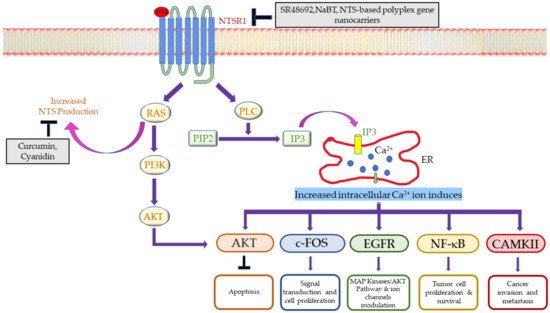
Neurotensin (NTS), together with its receptor, plays a pivotal role in stimulating oncogenesis and tumorigenesis in CRC cells and significantly influences many signaling pathways, such as Wnt/β-catenin, MAPK, PI3K/AKT, and Src/Raf dependent pathway. Growing evidence indicates that the NTSR1-NTS interaction activates RAS and PLC, further stimulating other downstream signaling processes. Remarkably, activation of the RAS gene induces an increase in the production of NTS, which exhibits autocrine and paracrine properties and simultaneously upregulates the AKT gene, which has been found to inhibit apoptosis in colon cells [85,86,87][2][3][4]. On the other hand, PLC plays a critical role in converting PIP2 to IP3, which increases calcium ion production. The increased concentration of calcium ions stimulates several downstream regulatory genes, such as AKT, c-Fos, EGFR, NF-κB, and CAMKII, which further promote various cellular metabolic processes, such as apoptosis, signal transduction, activation of MAP Kinase, AKT pathway, ion channels modulation, tumor cell proliferation and survival, cell invasion and metastasis. The regulatory role of NTS in promoting CRC through signaling processes has been depicted in Figure 1.
Figure 1. Signaling cascade driven by NTS interacting with NTSR1. This complex is involved in the gene regulation of various biological processes via activating the RAS-PI3K-AKT and PLC-IP3-Ca2+, finally contributing to cancer progression. Abbreviations: NTSR1: neurotensin receptor type 1; PI3K: phosphatidylinositol-3-kinase; PLC: protein lipase C; PIP2: phosphatidylinositol-4,5-bisphosphate; IP3: inositol trisphosphate; ER: endoplasmic reticulum; AKT, protein kinase B; EGFR: epidermal growth factor receptor; NF-ĸB: nuclear factor of kappa-light-chain-enhancer of activated B-cells; CAMKII: calcium/calmodulin-dependent protein kinase II.
Researchers highlighted the elevated expression of more than 40% of NTS receptors in human CRC cell lines and activation of the Wnt/APC signaling pathway, which led to the upregulation of their oncogenic activity. It is clear that the NTS-NTSR1 complex in CRC cell lines (HT29, HCT116, SW620, SW480) raises the possibility that this complex increases the risk of CRC progression [122][39]. Thus, the expression of NTS/NTSR1 directly influences CRC progression and development and can serve as a potential molecular biomarker in CRC diagnosis and prognosis. Several treatments have already been proposed for targeting the expression of NTS/NTSR in CRC. Sodium butyrate (NaBT) was reported to have a beneficial effect in downregulating the NTS and NTSR activity [123][40]. NaBT is a histone deacetylase inhibitor that directly hinders the functional activity of NTSR mRNA, protein, and promoter function, thereby suspending the growth and proliferation of CRC cells and increasing their apoptosis. In another study, Iwase et al. 1996 identified a potent NTSR antagonist, SR48692, which downregulates the expression of NTS and inhibits tumor growth and development [124][41]. Concurrent work by Maoret et al. also considered a similar approach based on a non-peptidic NTSR antagonist, i.e., SR 48692. In vitro experiments revealed that SR48692 markedly reduces colony formation by interacting with the NTSR expressed in colon cancer cells [109][27]. Further support for this positive result was obtained in vivo. Treatment with SR 48,692 (1.7 μmol/kg every 24 h) reduced the tumor formed by xenografted SW480 and HCT116 cells in nude mice, suggesting that SR 48,692 deserves further attention as it could be an antagonist with potential anticancer activity in the treatment of colon cancer. Another therapeutic approach exploited the use of various antioxidants and dietary compounds on the activity and function of NTS in CRC. For example, natural products, such as curcumin, effectively inhibited the secretion of NTS-mediated IL-8 protein and downregulated the migration activity of colon cells [125][42]. Cyanidin, another dietary product, has also been shown to have an inhibitory effect on NTS and other pathways related to cancer cell metabolism induced by the epidermal growth factor (EGF) [126][43]. A nanotechnological approach proposed by Hernandez and co-workers exploited NTS-based polyplex gene nanocarriers targeting specifically the NTSR, demonstrating its feasibility as an effective therapeutic approach for treating CRC [127][44].

2.3. Vasoactive Intestinal Peptide

Vasoactive intestinal peptide (VIP) has been reported to contribute significantly to CRC progression and development, although in some cases it inhibited it. Nevertheless, VIP antagonists have been used as potential inhibitors to limit the neoplastic progression of several cancer cell lines. Levy and colleagues demonstrated the in vitro antineoplastic activity of VIP hybrid antagonists, such as NTS6–11VIP7–28, in CRC cell lines (HCT-15) expressing functional VIP receptors [128][45]. NTS6–11VIP7–28 was able to inhibit cancer growth and proliferation at nanomolar concentrations, showing its potential as a potent preventing agent in cancer therapy and confirming that the VIP hybrid antagonist could be efficiently utilized as a potential therapeutic approach for treating and preventing CRC.

2.4. Substance P

Substance P (SP) supports the proliferation and development of cancer cells in CRC, mainly due to the activation of NK1R with the help of various proinflammatory cytokines. Application of Spantide 1, an antagonist of NK1R, resulted in a decreased expression of cancer cells in CRC [94][11]. Aprepitant, another NK1R antagonist, also significantly downregulated cellular growth and proliferation of two colon cancer cell lines, i.e., LiM6 and DLD1 [90][7]. Inactivation of the SP/NK1R signaling pathways led to aberrant inhibition of the Wnt signaling pathway, thus resulting in the downregulation of various cellular processes [90,91][7][8]. However, SP/NK2R signaling in CRC has not been explored well yet [129][46]. The influential work of Xiang and colleagues gave rise to a renewed interest in understanding the higher expression of NK2R, which was demonstrated to be associated with tumorigenesis, metastasis, and poor survival of CRC patients. The higher expression of NK2R in colon cancer was observed due to interferon (IFNα/β) stimulation and polyinosinic-polycytidylic acid (poly I:C) administration in vitro and in vivo, respectively. However, the use of a potent and selective antagonist (GR 159897) against the NK2R, inhibited the tumor cell proliferation in vitro and the tumor formation in cancer-bearing mice, suggesting it is an optimistic target in patients with colon cancer [130][47].

2.5. Neuropeptide Y

Neuropeptide Y is secreted from tumor cells and acts through multiple receptors, especially the Y2 receptor (Y2R). It mediates proliferation and angiogenesis during cancer development. However, in some cases, it mediated chemoresistance by virtue of its Y5 receptor under various pathological conditions. Consequently, it was observed that altered expression of NPY and its receptors are directly correlated with poor clinical manifestations, low survival, and enhanced cell proliferation in different types of cancer, suggesting NPY as a remarkable drug target [131][48]. Overexpression of NPY activates the Y2 receptor on colonic endothelial cells and has a direct impact on angiogenesis and tumor growth following activation of the ERK/MAPK pathway in colon adenocarcinoma. However, it was demonstrated that the use of a specific Y2R antagonist inhibited the NPY-induced angiogenesis and orthotopic HT29 tumor growth in colon adenocarcinoma [132][49].

2.6. Orexins

Orexin-A and orexin-B are two new NPs that interact with their respective receptors (OX1R and OX2R) and are involved in different pathophysiologic processes, including inflammation, ulcerative colitis, and cancer [133,134][50][51]. Previous studies have demonstrated a strong and consistent link between OX1R and OX2R expression in cancer. The high prevalence of OX1R in GI cancer is responsible for metastasis to the lung and liver from the colon [135][52]. It is noteworthy that orexins and their receptors’ antagonists were found to exert an antitumor effect on the growth of colon cancer cells, possibly due to the induction of mitochondrial apoptosis [136][53]. Circumstantial evidence supported that orexin treatment reduces the cell proliferation in colon cancer cell lines (HT-29, Caco-2, and LoVo) by promoting apoptosis, and diminishes the tumor growth in tumor-bearing mice and xenografted tumor in nude mice. However, the antitumor activity of orexin can be reversed by using suitable inhibitors, such as NSC-87877 and PD169316 [137][54].

References

  1. Schuller, H.M. Neurotransmission and cancer: Implications for prevention and therapy. Anti-Cancer Drugs 2008, 19, 655–671.
  2. Itoh, N.; Semba, S.; Ito, M.; Takeda, H.; Kawata, S.; Yamakawa, M. Phosphorylation of Akt/PKB is required for suppression of cancer cell apoptosis and tumor progression in human colorectal carcinoma. Cancer 2002, 94, 3127–3134.
  3. Dupouy, S.; Mourra, N.; Gompel, A.; Alifano, M.; Forgez, P. The potential use of the neurotensin high affinity receptor 1 as a biomarker for cancer progression and as a component of personalized medicine in selective cancers. Biochimie 2011, 93, 1369–1378.
  4. Qiu, S.; Pellino, G.; Fiorentino, F.; Rasheed, S.; Darzi, A.; Tekkis, P.; Kontovounisios, C. A Review of the Role of Neurotensin and Its Receptors in Colorectal Cancer. Gastroenterol. Res. Pract. 2017, 2017, 6456257.
  5. Tang, B.; Yong, X.; Xie, R.; Li, Q.-W.; Yang, S.-M. Vasoactive intestinal peptide receptor-based imaging and treatment of tumors (Review). Int. J. Oncol. 2014, 44, 1023–1031.
  6. Zawilska, J.B.; Niewiadomski, P.; Nowak, J.Z. Receptors for vasoactive intestinal peptide and pituitary adenylate cyclase-activating polypeptide in the goose cerebral cortex. Pol. J. Pharmacol. 2004, 56, 203–211.
  7. Garnier, A.; Vykoukal, J.; Hubertus, J.; Alt, E.; Von Schweinitz, D.; Kappler, R.; Berger, M.; Ilmer, M. Targeting the neurokinin-1 receptor inhibits growth of human colon cancer cells. Int. J. Oncol. 2015, 47, 151–160.
  8. Mehboob, R.; Gilani, S.A.; Hassan, A.; Tanvir, I.; Javaid, S.; Khalid, S.; Hasan, S.; Waseem, H.; Alwazzan, A.; Munoz, M. Prognostic Significance of Substance P/Neurokinin 1 Receptor and Its Association with Hormonal Receptors in Breast Carcinoma. Biomed. Res. Int. 2020, 2021, 5577820.
  9. Bajo, A.M.; Schally, A.V.; Groot, K.; Szepeshazi, K. Bombesin antagonists inhibit proangiogenic factors in human experimental breast cancers. Br. J. Cancer 2004, 90, 245–252.
  10. Chanda, N.; Kattumuri, V.; Shukla, R.; Zambre, A.; Katti, K.; Upendran, A.; Kulkarni, R.R.; Kan, P.; Fent, G.M.; Casteel, S.W.; et al. Bombesin functionalized gold nanoparticles show in vitro and in vivo cancer receptor specificity. Proc. Natl. Acad. Sci. USA 2010, 107, 8760–8765.
  11. Moreno, P.; Ramos-Álvarez, I.; Moody, T.W.; Jensen, R.T. Bombesin related peptides/receptors and their promising therapeutic roles in cancer imaging, targeting and treatment. Expert Opin. Ther. Targets 2016, 20, 1055–1073.
  12. Moody, T.W.; Lee, L.; Ramos-Alvarez, I.; Iordanskaia, T.; Mantey, S.A.; Jensen, R.T. Bombesin Receptor Family Activation and CNS/Neural Tumors: Review of Evidence Supporting Possible Role for Novel Targeted Therapy. Front. Endocrinol. 2021, 12, 728088.
  13. Li, J.; Tian, Y.; Wu, A. Neuropeptide Y receptors: A promising target for cancer imaging and therapy. Regen. Biomater. 2015, 2, 215–219.
  14. Tilan, J.; Kitlinska, J. Neuropeptide Y (NPY) in tumor growth and progression: Lessons learned from pediatric oncology. Neuropeptides 2016, 55, 55–66.
  15. Galli, S.; Naranjo, A.; Van Ryn, C.; Tilan, J.U.; Trinh, E.; Yang, C.; Tsuei, J.; Hong, S.-H.; Wang, H.; Izycka-Swieszewska, E.; et al. Neuropeptide Y as a Biomarker and Therapeutic Target for Neuroblastoma. Am. J. Pathol. 2016, 186, 3040–3053.
  16. Martinez, A.; Miller, M.J.; Unsworth, E.J.; Siegfried, J.M.; Cuttitta, F. Expression of adrenomedullin in normal human lung and in pulmonary tumors. Endocrinology 1995, 136, 4099–4105.
  17. Rocchi, P.; Boudouresque, F.; Zamora, A.J.; Muracciole, X.; Lechevallier, E.; Martin, P.-M.; Ouafik, L.H. Expression of adrenomedullin and peptide amidation activity in human prostate cancer and in human prostate cancer cell lines. Cancer Res. 2001, 61, 1196–1206.
  18. Michelsen, J.; Thiesson, H.; Walter, S.; Ottosen, P.D.; Skøtt, O.; Jensen, B.L. Tissue expression and plasma levels of adrenomedullin in renal cancer patients. Clin. Sci. 2006, 111, 61–70.
  19. Nakata, T.; Seki, N.; Miwa, S.; Kobayashi, A.; Soeda, J.; Nimura, Y.; Kawasaki, S.; Miyagawa, S. Identification of genes associated with multiple nodules in hepatocellular carcinoma using cDNA microarray: Multicentric occurrence or intrahepatic metastasis? Hepatogastroenterology 2008, 55, 865–872.
  20. Tsang, W.Y.; Chan, J.K. Neural invasion in intraductal carcinoma of the breast. Hum. Pathol. 1992, 23, 202–204.
  21. Wang, J.; He, Y. Diagnostic role of NPY methylation in patients with colorectal cancer. JUSTC 2022, 52, 2.
  22. Souazé, F.; Viardot-Foucault, V.; Roullet, N.; Toy-Miou-Leong, M.; Gompel, A.; Bruyneel, E.; Comperat, E.; Faux, M.C.; Mareel, M.; Rostène, W.; et al. Neurotensin receptor 1 gene activation by the Tcf/β-catenin pathway is an early event in human colonic adenomas. Carcinogenesis 2006, 27, 708–716.
  23. Dupouy, S.; Viardot-Foucault, V.; Alifano, M.; Souazé, F.; Plu-Bureau, G.; Chaouat, M.; Lavaur, A.; Hugol, D.; Gespach, C.; Gompel, A.; et al. The neurotensin receptor-1 pathway contributes to human ductal breast cancer progression. PLoS ONE 2009, 4, e4223.
  24. Munoz, M.; Covenas, R. Involvement of substance P and the NK-1 receptor in cancer progression. Peptides 2013, 48, 1–9.
  25. Muñoz, M.; Coveñas, R. Involvement of substance P and the NK-1 receptor in human pathology. Amino Acids 2014, 46, 1727–1750.
  26. Muñoz, M.; Coveñas, R. Neurokinin-1 receptor antagonists as antitumor drugs in gastrointestinal cancer: A new approach. Saudi J. Gastroenterol. Off. J. Saudi Gastroenterol. Assoc. 2016, 22, 260.
  27. Maoret, J.J.; Anini, Y.; Rouyer-Fessard, C.; Gully, D.; Laburthe, M. Neurotensin and a non-peptide neurotensin receptor antagonist control human colon cancer cell growth in cell culture and in cells xenografted into nude mice. Int. J. Cancer 1999, 80, 448–454.
  28. Brzozowska, M.; Całka, J. Occurrence and distribution of galanin in the physiological and inflammatory states in the mammalian gastrointestinal tract. Front. Immunol. 2021, 11, 602070.
  29. Zhang, L.; Fang, P.; Chai, C.; Shao, L.; Mao, H.; Qiao, D.; Kong, G.; Dong, X.; Shi, M.; Zhang, Z. Galanin expression is down-regulated in patients with gastric cancer. J. Int. Med. Res. 2019, 47, 1241–1249.
  30. Kwiatkowski, P.; Godlewski, J.; Kieżun, J.; Kraziński, B.E.; Kmieć, Z. Colorectal cancer patients exhibit increased levels of galanin in serum and colon tissues. Oncol. Lett. 2016, 12, 3323–3329.
  31. Kiezun, J.; Godlewski, J.; Krazinski, B.E.; Kozielec, Z.; Kmiec, Z. Galanin Receptors (GalR1, GalR2, and GalR3) Expression in Colorectal Cancer Tissue and Correlations to the Overall Survival and Poor Prognosis of CRC Patients. Int. J. Mol. Sci. 2022, 23, 3735.
  32. Talaat, I.; Yakout, N.; Soliman, A.; Venkatakhalam, T.; Vinod, A.; Eldohaji, L.; Nair, V.; Hareedy, A.; Kandil, A.; Abdel-Rahman, W. Evaluation of Galanin Expression in Colorectal Cancer: An Immunohistochemical and Transcriptomic Study. Front. Oncol. 2022, 12, 877147.
  33. Srivastava, A.; Rai, S.; Singh, M.P.; Srivastava, S. Computational Intelligence-Based Gene Expression Analysis in Colorectal Cancer: A Review. In Computational Intelligence in Oncology; Studies in Computational, Intelligence; Raza, K., Ed.; Springer: Singapore, 2022; Volume 1016, pp. 387–410.
  34. Singh, M.P.; Rai, S.; Singh, N.K.; Srivastava, S. Transcriptomic landscape of early age onset of colorectal cancer identifies novel genes and pathways in Indian CRC patients. Sci. Rep. 2021, 11, 11765.
  35. Kasprzak, A.; Adamek, A. The Neuropeptide System and Colorectal Cancer Liver Metastases: Mechanisms and Management. Int. J. Mol. Sci. 2020, 21, 3494.
  36. Reubi, J.C.; Wenger, S.; Schmuckli-Maurer, J.; Schaer, J.-C.; Gugger, M. Bombesin receptor subtypes in human cancers: Detection with the universal radioligand 125I--bombesin(6–14). Clin. Cancer Res. 2002, 8, 1139–1146.
  37. Srivastava, A.; Rai, S.; Bisht, D.; Sachan, M.; Jit, B.P.; Srivastava, S. Targeting the altered tyrosine kinases in colorectal cancer: From inhibitors to drugs. In Protein Kinase Inhibitors: From Discovery to Therapeutics; Hassan, I., Noor, S., Eds.; Academic Press: London, UK, 2022; pp. 361–391.
  38. Sancho, V.; Di Florio, A.; Moody, T.W.; Jensen, R.T. Bombesin receptor-mediated imaging and cytotoxicity: Review and current status. Curr. Drug Deliv. 2011, 8, 79–134.
  39. Maoret, J.-J.; Pospaï, D.; Rouyer-Fessard, C.; Couvineau, A.; Laboisse, C.; Voisin, T.; Laburthe, M. Neurotensin receptor and its mRNA are expressed in many human colon cancer cell lines but not in normal colonic epithelium: Binding studies and RT-PCR experiments. Biochem. Biophys. Res. Commun. 1994, 203, 465–471.
  40. Wang, X.; Jackson, L.N.; Johnson, S.M.; Wang, Q.; Evers, B.M. Suppression of Neurotensin Receptor Type 1 Expression and Function by Histone Deacetylase Inhibitors in Human Colorectal Cancers. Mol. Cancer Ther. 2010, 9, 2389–2398.
  41. Iwase, K.; Evers, B.M.; Hellmich, M.R.; Kim, H.J.; Higashide, S.; Gully, D.; Townsend, C.M., Jr. Indirect inhibitory effect of a neurotensin receptor antagonist on human colon cancer (LoVo) growth. Surg. Oncol. 1996, 5, 245–251.
  42. Wang, X.; Wang, Q.; Ives, K.L.; Evers, B.M. Curcumin inhibits neurotensin-mediated interleukin-8 production and migration of HCT116 human colon cancer cells. Clin. Cancer Res. 2006, 12, 5346–5355.
  43. Briviba, K.; Abrahamse, S.L.; Pool-Zobel, B.L.; Rechkemmer, G. Neurotensin-and EGF-induced metabolic activation of colon carcinoma cells is diminished by dietary flavonoid cyanidin but not by its glycosides. Nutr. Cancer 2001, 41, 172–179.
  44. Hernandez, M.E.; Rembao, J.D.; Hernandez-Baltazar, D.; Castillo-Rodriguez, R.A.; Tellez-Lopez, V.M.; Flores-Martinez, Y.M.; Orozco-Barrios, C.E.; Rubio, H.A.; Sánchez-García, A.; Ayala-Davila, J.; et al. Safety of the intravenous administration of neurotensin-polyplex nanoparticles in BALB/c mice. Nanomedicine 2014, 10, 745–754.
  45. Levy, A.; Gal, R.; Granoth, R.; Dreznik, Z.; Fridkin, M.; Gozes, I. In vitro and in vivo treatment of colon cancer by VIP antagonists. Regul. Pept. 2002, 109, 127–133.
  46. Chen, X.-Y.; Ru, G.-Q.; Ma, Y.-Y.; Xie, J.; Chen, W.-Y.; Wang, H.-J.; Wang, S.-B.; Li, L.; Jin, K.-T.; He, X.-L. High expression of substance P and its receptor neurokinin-1 receptor in colorectal cancer is associated with tumor progression and prognosis. OncoTargets Ther. 2016, 9, 3595.
  47. Xiang, H.; Toyoshima, Y.; Shen, W.; Wang, X.; Okada, N.; Kii, S.; Sugiyama, K.; Nagato, T.; Kobayashi, H.; Ikeo, K. IFN-α/β-mediated NK2R expression is related to the malignancy of colon cancer cells. Cancer Sci. 2022, 113, 2513.
  48. Medeiros, P.J.; Al-Khazraji, B.K.; Novielli, N.M.; Postovit, L.M.; Chambers, A.F.; Jackson, D.N. Neuropeptide Y stimulates proliferation and migration in the 4T1 breast cancer cell line. Int. J. Cancer 2012, 131, 276–286.
  49. Chakroborty, D.; Goswami, S.; Fan, H.; Frankel, W.L.; Basu, S.; Sarkar, C. Neuropeptide Y, a paracrine factor secreted by cancer cells, is an independent regulator of angiogenesis in colon cancer. Br. J. Cancer 2022, 1–10.
  50. Couvineau, A.; Nicole, P.; Gratio, V.; Voisin, T. The Orexin receptors: Structural and anti-tumoral properties. Front. Endocrinol. 2022, 13, 931970.
  51. Messal, N.; Fernandez, N.; Dayot, S.; Gratio, V.; Nicole, P.; Prochasson, C.; Chantret, I.; LeGuilloux, G.; Jarry, A.; Couvelard, A. Ectopic expression of OX1R in ulcerative colitis mediates anti-inflammatory effect of orexin-A. Biochim. Biophys. Acta (BBA)-Mol. Basis Dis. 2018, 1864, 3618–3628.
  52. Alain, C.; Pascal, N.; Valérie, G.; Thierry, V. Orexins/Hypocretins and Cancer: A Neuropeptide as Emerging Target. Molecules 2021, 26, 4849.
  53. Rouet-Benzineb, P.; Rouyer-Fessard, C.; Jarry, A.; Avondo, V.; Pouzet, C.; Yanagisawa, M.; Laboisse, C.; Laburthe, M.; Voisin, T. Orexins acting at native OX1 receptor in colon cancer and neuroblastoma cells or at recombinant OX1 receptor suppress cell growth by inducing apoptosis. J. Biol. Chem. 2004, 279, 45875–45886.
  54. Voisin, T.; El Firar, A.; Fasseu, M.; Rouyer-Fessard, C.; Descatoire, V.; Walker, F.; Paradis, V.; Bedossa, P.; Henin, D.; Lehy, T. Aberrant Expression of OX1 Receptors for Orexins in Colon Cancers and Liver Metastases: An Openable Gate to ApoptosisOrexin Receptor in Colon Cancers and Liver Metastases. Cancer Res. 2011, 71, 3341–3351.
More
ScholarVision Creations