Methane (CH
4) is one of the most important greenhouse gasses; its negative effect on global warming is 21 times greater than that of carbon dioxide (CO
2)
[1]. In addition, livestock is the human activity that generates the most CH
4, as ruminants emit large amounts during their digestive processes. This gas is formed in the forestomach (rumen) of ruminants by methanogenic archaea
[2]. During normal rumen function, plant material is degraded to produce volatile fatty acids, ammonia, hydrogen (H
2), and CO
2. Rumen methanogens principally consume H
2 to reduce CO
2 to CH
4 [3]. Cattle, buffalo, and small ruminants release the equivalent of 2448 million tons of CO
2 from both enteric processes and manure fermentation
[4]. Within the farm environment, enteric fermentation is the most important source of CH
4 emissions
[5]. Thus, enteric CH
4 generated in the gastrointestinal tracts of livestock is the single largest source of anthropogenic CH
4 [6]. In the rumen, numerous prokaryotic (bacteria and archaea) and eukaryotic microorganisms (protozoa and fungi) work together to degrade the feedstuff consumed by the host ruminant
[7]. In fact, on a well-managed confinement farm, enteric fermentation contributes about 45% of the total emission of greenhouse gases by the whole system. On more extensive grazing farms, these greenhouse-gas emissions could be even higher. For example, increased milk production has a positive correlation with CH
4 emission
[8]. Given that the livestock sector is one of the fastest-growing parts of the worldwide agricultural economy
[9], the demand for milk and dairy products is expected to increase in coming decades, and thus so too are the CH
4 emissions. It is therefore of utmost importance to find ways to mitigate the CH
4 emissions from enteric fermentation. Mitigation approaches targeted at reducing CH
4 must consider their effects on both enteric and manure fermentation, which account for approximately 90% and 10% of CH
4 emissions, respectively
[6]. Common approaches to reduce CH
4 emissions in ruminants include dietary manipulation, drugs to reduce or control the quantity of methanogenic microorganisms in the gut, and/or vaccination. However, current strategies to inhibit methanogen activities in the rumen typically fail or have limited success due to low efficacy, poor selectivity, microorganism resistance, toxicity, or side effects of the compounds or drugs in the host species
[3]. Dietary modification is the most-used strategy to reduce CH
4 in ruminants, taking into account that different concentrates, subproducts, and/or forage combinations can reduce the quantity of CH
4 production from the rumen
[10][11][12][10,11,12], e.g., Goetsch
[13] theorized that plant secondary metabolites could decrease CH
4 emission, permitting the use of H
2 to increase propionate production.
2. Antimethanogen Vaccines to Reduce CH4 in Ruminants
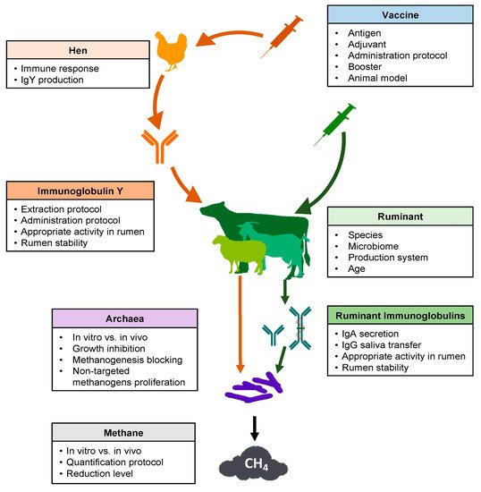
Several key points should be considered in the development of a successful strategy regarding the use of vaccines to reduce methane production from ruminal fermentation (
Figure 1). Many articles and reviews have cited this possibility
[16][17][18][26,30,65]. However, experimental research carried out between 1995 and 2020 was scarce in the consulted database (
Table 1).
Figure 1. Schematic overview of key points to consider for the use of vaccines to decrease methane emissions from ruminal fermentation.
Table 1.
Summary of experimental designs used in research into vaccination for mitigating methane in ruminants.
Several problems arose when comparing studies to assess the possibilities of using vaccines for this purpose. Concerning experimental design, as expected, the chosen antigens have developed along with the new technologies in the last 25 years, from whole methanogen cells to recombinant proteins from specific enzymes involved in CH
4 production. Additionally, the different adjuvants and vaccination protocols used (
Table 1) made it difficult to compare results. For example, Wedlock et al.
[26][53] and Subharat et al.
[27][66] both utilized recombinant glycosyl transferase protein (rGT2) as antigen, but the former with saponins as adjuvant and an intramuscular administration route in sheep as experimental animals, while the second was subcutaneous using Montanide in 5 month old calves. Additionally, those studies evaluated different immunoglobulins (IgG, IgA, and IgY) and samples (blood, saliva, and rumen), or analyzed the effect on CH
4 production using different approaches (in vitro, in vivo).
The most frequently used experimental animal model was the sheep, which was used in 8 out of 11 studies. One of the remaining studies used cattle and another used goats. Finally, a study proposed passive immunization producing antimethanogen Igs in hens. This made it difficult to compare research in order to draw solid conclusions. Patil et al.
[30][67] assayed the immune response of sheep, cattle, and goats against four different serotypes of Foot and mouth disease virus at different times postvaccination. The cows showed higher levels of neutralizing antibodies than small ruminants for all tested virus serotypes. Lobato et al.
[31][68] compared vaccination with recombinant toxin of
Clostridium perfringens in the three common livestock ruminant species. In this study, sheep showed the highest antibody level, cattle the lowest, and goats intermediate. Moreira et al.
[32][69] tested three recombinant vaccines against alpha, beta, and epsilon toxins of
C. perfringens in the same three species. They found an interaction between antigens and species. There were no differences between species, except for with epsilon toxin. In the latter, cattle showed the highest antitoxin levels, with no differences between sheep and goats. In the same way, each species had a different response to each recombinant toxin, whereby all these animals had higher values against beta and lower against alpha toxin. Iqbal et al.
[33][70] observed that ruminal bacterial, methanogen, and protozoal communities were different between cattle and buffalo, although
Methanobrevibacter was the major genus for both species. These studies show that the animal model selected has an interaction with the antigen used. Obviously, small ruminants are cheaper animal models than cattle, and have fast growth and immune maturity. For these reasons, the use of goats and sheep in the early stages of vaccine development is more practical. However, the novel antigen must also be tested in the species for which it is being developed.
Additionally, animal age was another source of variation, with vaccinated sheep ranging from 3–5 months to 5 years old. It is well known that lambs are more susceptible to infectious diseases than adult sheep, and their immune resistance progressively increases during the first year of life
[34][71]. According to Nguyen et al.
[35][72], who compared 3 months old lambs with 2–5 years old sheep following a single intravenous injection of chicken erythrocytes, the adults had higher antibody titers than the young animals. This author affirmed that the antibody response of lambs reached the adult level at age 7–8 months and sex was not a variable that influenced this humoral response. Similarly, Watson et al.
[34][71] assayed the antibody production of weaners and adult sheep against
Brucella abortus. They reported that adults always showed a higher level of antibodies than weaners. Additionally, those authors found that both CD4+ and CD8+ in lymph and blood were higher in adults than in weaners, but B cells are lower in adult than in weaners’ lymph, with no difference in blood between ages. The authors suggested that B cells are not completely functional in younger animals, leading to the lower antibody response. Shu et al.
[36][73] worked on a vaccine against
Streptococcus bovis plus Freund’s adjuvant, reporting a lower antibody concentration than the previous studies in sheep. They tentatively attributed this difference to the age of the animals: 6 months old for Gill et al.
[37][74], 1 year old for Shu et al.
[38][75], and 2 years old in Shu et al.
[36][73], where older animals showed higher antibody levels. However, methanogen vaccines in young animals are a very interesting target, because early programming of rumen microbiota using vaccines could be a better solution in comparison to adult animal vaccines. The rumen microbiota is established early in ruminant life, and it is possible to mold it through diet around weaning time, with a long-lasting effect
[39][76]. De Barbieri et al.
[40][77] found that rumen bacterial communities can change in both mothers and lambs after oral rumen inoculation in the neonatal period or first weeks of life.
The choice of the antigen to be inoculated is a key aspect for the development of a vaccine against methanogenic archaea in the rumen. Different approaches have been used to target methanogens (
Table 1). The first strategy was to vaccinate the animals with whole cells of different archaeal species found in the rumen. In some studies, they specified that the methanogens had previously been killed by formaldehyde
[19][20][21][23][78,79,80,81] or freeze-dried
[22][82]. Baker and Perth
[19][78] used a mix of ten strains of
Methanobrevibacter ruminantium,
M. arboriphilus,
M. smithii,
Methanobacter formicium, and
Methanosarcina barkeri. Wright
[20][79] checked 16S rDNA clone libraries from Australian sheep rumen samples. Based on that information, they chose one vaccine design with three strains of
Methanobrevibacter spp. (two of them isolated in their lab in Australia) and another vaccine with seven strains from the four
Methanobrevibacter species,
Methanomicrobium mobile,
M. barkeri, and
Methanobacterium formicicum. Despite promising results by Wright
[20][79], Clark et al.
[21][80] tried to replicate them using the same mixture of three methanogens, alongside a combination of this mix with methanogenic material isolated from New Zealand sheep. Williams et al.
[23][81] used whole cells of three
Methanobrevibacter strains,
Methanomicrobium mobile, and
Methanosphaera stadtmaniae, which altogether comprised more than half of all the methanogen strains detected. Cook et al.
[22][82] used
Methanobrevibacter ruminantium,
M. smithii, and
Methanosphaera stadtmaniae, each in an independent hen group. They compared the in vitro effect of semipurified IgY and freeze-dried egg yolk from hens vaccinated with each archaeal species and a combination of the three.
Another strategy, derived from the first, was to use cell components as antigens. Wedlock et al.
[24][83] compared the use of whole cells with cytoplasmic and wall-fraction proteins from
M. ruminantium. In parallel, Leahy et al.
[25][84] published the genome sequence of
M. ruminantium; based on this sequence, these researchers chose nine peptides from extracellular regions of the cited archaea. Those peptides were synthesized and joined to keyhole limpet hemocyanin (KHL), to be used as antigens. Later, Wedlock et al.
[26][53] compared cytoplasmic and wall-fraction proteins with seven peptides from the extracellular domain of SecE and rGT2. The latter protein was used by Subharat et al.
[27][66] and Subharat et al.
[29][85] to vaccinate cattle and sheep. Zhang et al.
[28][86] used the protein EhaF from
M. ruminantium M1, which was one of the potential antigen candidates identified by Leahy et al.
[25][84], with a key function in hydrogenotrophic methanogenesis.
Obviously, appropriate adjuvants must be selected for successful vaccine performance. This choice is based mainly on the animal species and antigen used. The experiments compiled herein show how adjuvant use has developed over time, as new experience is acquired. Four out of ten ruminant experiments and the one with hens added complete/incomplete Freund’s adjuvant (FCA/FIA). Another two used saponins, and two recent studies used Montanide ISA. Shu et al.
[36][73] compared the immune response to
S. bovis vaccine with six different adjuvants (FCA, FIA, QuilA, dextran sulphate, alum, Gerbu). They found that FCA produced the largest quantity of blood antibodies in sheep. Using antimethanogen vaccines, two studies compared the efficacy of different adjuvants. Subharat et al.
[29][85] contrasted four adjuvants (saponin, chitosan, lipid nanoparticles, and Montanide ISA). They reported that Montanide ISA61 produced the most IgG and IgA in saliva and serum. Subharat et al.
[27][66] had previously affirmed that this Montanide with and without monophosphoryl lipid A was able to induce a strong humoral response in both IgA and IgG. The most usual administration route was subcutaneous in ruminants (six out of eleven); intramuscular and intradermal were the next most frequently applied in ruminants (both used in two experiments), and Baker and Perth
[19][78] used intraperitoneal. The route in hens was intramuscular in the hen breast. Intramuscular and subcutaneous administration routes were the most common, although it has been suggested that intradermal injection could improve the mucosal response
[41][87]. This is of great interest concerning the present topic. More research is necessary about the antigen–adjuvant–administration route combinations able to achieve a better combined response.
Regarding the booster and booster time, a significant variation in both number and period is shown in
Table 1. Of the vaccination schedules, the most frequently used was one booster (six out of twelve studies) between 21 and 42 days postprimary, followed by two boosters (three out of twelve). The second vaccination given by Wright et al.
[20][79] was not considered a booster because those authors decided to administer it when they observed low antibody levels, and neither was the third vaccination by Subharat et al.
[29][85], since they tested only one group of animals to determine antibody longevity and the effect of boosting. Examining the results, administration of only one or two boosters appears insufficient to provide long-term immunity. For example, Williams et al.
[23][81] reported that one booster 28 days after primary provided a peak at Day 55 after primary, but the titer decreased by Day 99. Using two boosters, Subharat et al.
[29][85] achieved similar results, with a peak at Day 42 after the primary and the titer decreased until Day 133, when the animals were revaccinated and their specific antibodies titers increased. Those results indicate that a booster is necessary to reinforce antibody secretion. None of the other available studies elucidated the issue in this sense, despite this being a very important piece of knowledge to support this procedure for CH
4 mitigation.
The time of sample collection to evaluate the immune response was another source of variation. Some authors decided to take only one sample after vaccination to quantify the specific antibodies
[24][28][83,86], and this did not permit assessment of the specific antibodies’ secretion curves. Therefore, it is not possible to elucidate whether the curves were in their increasing, peak, or decreasing phases. In other studies, which measured immunoglobulins (Igs), the sampling time allowed analysis of the curve and also of the different phases of the antibody curves. Lobato et al.
[31][68] tested a toxin vaccine on sheep, goats, and cattle with a booster on Day 28 after the primary. They reported that no antitoxin antibodies were detected on Day 0. On Day 42, 40% of goats, 60% of sheep, and 80% of cattle had titers lower than 1 IU/mL. On Day 56, all animals had titers equal to or higher than 5.8 IU/mL; sheep had the highest values, followed by goats and cattle.

