You're using an outdated browser. Please upgrade to a modern browser for the best experience.
Mesenchymal Stem Cells Studies in the Goat Model: Comparison
Please note this is a comparison between Version 2 by Camila Xu and Version 1 by Isabel R. Dias.

This topic aims to compile the works published in the scientific literature, over the last two decades, that use the goat as an animal model in preclinical studies using stem cells, alone or associated with biomaterials, for the treatment of injury or disease in divers organ systems. Stem cells can be classified as embryonic stem cells (ESCs), induced pluripotent stem cells (iPSCs), and mesenchymal stem cells (MSCs), which differ in origin, plasticity, differentiation potential, and risk of tumorigenesis. MSCs have been the most studied cells, with excellent and safe results in multiple areas. In the area of tissue engineering and regenerative medicine, the caprine model is particularly used in studies using stem cells in the musculoskeletal system but, although in a more limited way, also in the field of dermatology, ophthalmology, dentistry, pneumology, cardiology, and urology.

  • mesenchymal stem cells
  • caprine model
  • MSCs

1. Introduction

Research in stem cell therapy continues to grow, with promising results and increasing expectations in the scientific community. Stem cells can be classified as embryonic stem cells (ESCs), induced pluripotent stem cells (iPSCs), and mesenchymal stem cells (MSCs), which differ in origin, plasticity, differentiation potential, and risk of tumorigenesis [1,2,3,4,5][1][2][3][4][5]. MSCs have been the most studied cells, with excellent and safe results in multiple areas. The therapy relies on the anti-inflammatory, immunosuppressive and regenerative properties of these cells [1,6][1][6]. Nowadays, different diseases in humans and animals have already been treated with cell-based therapies, mainly to regenerate damaged tissue or reduce inflammation. Nevertheless, there are still some concerns regarding the use of this stem cell therapy and unwanted side effects due to the migration of transplanted cells, as well as insufficient cell survival upon transplantation [7,8][7][8]. A solution to this problem might lie in the improvement of animal models and the selection of appropriate methods for grafting and transplantation, facilitating the eventual use of stem cells in clinical practice [8,9,10,11,12][8][9][10][11][12].
Animal models have been increasingly relevant for human medicine, namely to understand the pathophysiology of diseases and for the development and testing of new therapies [13,14][13][14]. The most frequently used animal models are represented by rodents (mice, rats, hamsters, guinea pigs) and lagomorphs (rabbits). Relatively to laboratory animal models, they are generally used and highly contribute to pilot and proof of concept in biomedical research studies. However, they have some important limitations, since they differ substantially from humans in body, organ size, and life expectancy, resulting in huge metabolic, physiological and behavioral differences [13,14][13][14]. To overcome this issue, after the first studies were carried out on laboratory animals and to complement them, other larger domestic animal species have been used, such as dogs, pigs, small ruminants (sheep, goats), and horses [14], with promising results. These preclinical translational studies’ results are closer to those expected in human patients. The term “large animal” is used to define animal species that are not included in the laboratory animal models, such as rodents and rabbits [14,15][14][15]. As gene sequencing and manipulation tools are now accessible to farm animals (cattle, pigs, sheep, and goats), genetically modified livestock models are available for biomedical research [13].
The use of large animals in these research studies presents great advantages related to their anatomy and dimensions, as they are larger than conventional laboratory animals, being quite similar to humans in most physiological systems [9,16][9][16]. Particularly, regarding small ruminants, they are recognized animal models for preclinical and translational studies for human biomedical research. They have the great advantage of being cooperative and compliant, easily available, easy to handle and house, and relatively low cost. They also do not require excessive care with their feeding and sanitary prophylaxis. Their physical stature and weight allow biomedical research to be conducted in more realistic clinical situations. They are livestock species, so their use in experimental research is relatively well accepted by the general society. Sheep have been widely used for biomedical research purposes, resulting in numerous scientific articles and very recent reviews. Comparatively, the goat remains largely unexplored in terms of scientific publications. In biomedical research the goat model has been used mainly for the study of human diseases (chronic rheumatoid arthritis, congenital myotonia, cardiovascular conditions such as atrial fibrillation, and Q fever), as antibody producer (immunological research, immunotherapy, diagnosis), orthopedic, reproduction and cancer research, genome engineering for the production of pharmaceutical proteins, generation of models of human diseases and hosts for the growth of human organs, among other fields. Small ruminants (mainly sheep and goats) have recently been used, namely for regenerative therapy studies with stem cells, in orthopedic and myocardial infarction research [17,18,19][17][18][19]. Nevertheless, large animals still represent less than 0.5% of animals used in stem cell research, mainly due to poor knowledge of the processes responsible for maintaining the pluripotency of stem cells [20].

2. Overview of Mesenchymal Stem Cells

Mesenchymal stem cells (MSCs) are undifferentiated cells of non-hematopoietic origin, with self-renewal capacity, located in various adult or extra-embryonic tissues. These cells are classified as multipotential, meaning they are capable to differentiate into multiple cell lines, namely cardiomyocytes, chondroblasts, endothelial cells, hepatocytes, myocytes, neuronal cells, osteoblasts, and tenocytes, among others [1,2][1][2]. MSCs can be obtained from embryonic cells in the first stages of embryo development before its implantation in the uterus, and in adults, they can be isolated from various tissues, such as umbilical cord, placenta, amnion, bone marrow (BMSCs), adipose tissue (ASCs), dental pulp and periosteum [37,38,39][21][22][23]. The diversity of sources of stem cells or MSCs and the wide range of potential applications of these cells lead to a challenge in selecting a suitable cell type for cell therapy [8]. In veterinary medicine, MSCs are collected mainly from bone marrow (BM) or adipose tissue (AT), since these tissues are easier to obtain [2,4,40][2][4][24]. Depending on how MSCs are obtained, therapy with these cells can be autologous, if they are obtained from the same animal, allogeneic if the donor is another individual from the same species, or xenogeneic if the donor is from a different species. Regarding the route of administration, they can be applied locally, systemically, generally intravenously, or both, depending on the disease [37][21]. These cells have the ability to differentiate into the target cell type allowing the regeneration of the injured area and also have great immunomodulatory potential. This immunomodulation is due to paracrine effects (by secreting different cytokines and growth and differentiation factors (GDFs) to adjacent cells) and by cell-to-cell contact, leading to vascularization, cell proliferation in injured tissues, and reducing inflammation [1,6,37,41][1][6][21][25]. Different diseases in animals have been treated using cell-based therapy, mainly to regenerate damaged tissue or reduce inflammation. These cells are considered “immune privileged” as they do not express histocompatibility complex class II (MHC-II) and costimulatory molecules (such as CD40, CD80, and CD86), allowing allogeneic therapy, as they escape the recognition and action of T cells and NK receptors [1,2,6,42][1][2][6][26]. MSCs can be applied in a wide range of clinical specialties, such as traumatology and orthopedic, ophthalmology, neurology, internal medicine, dermatology, and immunopathology, among others. Figure 1 shows the medical specialties studied for MSCs therapy in the goat model. The applications of these therapies in the veterinary medicine field include not only their clinical use in domestic animals, but also the translation of the results, as a preclinical model, for human medicine [14]. These cells show great resistance to cryopreservation, allowing the creation of cell banks for later use and the selection of the best donors by previously evaluating their MSCs in vitro [2].
Figure 1.
Clinical applications of MSCs studied in the goat model, according to medical specialties.

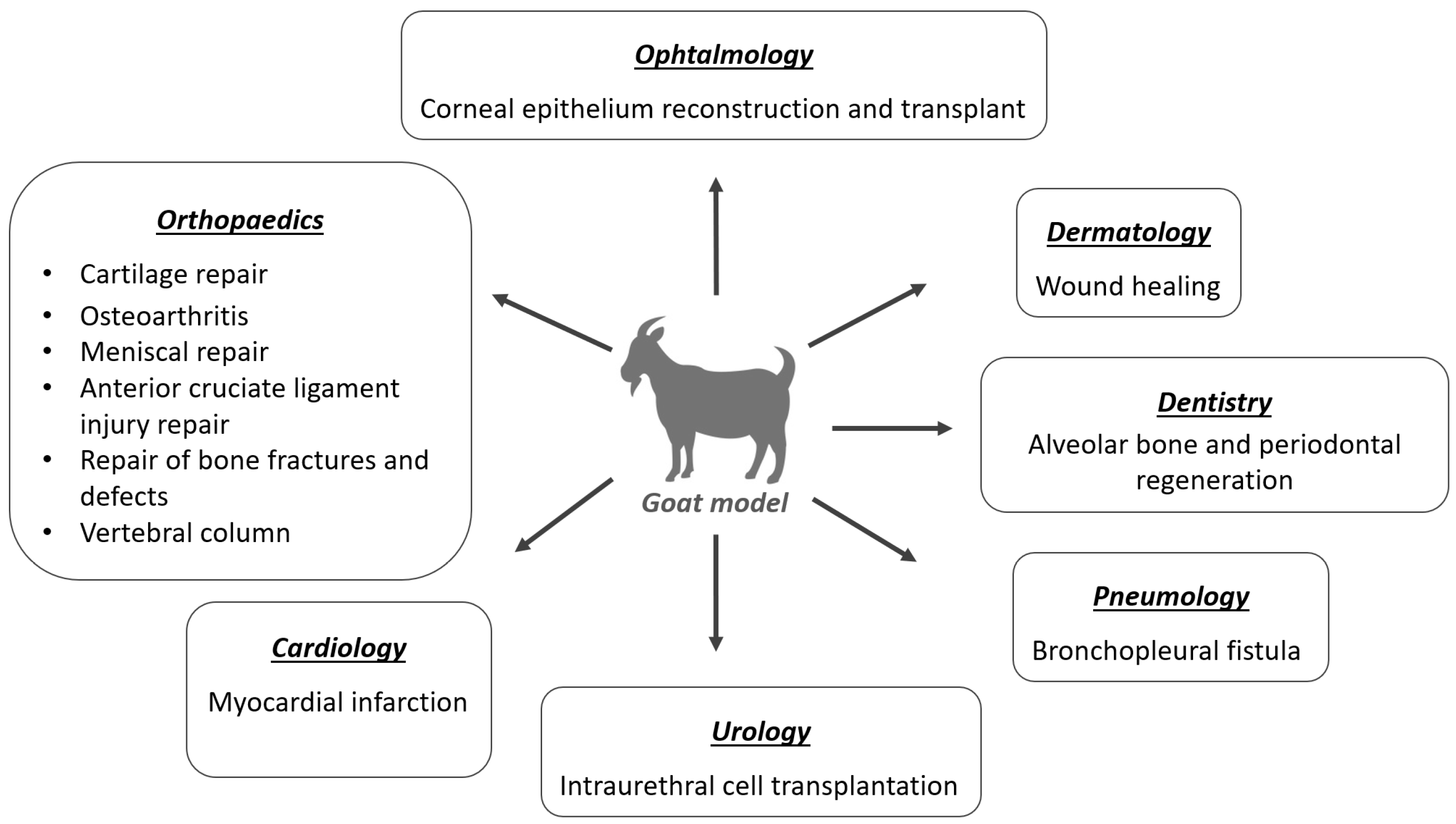
3. Application of MSCs in the Goat Model

3.1. Orthopedics

MSCs are an alternative source of cells for cartilage, muscle, tendon, and bone regeneration, as they are easily accessible and can be harvested from different tissues, having a great capacity to proliferate and differentiate into other sorts of cells in the body, such as osteoblasts and chondroblasts [37,46,47][21][27][28]. Caprine models have been used in preclinical and translational research studies to assess new approaches, resorting to MSCs for cartilage tissue engineering aiming for regenerative joint resurfacing, osteoarthritis treatments, bone fractures and defects, menisci and anterior cruciate ligament injury repair studies. Additionally, several studies have been performed on the goat at the spine level, namely for research on new treatments for intervertebral disc (IVD) disease.

3.2. Dermatology

Chronic cutaneous wounds and ulcers represent a therapeutic challenge, due to the difficulty of clinical management, high recumbence rate, and scar formation, both in human and veterinary medicine. Its incidence has increased due to population aging, diabetes, obesity, and concomitant diseases [2].
MSCs present an important role in all phases of tissue repair: inflammation, proliferation, and remodeling [2,3][2][3]. These cells also promote angiogenesis and show evidence of antimicrobial properties. A failure in the angiogenesis process during wound healing can induce the development of chronic wounds [2,3][2][3]. MSCs have already been shown to promote wound healing through a paracrine molecular cascade in goat models [128,129][29][30]. Furthermore, MSCs may have direct involvement in various stages of the wound healing process that need to be further explored [129][30].
Although goats have not generally been used as research models for cutaneous wound healing, they are a good choice as they have a mild temperament and a good anatomical skin surface available for creating lesion models of different shapes and sizes [16].
Studies with the goat model for wound healing have used different sources of MSCs with excellent results. Pratheesh et al. (2017) showed evidence of better wound healing with MSCs from the amniotic fluid origin than caprine BMSCs, by revealing greater epithelialization, neovascularization, and collagen development in the histomorphometric analysis [130][31]. Azari et al. (2011) also showed the re-epithelization capacity of transplanted Wharton’s jelly MSCs from caprine umbilical cords, revealing complete re-epithelization of cutaneous wounds in 7 days [131][32].
In addition, MSCs showed great capacity for wound regeneration and reduced healing time and plasticity, as they are capable of converting into cells of different tissues. A study carried out by Yang et al. (2007) with goats confirmed that epidermal adult stem cells can differentiate into different functional cells in vivo or in vitro, demonstrating the plasticity of stem cells [132][33].

3.3. Ophthalmology

In recent years, ophthalmologists have placed a great focus on stem cells to treat various traumatic and degenerative disorders due to their unique biologic properties [133][34]. The cornea is a protective barrier and is formed by three layers with different germinal origins: the epithelium (originated from superficial ectoderm) and the stroma and endothelium (originated from neural crest cells) [134][35]. Experimental studies have proved that there is a variety of stem cells present in each of these layers [134,135][35][36]. For example, limbal stem cells (residing in the limbus) maintain epithelial homeostasis and regenerate the cornea, with epithelial cell deficiency being the leading cause of blindness worldwide [134][35].
Stem cells have high potential in the treatment of eye diseases characterized by permanent cell loss, such as glaucoma, age-related macular degeneration, photoreceptor cell degeneration, hereditary retinopathy, and mechanical and ischemic retinal injuries [134,136,137][35][37][38]. The eyes of small ruminants are anatomically different from human eyes [16,138][16][39], however the resemblance in structure, size with, some properties and parameters to the human eye made possible to use these models successfully. Goats have been used mostly to study corneal epithelium reconstruction and transplant.
Studies have shown that epidermal adult stem cells (EpiASCs) from goat ear skin can be used to successfully repair damaged cornea with total limbal stem cells (LSCs) deficiency [139,140][40][41]. Moreover, these results demonstrated that EpiASCs can be induced to differentiate into corneal epithelial cell types in vivo in a corneal microenvironment, and had the skill to trigger corneal genetic programs [139][40].
In a study carried out by Mi et al. (2008), cryopreserved limbal corneal stem cells were applied in goats with damaged cornea with excellent results. The therapeutic effect of transplantation may be associated with the inhibition of inflammation-related angiogenesis after transplantation of cryopreserved LSCs [141][42].

3.4. Dentistry

There are only a few published studies with goats for stem cell therapy in dentistry, probably due to the anatomical differences between human and goat dentition. Because it is a ruminant species, it only has incisors in the mandible, being absent in the upper jaw. It has no canines and only premolars and molars, the latter fulfills the function of rumination of plant foods. Stem cell research in dentistry aims at the regeneration of damaged tissues such as periodontal tissues, dentin, pulp, and resorbed roots and the repair of endodontic iatrogenic perforations [142][43]. It is mainly used for periodontal regeneration and in association with biomaterials to optimize tooth regeneration in the goat model [143,144][44][45]. Undifferentiated MSCs are able to differentiate, providing the three critical tissues essential for periodontal tissue regeneration: cementum, bone, and periodontal ligaments, making stem cells a new approach for periodontal tissue regeneration [145][46]. The association of MSCs and fibrin glue using particulate mineralized bone has also shown promising results for vertical bone augmentation in animal models [146][47].
There is a growing need to use dental implants and improve their function to enhance normal dental physiology and proprioception [142][43]. An osseointegrated implant can closely resemble a natural tooth. Nevertheless, the absence of periodontal ligament and connective tissue results in important differences in implant adaptation to occlusal forces [145][46]. Several studies for tooth regeneration with scaffolds based on biomaterials and stem cells have shown very positive effects on regeneration [143,144,145,147][44][45][46][48]. Dense collagen gel scaffolds seeded on MSCs and nanostructured titanium surfaces have increased interest in bone regeneration due to the good osteointegration effect [148,149][49][50].
The placement of implants can be problematic, as, after a tooth loss, anatomical pneumatization of the maxillary sinuses can occur, as well as atrophy of the alveolar ridge, limiting the bone volume available to the implant placement. Maxillary sinus floor elevation is one of the preferred surgery options to solve this problem, where bone graft material is placed in the maxillary sinus to provide adequate support to the implants [143][44]. Zou et al. (2012) associated this grafting material with BMSCs and calcium phosphate cementum, promoting earlier bone formation and mineralization, and maintaining the height and volume of the augmented maxillary sinus in a goat model [143][44].
Bangun et al. (2021) proved that MSCs can improve earlier bone repopulation and complete faster bone regeneration in tissue-engineered bone grafts, supported by the paracrine activity of the resident stem cells [144][45].
It is also known that TGF-β1 plays an important role during tooth formation, this GDF can directly induce the differentiation of odontoblast-like cells, and positively regulate the secretion of matrix components in the dentin-pulp complex, being a potential therapy to induce tissue formation after dental pulp capping treatments [150][51].

3.5. Pneumology

Stem cells have been a promising therapy for asthma non-responsive to conventional therapy [1,42][1][26], as a potential treatment for destructive lung diseases including chronic obstructive pulmonary disease [151][52], and as a treatment for bronchopleural fistula (BPF), due to its plasticity and ability to differentiate into different cells [152][53].
Goats have been successfully used in a bronchopleural fistula model. In cases of lung cancer with limited disease, the most effective method of controlling the primary tumor is surgical resection, as it offers the best chance of cure. Pulmonary resection can lead to the development of a pathological connection between the airway (bronchus) and the pleural space, known as a post-resection BPF [152][53]. This research proved that bronchoscopic-guided transplantation of BMSCs successfully closes bronchopleural fistula by extraluminal fibroblast proliferation and collagenous matrix development [152][53].

3.6. Cardiology

The goat seems to be the ideal model for cardiovascular diseases, due to its anatomical dimensions and physiological similarities to the human heart. Additionally, parameters such as heart rate, coronary architecture, and capillary density are more similar to those of man [9,14][9][14].
One of the main causes of morbidity and mortality in cardiovascular diseases is myocardial infarction, characterized by the ischemic lesions of cardiac muscle tissue due to an occlusion of one of the coronary arteries or one of its branches by a thrombus [17]. This disease is increasing and gaining more importance due to the general aging of the population and the change in lifestyle [17,18][17][18].
The goat has been studied for the treatment of myocardial infarction through in vitro and in vivo studies. So far, several different sources of adult stem cells have been identified as a healing approach for infarcted myocardium. The most promising stem cells that have so far shown the best results in these studies are BMSCs. A study carried out by Liao et al. (2006) uses BMSCs enriched by small intestinal submucosal (SIS) films to treat myocardial infarction in goat models, the MSCs-SIS film was implanted and sutured in the infarct area. The obtained results revealed that this therapy can prevent ventricular chamber dilatation and can improve myocardium contractility, cardiac function, and collateral perfusion [18].
Another promising source of stem cells is glandular stem cells that can be easily extracted from exocrine glands, such as the salivary glands or the pancreas [17,153,154][17][54][55]. Maass et al. (2009) showed that glandular stem cells obtained from the submandibular gland can spontaneously differentiate into cardiac-like mesodermal cells in vitro. These results suggest that implanting these cells directly into infarcted myocardium can improve heart regeneration [17].

3.7. Urology

Cell-based therapy is emerging as a great alternative in the treatment of stress urinary incontinence. Goats are also an ideal model to study this subject, as the female caprine urethra has similar parameters to those reported in humans, by measuring the urethral pressure profile, making them a suitable experimental animal for testing intraurethral cell transplantation [155][56].
Burdzinska et al. (2017) demonstrated that caprine muscle-derived cells (MDCs) and MSCs can be expanded in vitro and applied for intraurethral injections [155][56]. In 2018, Burdzinska proved that both MSCs and MDCs collaborated in the formation of striated muscle when they were transplanted directly into the external urethral sphincter. This restudyearch suggests that MDC-MSC co-transplantation improves urethral closure better than if transplantation of each cell population is performed alone [156][57].

References

  1. Dias, I.E.; Pinto, P.O.; Barros, L.C.; Viegas, C.A.; Dias, I.R.; Carvalho, P.P. Mesenchymal stem cells therapy in companion animals: Useful for immune-mediated diseases? BMC Vet. Res. 2019, 15, 358.
  2. Jiménez, A.; Guerrero, F. Células madre mesenquimales como nueva terapia en dermatología: Conceptos básicos. Rev. Clínica Dermatol. Vet. 2017, 9, 8–18.
  3. Ogliari, K.S.; Marinowic, D.; Brum, D.E.; Loth, F. Stem cells in dermatology. An. Bras. Dermatol. 2014, 89, 286–291.
  4. Arnhold, S.; Elashry, M.I.; Klymiuk, M.C.; Wenisch, S. Biological macromolecules and mesenchymal stem cells: Basic research for regenerative therapies in veterinary medicine. Int. J. Biol. Macromol. 2019, 123, 889–899.
  5. Spencer, N.D.; Gimble, J.M.; Lopez, M.J. Mesenchymal stromal cells: Past, present, and future. Vet. Surg. 2011, 40, 129–139.
  6. Peroni, J.F.; Borjesson, D.L. Anti-Inflammatory and Immunomodulatory Activities of Stem Cells. Vet. Clin. N. Am.-Equine Pract. 2011, 27, 351–362.
  7. Mastrolia, I.; Foppiani, E.M.; Murgia, A.; Candini, O.; Samarelli, A.V.; Grisendi, G.; Veronesi, E.; Horwitz, E.M.; Dominici, M. Challenges in Clinical Development of Mesenchymal Stromal/Stem Cells: Concise Review. Stem Cells Transl. Med. 2019, 8, 1135–1148.
  8. Rajabzadeh, N.; Fathi, E.; Farahzadi, R. Stem cell-based regenerative medicine. Stem Cell Investig. 2019, 6, 18.
  9. Harding, J.; Roberts, R.M.; Mirochnitchenko, O. Large animal models for stem cell therapy. Stem Cell Res. Ther. 2013, 4, 23.
  10. Gugjoo, M.B.; Amarpal; Fazili, M.U.R.; Shah, R.A.; Mir, M.S.; Sharma, G.T. Goat mesenchymal stem cell basic research and potential applications. Small Rumin. Res. 2020, 183, 106045.
  11. Almarza, A.J.; Brown, B.N.; Arzi, B.; Ângelo, D.F.; Chung, W.; Badylak, S.F.; Detamore, M. Preclinical Animal Models for Temporomandibular Joint Tissue Engineering. Tissue Eng.-Part B Rev. 2018, 24, 171–178.
  12. Harness, E.M.; Mohamad-Fauzi, N.B.; Murray, J.D. MSC therapy in livestock models. Transl. Anim. Sci. 2022, 6, txac012.
  13. Polejaeva, I.A.; Rutigliano, H.M.; Wells, K.D. Livestock in biomedical research: History, current status and future prospective. Reprod. Fertil. Dev. 2016, 28, 112–124.
  14. Hotham, W.E.; Henson, F.M.D. The use of large animals to facilitate the process of MSC going from laboratory to patient—‘Bench to bedside’. Cell Biol. Toxicol. 2020, 36, 103–114.
  15. Thomas, B.; Bhat, K.; Mapara, M. Rabbit as an animal model for experimental research. Dent. Res. J. 2012, 9, 111.
  16. Alvites, R.D.; Branquinho, M.V.; Sousa, A.C.; Lopes, B.; Sousa, P.; Mendonça, C.; Atayde, L.M.; Maurício, A.C. Small ruminants and its use in regenerative medicine: Recent works and future perspectives. Biology 2021, 10, 249.
  17. Maass, A.; Kajahn, J.; Guerleyik, E.; Guldner, N.W.; Rapoport, D.H.; Kruse, C. Towards a pragmatic strategy for regenerating infarcted myocardium with glandular stem cells. Ann. Anat. 2009, 191, 51–61.
  18. Liao, B.; Deng, L.; Wang, F. Effects of bone marrow mesenchymal stem cells enriched by small intestinal submucosal films on cardiac function and compensatory circulation after myocardial infarction in goats. Chin. J. Reparative Reconstr. Surg. 2006, 20, 1248–1252.
  19. An, Y.H.; Friedman, R.J. Animal Selections in Orthopaedic Research. In Animal Models in Orthopaedic Research; Routledge: London, UK, 1999; pp. 39–57. ISBN 9780429173479.
  20. Madeja, Z.E.; Pawlak, P.; Piliszek, A. Beyond the mouse: Non-rodent animal models for study of early mammalian development and biomedical research. Int. J. Dev. Biol. 2019, 63, 187–201.
  21. Dias, I.E.; Cardoso, D.F.; Soares, C.S.; Barros, L.C.; Viegas, C.A.; Carvalho, P.P.; Dias, I.R. Clinical application of mesenchymal stem cells therapy in musculoskeletal injuries in dogs—A review of the scientific literature. Open Vet. J. 2021, 11, 188–202.
  22. Klingemann, H.; Matzilevich, D.; Marchand, J. Mesenchymal stem cells—Sources and clinical applications. Transfus. Med. Hemotherapy 2008, 35, 272–277.
  23. Webster, R.A.; Blaber, S.P.; Herbert, B.R.; Wilkins, M.R.; Vesey, G. The role of mesenchymal stem cells in veterinary therapeutics—A review. N. Zeal. Vet. J. 2012, 60, 265–272.
  24. Ayala-Cuellar, A.P.; Kang, J.; Jeung, E.; Choi, K. Roles of Mesenchymal Stem Cells in Tissue Regeneration and Immunomodulation. Biomol. Ther. 2019, 27, 25–33.
  25. Torres-Torrillas, M.; Rubio, M.; Damia, E.; Cuervo, B.; del Romero, A.; Peláez, P.; Chicharro, D.; Miguel, L.; Sopena, J.J. Adipose-derived mesenchymal stem cells: A promising tool in the treatment of musculoskeletal diseases. Int. J. Mol. Sci. 2019, 20, 3105.
  26. Quimby, J.M.; Borjesson, D.L. Mesenchymal stem cell therapy in cats: Current knowledge and future potential. J. Feline Med. Surg. 2018, 20, 208–216.
  27. Freitag, J.; Bates, D.; Boyd, R.; Shah, K.; Barnard, A.; Huguenin, L.; Tenen, A. Mesenchymal stem cell therapy in the treatment of osteoarthritis: Reparative pathways, safety and efficacy—A review. BMC Musculoskelet. Disord. 2016, 17, 230.
  28. Reissis, D.; Tang, Q.O.; Cooper, N.C.; Carasco, C.F.; Gamie, Z.; Mantalaris, A.; Tsiridis, E. Current clinical evidence for the use of mesenchymal stem cells in articular cartilage repair. Expert Opin. Biol. Ther. 2016, 16, 535–557.
  29. Shukla, A.; Choudhury, S.; Chaudhary, G.; Singh, V.; Prabhu, S.N.; Pandey, S.; Garg, S.K. Chitosan and gelatin biopolymer supplemented with mesenchymal stem cells (Velgraft®) enhanced wound healing in goats (Capra hircus): Involvement of VEGF, TGF and CD31. J. Tissue Viability 2021, 30, 59–66.
  30. Pratheesh, M.D.; Gade, N.E.; Nath, A.; Dubey, P.K.; Sivanarayanan, T.B.; Madhu, D.N.; Sreekumar, T.R.; Amarpal; Saikumar, G.; Sharma, G.T. Evaluation of persistence and distribution of intra-dermally administered PKH26 labelled goat bone marrow derived mesenchymal stem cells in cutaneous wound healing model. Cytotechnology 2017, 69, 841–849.
  31. Pratheesh, M.D.; Dubey, P.K.; Gade, N.E.; Nath, A.; Sivanarayanan, T.B.; Madhu, D.N.; Somal, A.; Baiju, I.; Sreekumar, T.R.; Gleeja, V.L.; et al. Comparative study on characterization and wound healing potential of goat (Capra hircus) mesenchymal stem cells derived from fetal origin amniotic fluid and adult bone marrow. Res. Vet. Sci. 2017, 112, 81–88.
  32. Azari, O.; Babaei, H.; Derakhshanfar, A.; Nematollahi-Mahani, S.N.; Poursahebi, R.; Moshrefi, M. Effects of transplanted mesenchymal stem cells isolated from Wharton’s jelly of caprine umbilical cord on cutaneous wound healing; Histopathological evaluation. Vet. Res. Commun. 2011, 35, 211–222.
  33. Yang, X.; Qu, L.; Wang, X.; Zhao, M.; Li, W.; Hua, J.; Shi, M.; Moldovan, N.; Wang, H.; Dou, Z. Plasticity of Epidermal Adult Stem Cells Derived from Adult Goat Ear Skin. Mol. Reprod. Dev. 2007, 74, 386–396.
  34. Holan, V.; Trosan, P.; Cejka, C.; Javorkova, E.; Zajicova, A.; Hermankova, B.; Chudickova, M.; Cejkova, J. A Comparative Study of the Therapeutic Potential of Mesenchymal Stem Cells and Limbal Epithelial Stem Cells for Ocular Surface Reconstruction. Stem Cells Transl. Med. 2015, 4, 1052–1063.
  35. Zakirova, E.Y.; Valeeva, A.N.; Aimaletdinov, A.M.; Nefedovskaya, L.V.; Akhmetshin, R.F.; Rutland, C.S.; Rizvanov, A.A. Potential therapeutic application of mesenchymal stem cells in ophthalmology. Exp. Eye Res. 2019, 189, 107863.
  36. Amano, S.; Yamagami, S.; Mimura, T.; Uchida, S.; Yokoo, S. Corneal Stromal and Endothelial Cell Precursors. Cornea 2006, 25, S73–S77.
  37. Joe, A.W.; Gregory-Evans, K. Mesenchymal stem cells and potential applications in treating ocular disease. Curr. Eye Res. 2010, 35, 941–952.
  38. Öner, A. Stem cell treatment in retinal diseases: Recent developments. Turk. J. Ophthalmol. 2018, 48, 33–38.
  39. Banks, M.S.; Sprague, W.W.; Schmoll, J.; Parnell, J.A.Q.; Love, G.D. Why do animal eyes have pupils of different shapes? Sci. Adv. 2015, 1, e1500391.
  40. Zhang, P.; Ma, X.-Y.; Huang, D.-T.; Yang, X.-Y. The capacity of goat epidermal adult stem cells to reconstruct the damaged ocular surface of total LSCD and activate corneal genetic programs. J. Mol. Histol. 2020, 51, 277–286.
  41. Yang, X.; Moldovan, N.I.; Zhao, Q.; Mi, S.; Zhou, Z.; Chen, D.; Gao, Z.; Tong, D.; Dou, Z. Reconstruction of damaged cornea by autologous transplantation of epidermal adult stem cells. Mol. Vis. 2008, 14, 1064–1074.
  42. Mi, S.; Yang, X.; Zhao, Q.; Qu, L.; Chen, S.; Meek, K.M.; Dou, Z. Reconstruction of corneal epithelium with cryopreserved corneal limbal stem cells in a goat model. Mol. Reprod. Dev. 2008, 75, 1607–1616.
  43. Bansal, R.; Jain, A. Current overview on dental stem cells applications in regenerative dentistry. J. Nat. Sci. Biol. Med. 2015, 6, 29–34.
  44. Zou, D.; Guo, L.; Lu, J.; Zhang, X.; Wei, J.; Liu, C.; Zhang, Z.; Jiang, X. Engineering of bone using porous calcium phosphate cement and bone marrow stromal cells for maxillary sinus augmentation with simultaneous implant placement in goats. Tissue Eng.-Part A 2012, 18, 1464–1478.
  45. Bangun, K.; Sukasah, C.L.; Dilogo, I.H.; Indrani, D.J.; Siregar, N.C.; Pandelaki, J.; Iskandriati, D.; Kekalih, A.; Halim, J. Bone Growth Capacity of Human Umbilical Cord Mesenchymal Stem Cells and BMP-2 Seeded into Hydroxyapatite/Chitosan/Gelatin Scaffold in Alveolar Cleft Defects: An Experimental Study in Goat. Cleft Palate-Craniofacial J. 2021, 58, 707–717.
  46. Marei, M.K.; Saad, M.M.; El-ashwah, A.M.; El-backly, R.M.; Al-khodary, M.A. Experimental Formation of Periodontal Structure around Titanium Implants Utilizing Bone Marrow Mesenchymal Stem Cells: A Pilot Study. J. Oral Implant. 2009, 35, 106–129.
  47. Khojasteh, A.; Eslaminejad, M.B.; Nazarian, H.; Morad, G.; Dashti, S.G.; Behnia, H.; Stevens, M. Vertical bone augmentation with simultaneous implant placement using particulate mineralized bone and mesenchymal stem cells: A preliminary study in rabbit. J. Oral Implantol. 2013, 39, 3–13.
  48. Chang, C.C.; Lin, T.A.; Wu, S.Y.; Lin, C.P.; Chang, H.H. Regeneration of Tooth with Allogenous, Autoclaved Treated Dentin Matrix with Dental Pulpal Stem Cells: An In Vivo Study. J. Endod. 2020, 46, 1256–1264.
  49. Kunrath, M.; dos Santos, R.; de Oliveira, S.; Hubler, R.; Sesterheim, P.; Teixeira, E. Osteoblastic Cell Behavior and Early Bacterial Adhesion on Macro-, Micro-, and Nanostructured Titanium Surfaces for Biomedical Implant Applications. Int. J. Oral Maxillofac. Implant. 2020, 35, 773–781.
  50. Chamieh, F.; Collignon, A.M.; Coyac, B.R.; Lesieur, J.; Ribes, S.; Sadoine, J.; Llorens, A.; Nicoletti, A.; Letourneur, D.; Colombier, M.L.; et al. Accelerated craniofacial bone regeneration through dense collagen gel scaffolds seeded with dental pulp stem cells. Sci. Rep. 2016, 6, 38814.
  51. Zhang, W.; Walboomers, X.F.; Jansen, J.A. The formation of tertiary dentin after pulp capping with a calcium phosphate cement, loaded with PLGA microparticles containing TGF-β1. J. Biomed. Mater. Res.-Part A 2008, 85, 439–444.
  52. Hind, M.; Maden, M. Is a regenerative approach viable for the treatment of COPD? Br. J. Pharmacol. 2011, 163, 106–115.
  53. Petrella, F.; Toffalorio, F.; Brizzola, S.; De Pas, T.M.; Rizzo, S.; Barberis, M.; Pelicci, P.; Spaggiari, L.; Acocella, F. Stem cell transplantation effectively occludes bronchopleural fistula in an animal model. Ann. Thorac. Surg. 2014, 97, 480–483.
  54. Seaberg, R.M.; Smukler, S.R.; Kieffer, T.J.; Enikolopov, G.; Asghar, Z.; Wheeler, M.B.; Korbutt, G.; Van Der Kooy, D. Clonal identification of multipotent precursors from adult mouse pancreas that generate neural and pancreatic lineages. Nat. Biotechnol. 2004, 22, 1115–1124.
  55. Kruse, C.; Birth, M.; Rohwedel, J.; Assmuth, K.; Goepel, A.; Wedel, T. Pluripotency of adult stem cells derived from human and rat pancreas. Appl. Phys. A Mater. Sci. Process. 2004, 79, 1617–1624.
  56. Burdzinska, A.; Dybowski, B.; Zarychta-Wisniewska, W.; Kulesza, A.; Zagozdzon, R.; Gajewski, Z.; Paczek, L. The Anatomy of Caprine Female Urethra and Characteristics of Muscle and Bone Marrow Derived Caprine Cells for Autologous Cell Therapy Testing. Anat. Rec. 2017, 300, 577–588.
  57. Burdzinska, A.; Dybowski, B.; Zarychta-Wiśniewska, W.; Kulesza, A.; Butrym, M.; Zagozdzon, R.; Graczyk-Jarzynka, A.; Radziszewski, P.; Gajewski, Z.; Paczek, L. Intraurethral co-transplantation of bone marrow mesenchymal stem cells and muscle-derived cells improves the urethral closure. Stem Cell Res. Ther. 2018, 9, 239.
More
Academic Video Service