Tumour onset and development occur because of specific immune support. The immune system, which is originally able to perceive and eliminate incipient cancer cells, becomes suppressed and hijacked by cancer. For these purposes, tumour cells use extracellular vesicles (TEVs). Specific molecular composition allows TEVs to reprogram immune cells towards tumour tolerance. Circulating TEVs move from their site of origin to other organs, preparing “a fertile soil” for metastasis formation.
- extracellular vesicles
- exosomes
- tumour immune editing
1. Introduction
2. TEV and CTL Reprogramming
In healthy subjects, differentiated tumour-specific effector CTL enter the circulation, infiltrate the tumour, and attack tumour cells by interacting with a specific tumour antigen. CTL cytotoxicity is mediated by the release of pro-inflammatory cytokines, perforin and granzymes, or by direct contact with apoptosis-inducing molecules expressed by tumour cells [8,9][8][9]. Tumour antigen presentation by APC is crucial for CTL differentiation, while insufficient to sustain the cytotoxic immune response [10]. To drive specific antitumour activity, T-cells must be activated by “mature” APC. During differentiation, APC “capture” invading pathogens [11[11][12],12], neo-antigens [13], apoptotic cells [14] and EV [15]. These signals dictate APC specification towards pro-inflammatory or tolerant phenotypes, resulting in a specific T-cell commitment [16]. Since the role of DC in orchestrating the immune response has been extensively investigated, the mechanisms driving tumour tolerance described herein mainly refer to DC-mediated T-cell differentiation. The high expression of MHC, CCR7, co-stimulatory factors CD80, CD83 and CD86 identifies pro-inflammatory DC, also named “mature” DC [17,18,19][17][18][19]. Conversely, the low expression of these markers defines “immature” DC, which maintain the immune tolerance, suppress immune cells-mediated attack, and prevent autoimmunity. Physiologically, “immature” DC differentiation occurs during apoptosis, since apoptotic cells inhibit the expression of co-stimulatory molecules [14,20,21,22,23,24][14][20][21][22][23][24]. TEV hold endogenous tumour antigens; therefore, they can be considered for potential use in an anticancer vaccine [25]. However, they mainly act as immune suppressors. Several studies have demonstrated that in vivo TEV are mostly internalised by myeloid cells [26,27,28][26][27][28]. Once stimulated with TEV, DC, displaying an immature phenotype, prevent the immune attack and stimulate the expression of immune checkpoint molecules [7,27,29,30][7][27][29][30]. For example, Cytotoxic T Lymphocyte Antigen-4 (CTLA4) and Programmed cell Death protein 1 (PD-1) induce CTL anergy or promote regulatory T-cell differentiation (Treg) [31,32,33,34,35,36][31][32][33][34][35][36]. DC interact with thousands of T cells per hour [37], while their MHC+ EV can cross-activate additional T cells and amplify the “onco-message” [38,39,40][38][39][40]. Moreover, exhausted T cells [41], Treg [42,43][42][43] and other tumour-infiltrating immune cells release their own EV, further suppressing the immune response. Apart from tumour-infiltrating DC [44], the role of other myeloid cells (monocytes and macrophages) in tumours is extensively documented [45,46,47][45][46][47]. Indeed, macrophages trapping TEV can transfer their oncogenic cargo to fibroblasts and endothelial cells, resulting in cancerisation of adjacent tissue [48]. Since tumour-specific T-cell differentiation depends on the maturation state of APC, and APC are primary TEV targets, it is conceivable that TEV interaction with peripheral and tissue resident APC may be considered a driving event in tumour immune editing.3. TEV Characterisation
Tumour cells release different subpopulations of TEV according to their biogenesis and origin: microvesicles, exosomes, apoptotic bodies and oncosomes, to name a few. Microvesicles bud directly from the plasma membrane and are 0.1–1 μm in size. Exosomes include EV with different functions, usually formed from multivesicular bodies and with a diameter ranging from 30–120 nm. Dying cells release vesicular apoptotic bodies (50 nm–2 μm) that play a role in autoimmunity [49,50,51][49][50][51]. In cancer, membrane shedding can also give rise to large EV, termed oncosomes (1–10 μm) [52,53][52][53]. Oncosomes transfer tumour-promoting factors as oncoproteins, oncomiRNAs and tumour DNA [54]. In thies reviewe contents, the term EV will refer to a mixed population of small vesicles unless otherwise stated by the studies. The EV subpopulation shares several markers, including Toll-like receptors (TLRs), pathogen-associated molecular patterns (PAMPs) and internal damage-associated molecular patterns (DAMPs) [55[55][56][57][58],56,57,58], involved in myeloid cell targeting and immune regulation. Here in, we describs the TEV marker participation in tumour immune suppression (Figure 1 and Table 1).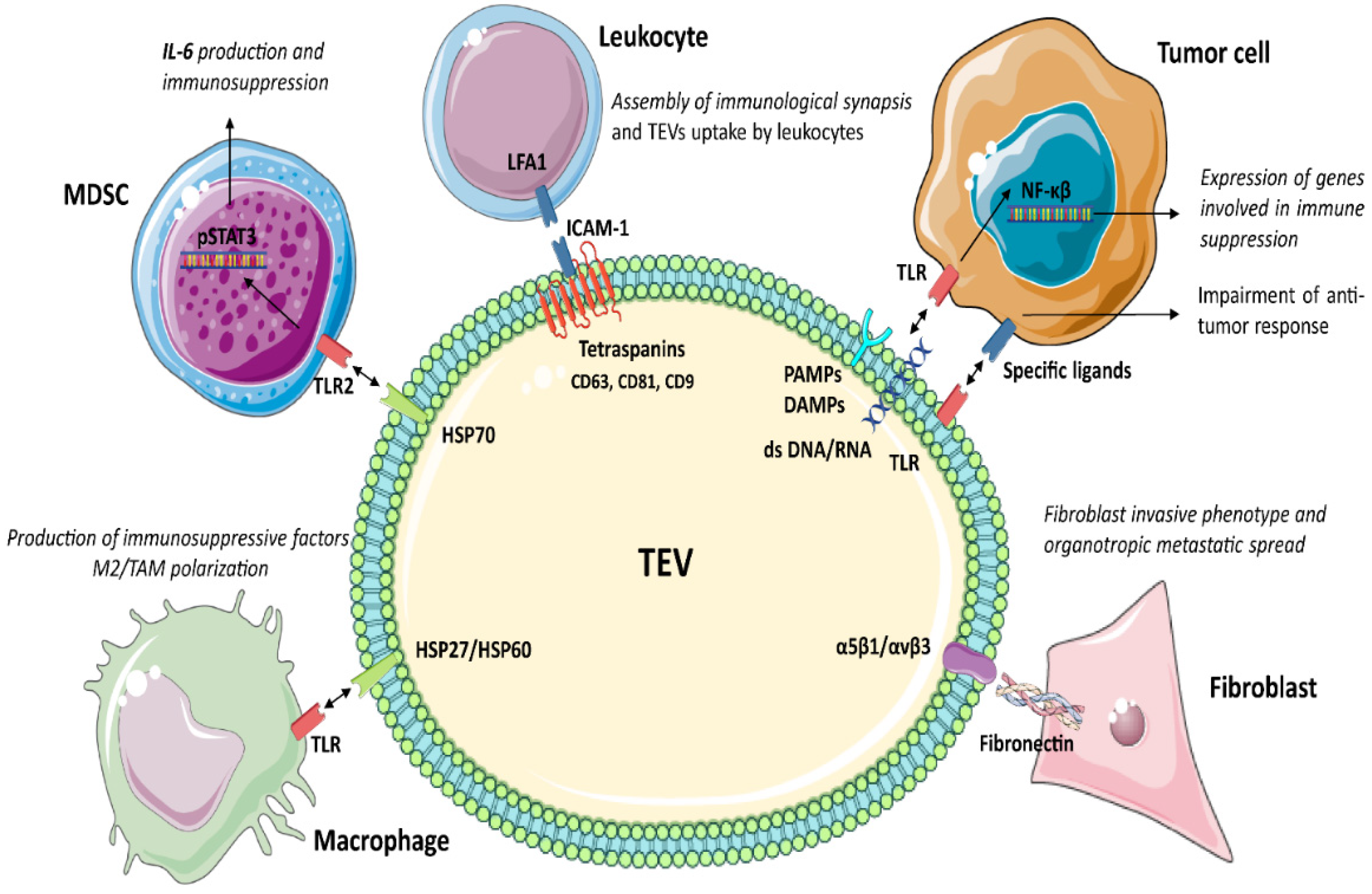
3.1. Tetraspanins
Tetraspanins (CD63, CD81, CD9, etc.) organise membrane tetraspanin-enriched microdomains involving selected transmembrane and cytosolic signalling proteins [59]. The tetraspanin-mediated sorting of specific adhesion receptors may influence EV uptake. For example, the adhesion molecules ICAM-1/LFA1 are involved in EV uptake by leukocytes [60,61,62][60][61][62]. Moreover, tetraspanins interact with ADAM10 and ADAM17 and control their sheddase activity [63]. In the endosomal compartment, active ADAM10 and ADAM17 enriched in EV cleave preproteins to release soluble molecules, as shown for TNFα in ovarian cancer and melanoma [64]. Tetraspanins also regulate sorting of MHC I and MHC II in EV [55], thereby regulating TEV immunogenicity. Several studies also provide evidence for the role of CD81 in the assembly of functional immunological synapses acting as T-cell co-stimulatory signals [65]. Therefore, tetraspanins, besides activating naive T-cells via the MHC complexes, also act as co-stimulatory signals.3.2. Toll-Like Receptors (TLRs)
TLRs belong to a family of pattern recognition receptors, which play a critical role in the innate immune responses by recognising several ligands also carried by TEV [66,67][66][67]. In cancer, these ligands include PAMPs, double-stranded RNA and/or DNA [68], and internal DAMPs, as fibrinogen, nuclear and cytosolic proteins, and Heat Shock Proteins (HSP) [69,70,71,72][69][70][71][72]. TLR1-2 and TLR4-6 proteins are localised on the surface membrane, whereas TLR3 and TLR7–9 are accumulated in the endosome or lysosome compartment, as well as in the endoplasmic reticulum [73]. Immune cells, epithelial [74], endothelial [75] and tumour cells [76] express TLRs [77]. TLR stimulation by TEV activates both the innate and adaptive immune responses and induces inflammatory signals in target cells. In cancer, TLR activation triggers an intracellular cascade, resulting in NF-κβ activation and the expression of genes involved in immune suppression [8,78][8][78]. Moreover, the expression of TLRs on TEV suggests that they can bind and transfer specific ligands to target cells, further impairing the antitumour immune response [39].3.3. Heat Shock Proteins (HSP)
HSP are involved in antiapoptotic signalling [79,80][79][80], EMT [81], tumour angiogenesis and invasion [82], as well as in drug resistance [83,84][83][84]. Immune cells express HSP receptors as TLRs, leading to TEV endocytosis, antigen cross-presentation, T-cell cross priming, and activation of the immune response. HSP90, HSP70, HSP27 are DAMPs members expressed by EV derived from cancerous [85] and non-cancerous cells. However, the number of circulating HSP70+ EV is higher in cancer patients than in healthy donors [85] and is correlated with metastases growth [86]. Proteomics revealed that TEV are enriched in HSP90 [87], HSP72 [88], HSP105 [89], HSP70 [90,91][90][91] and HSP86 [92]. These observations have suggested that for HSP soluble forms, TEV can act as immunoregulators. It has been shown that the interaction of HPS expressed by TEV and TLR2 or TLR4 on myeloid cells (DC or MDSC) triggers pSTAT3 expression and IL-6 production, thereby repressing the antitumour immune reaction [88,90][88][90]. Moreover, TEV released upon hypoxia were found to be enriched in HSP70 and HSP90 and involved in invasiveness and stemness properties of prostate cancer cells [91]. In addition, human-melanoma-derived TEV enriched in HSP86 interact with TLR4 on myeloid cells, resulting in PDL-1 upregulation and blockade of T-cell activation [92]. Meanwhile, the antitumour activity of TEV enriched in different HSP isoforms was also reported [93,94,95,96,97][93][94][95][96][97]. HSP70+ TEV secreted by human pancreatic and colon carcinoma cell lines induced NK [93,94][93][94] and Th1 reactivity [95], and converted Treg into Th17 cells [96]. This resulted in a strong antitumour immune response. Pretreatment of melanoma and colon carcinoma cells with recombinant HSP70 increased the release of HPS70+ TEV, which, in vivo, translates in a strong anticancer immune response [97]. The correlation of different circulating HSP in cancer patients with tumour progression [86], the relevance of HSP in immune regulation [98], as well as their enrichment in TEV [84] strongly support the contribute of HSP+ TEV in mediating tumour immune suppression. However, further studies are required to clarify this specific topic.3.4. Integrins (ITGA/ITGB)
Integrins are critical receptors for extracellular matrix proteins supporting cell adhesion and driving cell migration. α1β1 and α2β1 integrins are major collagen receptors, whereas α5β1 and αvβ3 integrins preferentially bind fibronectin. Fibronectin interacting with TEV-associated integrins drives the invasive phenotype to recipient fibroblasts [99]. TEV enriched in integrins support tumour spreading and metastasis formation [100,101,102,103][100][101][102][103]. Extracellular matrix proteins can link TEV and target cells as a bridge to induce specific biological responses. Indeed, it was shown that the expression of α6β1, α6β4 and αvβ5 in TEV dictates cell target specificity and organotropic metastasis [100]. Moreover, it has been shown that EV integrin cargo is involved in immune cell recruitment [104[104][105],105], suggesting that integrins in TEV may be instrumental to immune cell homing and specific targeting. Overall, TEV integrin content regulates their uptake [106], identifies specific secondary tissues [100], and attracts immune cells [107,108][107][108]. CD47 (Cluster of Differentiation 47) is an integrin-associated protein that is overexpressed by tumour cells and TEV and plays a role in immune regulation [109,110][109][110]. CD47 binds the SIgnal-Regulatory Protein alpha (SIRPα) [111], able to deliver inhibitory signals to macrophages. It was shown that downregulation of CD47 in TEV enhanced macrophages-mediated cancer cell phagocytosis [112].3.5. TEV as Immunosuppressive Shuttles
TEV hold immune checkpoint molecules and anti-inflammatory cytokines. Physiologically, the checkpoint machinery controls T-cell activation, counteracting unnecessary tissue damage and autoimmunity. In cancer, TEV enriched in immune checkpoint ligands bind T-cell-associated cognate receptors, thereby reducing tumour killing. Several immune checkpoint pathways are referred to as crucial for anticancer therapeutic approaches, and among them PD1/PD-L1, CTLA4/B7, TIM3, Trail and Fas/FasL have received particular attention [113] (Figure 2).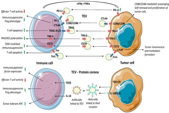
3.6. Programmed Death Receptor 1 (PD-1) and Its Ligand (PD-L1)
PD-1/PD-L1 signalling controls and impairs inflammation. Activated T cells express PD-1 and secrete inflammatory factors, such as IFNγ and TNFα, that in turn, upregulate PD-L1 in surrounding cells as well as in EV [114,115,116,117][114][115][116][117]. Several tumours hijack the PD-1/PD-L1 pathway by inducing the expression of PD-L1 in the TME compartment. Persistent PD-1/PD-L1 binding leads to T-cell exhaustion, inhibition of proliferation and T-cell apoptosis. Conversely, Treg (CD4+ Foxp3+) bearing PD-1 promotes naive CD4+ T cell differentiation towards Treg [118]. Thus, PD-1 expression not only suppresses effector T-cell function, but also dictates T-cell shift towards an immunosuppressive phenotype. The immunosuppressive role of PD-L1+ TEV was reported in head and neck squamous cell carcinoma [119], prostate cancer, melanoma [115] and non-small cell lung cancer [120]. PD-1 blocking antibodies have been shown to exert remarkable anticancer immune activity in PD-L1+ tumours [121]. However, TEV enriched in PD-L1 were described as both mediators of tumour immune evasion and predictors of anti-PD-1 treatment effectiveness [118,122][118][122].3.7. CTLA-4/B7/CD152
As PD-1, CTLA-4 is broadly engaged during tumour immune evasion to suppress CD4+ effector T-cells and boost Treg activity [123]. After CD80/CD86 binding, CTLA-4 suppresses CD8+ T cell proliferation, cell cycle progression and cytokine (IL-2, IFN-γ) production [124]. CTLA-4 competes with CD28 for CD80/CD86 ligands. CD28 activates and induces a T-cell pro-inflammatory phenotype, while CTLA-4, displaying high affinity for CD80/CD86, guarantees T-cell suppression [125]. All at once, stimulation of CTLA-4 on Treg leads to FoxP3 upregulation, thereby improving Treg functions [126]. In fact, anti-CTLA-4 antibodies reduce the number of Treg at the tumour site, inhibit their negative regulatory effects, and enhance the antitumour immune response [127]. Therefore, CTLA-4 may exert pleiotropic effects on target cells depending on the presence of discrete markers of activation. TEV carry both CTLA-4 and its ligands CD80/CD86 [127,128,129,130][127][128][129][130]. It has been proposed that the expression of CD80/CD86 in TEV interferes with CTL activity by binding to CTLA-4 [128,129][128][129]. Circulating TEV carrying CTLA-4 were also detected in cancer patients, and their concentration correlated with a poor outcome [127,130][127][130]. It has been proposed that CTLA-4 in TEV acts as a scavenger for the CD80/CD86 pro-apoptotic ligands, thereby protecting tumour cells. According to this possibility, TEV enriched in CTLA-4 regulate the PTEN/CD44 signalling pathway to promote proliferation, self-renewal and liver metastasis [131].3.8. T-Cell Immunoglobulin Domain and Mucin Domain 3 (TIM-3)
TIM-3, a transmembrane protein expressed by activated immune cells, suppresses pro-inflammatory cytokine secretion [132] and CTL function [133], thereby mediating immune suppression. It was shown that TEV are enriched in TIM-3 [134] and that the level of TIM-3 on circulating TEV positively correlated with cancer progression [135]. Moreover, TEV can transfer TIM-3 to macrophages, driving their M2 differentiation [134].3.9. Cluster of Differentiation 73 (CD73)
CD73 is a 5′-nucleotidase that converts AMP into the anti-inflammatory adenosine [136]. In cancer, free adenosine generated by CD73 inhibits cellular immune responses, thereby promoting tumour immune escape [137]. MDSC, Treg or DC expressing CD73 generate adenosine in tumours, which triggers the anti-inflammatory response via T-cells, macrophages, and natural killer cells (NK) [138,139][138][139]. Several studies have reported that the expression of CD73 in TEV also contributes to immunosuppression in different cancers [140,141][140][141]. TEV transfer CD73 to T-cells and inhibit their cell cycle entry and clonal proliferation by increasing adenosine [140]. Consistently, inhibition of TEV production or CD73 expression significantly inhibit tumour growth by restoring T-cell clonal proliferation [140]. It was also shown that CD73+ TEV upregulate the expression of IL-6, IL-10, TNFα and TGFβ1 in TAM by activating the NF-κB pathway [141]. Moreover, circulating CD73+ TEV increased the risk of lymph node metastases and was associated with a poor prognosis. In addition, loss of CD73+ TEV enhanced the effectiveness of anti-PD-1 treatment by rescuing CTL function [141].3.10. Fas/FasL
Like PD-L1, CTLA-4 and CD73, Fas/FasL is hijacked by cancer cells to suppress the immune response and induce tumour immune evasion. FasL (CD95) is the most studied mediator of T-cell apoptosis [142]. Fas+ T cells undergo apoptosis upon binding to FasL, which is expressed in APC, tumours, and other cell types. It was also shown that IL-10, VEGF and prostaglandin E2 (PGE2) enhance the expression of FasL in tumour cells [143]. The expression of the Fas competitor, c-FLIP, in Treg and tumour cells determines their resistance to FasL-mediated apoptosis [144,145,146][144][145][146]. In addition, in cancer cells, the binding of c-FLIP to FasL+ EV interferes with the activation of caspase-8, thereby promoting tumour invasiveness and metastasis formation [147]. Circulating TEV carrying both Fas and FasL were detected in cancer patients [148]. It has been proposed that TEV expressing Fas may act as FasL scavenger, protecting tumour cells from death-receptor-mediated apoptosis [148], while FasL+ TEV act as inducers of T-cell apoptosis [149,150][149][150]. In addition, patient-derived FasL+ TEV were shown to induce caspase-3 cleavage, cytochrome c release and the loss of mitochondrial membrane potential in target T-lymphocytes [151].3.12. Protein Corona and TEV Mechanism of Action
3.11. Protein Corona and TEV Mechanism of Action
Apart from membrane-associated proteins, such as PD-L1, CD73 and FasL, TEV are enriched in growth factors and cytokines involved in the immune regulation. These proteins can be part of “protein corona”, artificially linked to TEV [166–168], or naturally linked to their receptors, as shown for TGFβ1 [169].
Apart from membrane-associated proteins, such as PD-L1, CD73 and FasL, TEV are enriched in growth factors and cytokines involved in the immune regulation. These proteins can be part of “protein corona”, artificially linked to TEV [152][153][154], or naturally linked to their receptors, as shown for TGFβ1 [155].Several membrane receptors expressed in TEV can bind their ligands (integrins/extracellular matrix proteins; TLRs/HSP and nucleic acids; receptors/growth factors and cytokines). Interestingly, EV covered with protein corona were more effective in monocyte activation, compared to “pure” EV or protein aggregates (protein corona). This suggests that EV-mediated immune cell activation depends on both protein corona and EV cargo. Conversely, it has been shown that DC differentiation and maturation only depend on EV cargo [167,168]. These findings indicate that TEV-mediated immune regulation is much more complex than expected.
Several membrane receptors expressed in TEV can bind their ligands (integrins/extracellular matrix proteins; TLRs/HSP and nucleic acids; receptors/growth factors and cytokines). Interestingly, EV covered with protein corona were more effective in monocyte activation, compared to “pure” EV or protein aggregates (protein corona). This suggests that EV-mediated immune cell activation depends on both protein corona and EV cargo. Conversely, it has been shown that DC differentiation and maturation only depend on EV cargo [153][154]. These findings indicate that TEV-mediated immune regulation is much more complex than expected.3.13. TGFβ1
3.12. TGFβ1
TGFβ1 has been extensively investigated as an immunosuppressive factor in tumour settings. Webber and colleagues [169][155] showed the natural surface binding of TGFβ1 on TEV. TGFβ1, bound as a latent form, is able to activate the SMAD signalling pathway and gene expression in target cells [169][155]. TEV enriched in TGFβ1 were described in gastric [170][156], bladder [171][157], head and neck cancer [39], mesothelioma [172][158], breast [173][159], prostate and other cancers [169][155]. It was shown that TGFβ1+ TEV boost the expression of CTLA-4, FasL, IL-10 and other immunosuppressive proteins [174][160]. TGFβ1 acts on CTL by interfering with the expression of perforin, granzyme A and B, FasL and IFNγ [175][161], and by inducing Treg differentiation and IL-10 production [176,177][162][163].
3.14. Interleukin 10 (IL-10)
3.13. Interleukin 10 (IL-10)
IL-10 is a regulatory cytokine with pleiotropic effects in various cell types [178]. IL-10 can directly inhibit T-cell responses [179,180], thereby enhancing Treg differentiation [177,181,182]. More importantly, IL-10 inhibits DC and macrophage functions by suppressing inflammatory cytokine production and inhibiting the expression of MHC class II and the co-stimulatory-molecules CD80/CD86 [183]. In cancer, the co-expression of IL-10 and tumour antigens reprogram APC towards a tumour-tolerant phenotype.
IL-10 is a regulatory cytokine with pleiotropic effects in various cell types [164]. IL-10 can directly inhibit T-cell responses [165][166], thereby enhancing Treg differentiation [163][167][168]. More importantly, IL-10 inhibits DC and macrophage functions by suppressing inflammatory cytokine production and inhibiting the expression of MHC class II and the co-stimulatory-molecules CD80/CD86 [169]. In cancer, the co-expression of IL-10 and tumour antigens reprogram APC towards a tumour-tolerant phenotype. As TGFβ1, TEV enriched in IL-10 were detected in plasma of different cancer patients; however, the most relevant action performed by TEV is the induction of IL-10 and TFGβ1 in target cells via their RNA content [184–186][170][171][172].
4. TEV and Drug Resistance
During treatment, tumour cells develop resistance through different mechanisms specifically impairing drug efficacy. In particular, drug efflux and inactivation [226], pro-survival pathway activation [227], EMT [228], immune evasion and de-differentiation towards stem-like phenotype [229] are the most relevant mechanisms. Drug efflux was detected in many types of cancer cells and depends on several transporters [230]. Among them, P-glycoprotein (p-gp) is the most investigated in cancer [231]. Physiologically, p-gp pumps out a broad range of xenobiotics and manages the efflux of toxins in the intestine, liver or kidney. Different types of cancer also exploit p-gp to decrease local drug concentration. The overexpression of p-gp was shown in haematological malignancies [232], colorectal cancer [233] and others [234]. Mechanisms of TEV-mediated acquired drug resistance mainly rely on protein or RNA transfer from resistant cells to susceptible recipient cells, or by exporting cytotoxic substances. As shown for doxorubicin-resistant osteosarcoma, the transfer of p-gp by TEV allows recipient cells to remove anticancer drugs [235]. Moreover, it was shown that TEV could transfer miR-451 and miR-27a to increase p-gp expression [236,237].
TEV cargo also turns on pro-survival pathways in recipient cancer cells. In particular, IL-6 promotes drug resistance [238] by upregulating multidrug resistance-related genes (MDR1 and GSTpi), apoptosis inhibitory proteins (Bcl-2, Bcl-xL and XIAP) and activating the Ras/MEK/ERK and PI3K/Akt signalling pathway [239]. Indeed, several studies demonstrated that TEV-mediated IL-6 production is a common mechanism of drug resistance [240–242]. The in vivo blockade of the release of TEV from TAM was reported as the most relevant mechanism improving gemcitabine sensitivity. The authors also provided evidence that TEV miR-365 content was involved in gemcitabine resistance [243].
As shown by Pavlyukov and colleagues [244], drug resistance also relies on RNA splicing. They found that glioblastoma cells release TEV enriched in the splicing factor RBM11. RBM11 is generally upregulated in apoptotic tumour cells during anticancer therapies. Once transferred to recipient cells, RBM11 switches the splicing of MDM4 and Cyclin D1 genes towards more oncogenic isoforms [244].
TEV also regulate immune checkpoint molecules to drive immune evasion, leading to tumour resistance. The expression of CD28 and CD80/86, required for PD-1 or CTLA-4 immune checkpoint blockade [245,246], was found under the control of TEV-miRNA content. Indeed, it was shown that miR-424 in TEV suppresses the expression of CD28-CD80/86 in TILs and DC, leading to drug resistance [208].
TEV surface proteins can act as decoy receptors for antibodies-based therapy, thereby decreasing drug bioavailability. In B-cell lymphoma, the expression of CD20 in TEV protects lymphoma cells from rituximab [247]. Similarly, in breast cancer, HER2+ TEV modulate resistance to the anti-HER2 monoclonal antibody [248]. In melanoma patients, circulating PD-L1+ TEV early after immune checkpoint blockade classifies patients as resistant to anti-PD-1 therapy [122]. Indeed, PD-L1+ TEV act as scavengers for immunotherapeutics [114,122].
Therefore, TEV-mediated transfer of proteins or RNAs induces drug resistance by reprogramming tumour cells towards a highly metastatic phenotype or by inactivating immune cells.
5. TEV Released by TME Cells
Tumour tissue consists of cancer cells and recruited stromal, vascular and immune cells which, by releasing stimulatory growth factors, chemokines, and cytokines at the primary tumour site, promote angiogenesis and metastasis and suppress the anticancer immune response [249]. These cells in the TME include cancer-associated fibroblasts [240], tumour endothelial cells (TEC) [250], adipocytes [251,252], tumour-associated immune cells as TAM [253] and MDSC [254], DC [255], monocytes [256], CTL [257] and Treg [258], B cells [259], regulatory NK [260] and tumour-educated platelets [261]. Growing evidence indicates that cancer cells could recruit and reprogram all cell types. Notably, it was shown that TEV can induce tumour innervation [262] and that tumour-associated neuronal cells regulate tumour-infiltrated immune cell functions.
5.1. TEV Derived from Tumour-Educated Platelets
Activated platelets professionally release EV [264][212], which are the most abundant population of circulating EV. It has been shown that platelet concentration positively correlates with cancer growth [265][213]. Platelets and their EV are enriched in transcription factors, RNA and spliceosomes [266][214]. In cancer, megakaryocytes and platelets cross-talk with tumour via EV. This results in tumour-specific rearrangement of platelet transcriptome and proteome, leading to the establishment of the so-called so-called tumour-educated platelets [267–272][215][216][217][218][219][220]. TEV expressing P-selectin (CD62P) and integrin αIIβ3 [273,274][221][222] regulate the immune system, stimulate angiogenesis, promote metastasis and are involved in drug resistance by activating discrete signalling cascades [275,276][223][224]. In particular, TEV released from tumour-educated platelets stimulate MAPK p42/44 and AKT, increase the expression of MMP1, MMP9, VEGF and cyclin D2 [277][225] and induce EMT by activating TGFβ1 and NF-κB signalling [278][226].
5.2. TEV from TEC
TEV derived from TEC are involved in cancer development, metastatic spread and tumour immune editing [39,279–281][39][227][228][229]. TEC-derived TEV contain pro-oncogenic proteins and RNAs involved in immune regulation (TGFβ1, HLA-G, IL-6, M-CSF, as well as lncRNA MALAT1 and miR-24-3p) [39,279,280][39][227][228]. It has been shown that TEC-derived TEV enhance the expression of PD-L1 in tumour and myeloid cells, promote the formation of Treg and decrease T-cell cytotoxic activity [39,279,281][39][227][229]. Interestingly, stress conditions in the TME (hypoxia, inflammation, changes in the pH and many others) induce the release of TEV with specific tumour-promoting properties. In particular, IL-3 signalling in TEC rearranges TEV miRNA cargo, which control β-catenin and PD-L1 expression as well as the metastatic spread [279,281][227][229].
5.3. EV Derived from DC
Tumour-primed immune cells can secrete immunologically active TEV. Commonly, EV derived from DC (DC-EV) carry functional MHC-complexes, thereby educating additional APC to recognise specific antigens [282] and cross-reactivating CD8+ T cells [283]. DC-EV play a critical role in generating antitumour CD8+ T cells. Indeed, several clinical trials based on tumour-primed DC-TEV were performed to find an effective antitumour vaccine [284,285].
DC-EV function depends on their molecular composition, in particular the concentration of co-stimulatory molecules (CD86, CD80, CD40) and antigen-presenting molecules (MHC-II, MHC-I) [286,287]. It was previously described that TEV cargo (HSP, TLR, HLA g [29]; S100A8, S100A9, Annexin A1 [30]; PGE2 [288]; TGFβ1 [289]; or microRNAs [290]), suppress DC maturation, resulting in the loss of tumour cell recognition [29,30]. Moreover, once activated by TEV, DC release their own TEV conveying tumour-associated antigens [291], which translate in T-cell cross-activation, antigen presentation, and immune tolerance [292,293]. Overall, TEV released by “immature” tumour-primed DC support cancer outgrowth by reprogramming immune cells towards immune tolerance.
6. Conclusions
Early after EV discovery [294][242] it was reported that cancer cells release TEV enriched in proteins and lipids, contributing to tumour immune escape [295][243]. Soon after, the same group showed that TEV express antigens recapitulating their cell of origin and can exert both pro- or antitumour action [296][244]. Moreover, since MHC class II on TEV induces specific T-cell responses [297][245], TEV have been proposed for immunotherapeutic strategies. [4,282,298–301][4][230][239][246][247][248]. However, TEV-based anticancer vaccines still lack robust clinical benefits, since the complex TEV molecular composition mainly reprograms immune cells for tolerance [7,302][7][249]. After TEV-mediated activation, immune cells release their own TEV to amplify and spread the oncogenic signals. In fact, elevated circulating EV displaying oncogenic properties are commonly detected in cancer patients and contribute to drug resistance.
Overall, TEV may be considered a “complex system”, and prediction of their biological functions based on their individual molecular components may somehow be misleading. Therefore, efforts to develop methodologies to deeply investigate how their molecular components control the antitumour immune response in concert are warranted. If successful, EV engineering approaches would offer the chance to reshape the immune reaction into tumour recognition and eradication.
Overall, TEV may be considered a “complex system”, and prediction of their biological functions based on their individual molecular components may somehow be misleading. Therefore, efforts to develop methodologies to deeply investigate how their molecular components control the antitumour immune response in concert are warranted. If successful, EV engineering approaches would offer the chance to reshape the immune reaction into tumour recognition and eradication.
References
- Whiteside, T.L. Exosomes and tumor-mediated immune suppression. J. Clin. Invest. 2016, 126, 1216–1223.
- Hoshino, A.; Kim, H.S.; Bojmar, L.; Gyan, K.E.; Cioffi, M.; Hernandez, J.; Zambirinis, C.P.; Rodrigues, G.; Molina, H.; Heissel, S.; et al. Extracellular Vesicle and Particle Biomarkers Define Multiple Human Cancers. Cell 2020, 182, 1044–1061.e18.
- Slingluff, C.L., Jr.; Cox, A.L.; Stover, J.M., Jr.; Moore, M.M.; Hunt, D.F.; Engelhard, V.H. Cytotoxic T-Lymphocyte Response to Autologous Human Squamous Cell Cancer of the Lung: Epitope Reconstitution with Peptides Extracted from HLA-Aw681. Cancer Res. 1994, 54, 2731–2737.
- Wolfers, J.; Lozier, A.; Raposo, G.; Regnault, A.; Théry, C.; Masurier, C.; Flament, C.; Pouzieux, S.; Faure, F.; Tursz, T.; et al. Tumor-derived exosomes are a source of shared tumor rejection antigens for CTL cross-priming. Nat. Med. 2001, 7, 297–303.
- Echchakir, H.; Vergnon, I.; Dorothée, G.; Grunenwald, D.; Chouaib, S.; Mami-Chouaib, F. Evidence for in situ expansion of diverse antitumor-specific cytotoxic T lymphocyte clones in a human large cell carcinoma of the lung. Int. Immunol. 2000, 12, 537–546.
- Kambayashi, T.; Laufer, T.M. Atypical MHC class II-expressing antigen-presenting cells: Can anything replace a dendritic cell? Nat. Rev. Immunol. 2014, 14, 719–730.
- Costa-Silva, B.; Aiello, N.M.; Ocean, A.J.; Singh, S.; Zhang, H.; Thakur, B.K.; Becker, A.; Hoshino, A.; Mark, M.T.; Molina, H.; et al. Pancreatic cancer exosomes initiate pre-metastatic niche formation in the liver. Nat. Cell Biol. 2015, 17, 816–826.
- Andersen, M.H.; Schrama, D.; Thor Straten, P.; Becker, J.C. Cytotoxic T cells. J. Invest. Dermatol. 2006, 126, 32–41.
- Coulie, P.G.; Van den Eynde, B.J.; van der Bruggen, P.; Boon, T. Tumour antigens recognized by T lymphocytes, at the core of cancer immunotherapy. Nat. Rev. Cancer 2014, 14, 135–146.
- Arina, A.; Tirapu, I.; Alfaro, C.; Rodríguez-Calvillo, M.; Mazzolini, G.; Inogés, S.; López, A.; Feijoo, E.; Bendandi, M.; Melero, I. Clinical implications of antigen transfer mechanisms from malignant to dendritic cells: Exploiting cross-priming. Exp. Hematol. 2002, 30, 1355–1364.
- Wilson, N.S.; El-Sukkari, D.; Villadangos, J.A. Dendritic cells constitutively present self antigens in their immature state in vivo and regulate antigen presentation by controlling the rates of MHC class II synthesis and endocytosis. Blood 2004, 103, 2187–2195.
- Worbs, T.; Hammerschmidt, S.I.; Förster, R. Dendritic cell migration in health and disease. Nat. Rev. Immunol. 2017, 17, 30–48.
- Carreno, B.M.; Magrini, V.; Becker-Hapak, M.; Kaabinejadian, S.; Hundal, J.; Petti, A.A.; Ly, A.; Lie, W.-R.; Hildebrand, W.H.; Mardis, E.R.; et al. A dendritic cell vaccine increases the breadth and diversity of melanoma neoantigen-specific T cells. Science 2015, 348, 803–808.
- Albert, M.L.; Sauter, B.; Bhardwaj, N. Dendritic cells acquire antigen from apoptotic cells and induce class I-restricted CTLs. Nature 1998, 392, 86–89.
- Mallegol, J.; Van Niel, G.; Lebreton, C.; Lepelletier, Y.; Candalh, C.; Dugave, C.; Heath, J.; Raposo, G.; Cerf-Bensussan, N.; Heyman, M. T84-Intestinal Epithelial Exosomes Bear MHC Class II/Peptide Complexes Potentiating Antigen Presentation by Dendritic Cells. Gastroenterology 2007, 132, 1866–1876.
- Appleman, L.J.; Boussiotis, V.A. T cell anergy and costimulation. Immunol. Rev. 2003, 192, 161–180.
- Förster, R.; Schubel, A.; Breitfeld, D.; Kremmer, E.; Renner-Müller, I.; Wolf, E.; Lipp, M. CCR7 coordinates the primary immune response by establishing functional microenvironments in secondary lymphoid organs. Cell 1999, 99, 23–33.
- Ohl, L.; Mohaupt, M.; Czeloth, N.; Hintzen, G.; Kiafard, Z.; Zwirner, J.; Blankenstein, T.; Henning, G.; Förster, R. CCR7 governs skin dendritic cell migration under inflammatory and steady-state conditions. Immunity 2004, 21, 279–288.
- Steinman, R.M. The control of immunity and tolerance by dendritic cell. Pathol. Biol. 2003, 51, 59–60.
- Steinman, R.M.; Hawiger, D.; Liu, K.; Bonifaz, L.; Bonnyay, D.; Mahnke, K.; Iyoda, T.; Ravetch, J.; Dhodapkar, M.; Inaba, K.; et al. Dendritic cell function in vivo during the steady state: A role in peripheral tolerance. Ann. N. Y. Acad. Sci. 2003, 987, 15–25.
- Shiokawa, A.; Kotaki, R.; Takano, T.; Nakajima-Adachi, H.; Hachimura, S. Mesenteric lymph node CD11b- CD103+ PD-L1High dendritic cells highly induce regulatory T cells. Immunology 2017, 152, 52–64.
- Liu, K.; Iyoda, T.; Saternus, M.; Kimura, Y.; Inaba, K.; Steinman, R.M. Immune Tolerance After Delivery of Dying Cells to Dendritic Cells In Situ. J. Exp. Med. 2002, 196, 1091–1097.
- Stuart, L.M.; Lucas, M.; Simpson, C.; Lamb, J.; Savill, J.; Lacy-Hulbert, A. Inhibitory effects of apoptotic cell ingestion upon endotoxin-driven myeloid dendritic cell maturation. J. Immunol. Baltim. Md. 1950 2002, 168, 1627–1635.
- Wallet, M.A.; Sen, P.; Flores, R.; Wang, Y.; Yi, Z.; Huang, Y.; Mathews, C.E.; Earp, H.S.; Matsushima, G.; Wang, B.; et al. MerTK is required for apoptotic cell-induced T cell tolerance. J. Exp. Med. 2008, 205, 219–232.
- Patente, T.A.; Pinho, M.P.; Oliveira, A.A.; Evangelista, G.C.M.; Bergami-Santos, P.C.; Barbuto, J.A.M. Human Dendritic Cells: Their Heterogeneity and Clinical Application Potential in Cancer Immunotherapy. Front. Immunol. 2018, 9, 3176.
- Verweij, F.J.; Revenu, C.; Arras, G.; Dingli, F.; Loew, D.; Pegtel, D.M.; Follain, G.; Allio, G.; Goetz, J.G.; Zimmermann, P.; et al. Live Tracking of Inter-organ Communication by Endogenous Exosomes In Vivo. Dev. Cell 2019, 48, 573–589.e4.
- Hyenne, V.; Ghoroghi, S.; Collot, M.; Bons, J.; Follain, G.; Harlepp, S.; Mary, B.; Bauer, J.; Mercier, L.; Busnelli, I.; et al. Studying the Fate of Tumor Extracellular Vesicles at High Spatiotemporal Resolution Using the Zebrafish Embryo. Dev. Cell 2019, 48, 554–572.e7.
- Scott, A.; Sueiro Ballesteros, L.; Bradshaw, M.; Tsuji, C.; Power, A.; Lorriman, J.; Love, J.; Paul, D.; Herman, A.; Emanueli, C.; et al. In Vivo Characterization of Endogenous Cardiovascular Extracellular Vesicles in Larval and Adult Zebrafish. Arterioscler. Thromb. Vasc. Biol. 2021, 41, 2454–2468.
- Grange, C.; Tapparo, M.; Tritta, S.; Deregibus, M.C.; Battaglia, A.; Gontero, P.; Frea, B.; Camussi, G. Role of HLA-G and extracellular vesicles in renal cancer stem cell-induced inhibition of dendritic cell differentiation. BMC Cancer 2015, 15, 1009.
- Maus, R.L.G.; Jakub, J.W.; Nevala, W.K.; Christensen, T.A.; Noble-Orcutt, K.; Sachs, Z.; Hieken, T.J.; Markovic, S.N. Human Melanoma-Derived Extracellular Vesicles Regulate Dendritic Cell Maturation. Front. Immunol. 2017, 8, 358. Available online: https://www.frontiersin.org/article/10.3389/fimmu.2017.00358 (accessed on 27 May 2022).
- Kurts, C.; Kosaka, H.; Carbone, F.R.; Miller, J.F.A.P.; Heath, W.R. Class I–restricted Cross-Presentation of Exogenous Self-Antigens Leads to Deletion of Autoreactive CD8+ T Cells. J. Exp. Med. 1997, 186, 239–245.
- Manicassamy, S.; Pulendran, B. Dendritic cell control of tolerogenic responses. Immunol. Rev. 2011, 241, 206–227.
- Saito, M.; Nagasawa, M.; Takada, H.; Hara, T.; Tsuchiya, S.; Agematsu, K.; Yamada, M.; Kawamura, N.; Ariga, T.; Tsuge, I.; et al. Defective IL-10 signaling in hyper-IgE syndrome results in impaired generation of tolerogenic dendritic cells and induced regulatory T cells. J. Exp. Med. 2011, 208, 235–249.
- Sela, U.; Park, C.G.; Park, A.; Olds, P.; Wang, S.; Steinman, R.M.; Fischetti, V.A. Dendritic Cells Induce a Subpopulation of IL-12Rβ2-Expressing Treg that Specifically Consumes IL-12 to Control Th1 Responses. PLoS ONE 2016, 11, e0146412.
- Adler, A.J.; Marsh, D.W.; Yochum, G.S.; Guzzo, J.L.; Nigam, A.; Nelson, W.G.; Pardoll, D.M. CD4+ T cell tolerance to parenchymal self-antigens requires presentation by bone marrow-derived antigen-presenting cells. J. Exp. Med. 1998, 187, 1555–1564.
- Pardoll, D.M. The blockade of immune checkpoints in cancer immunotherapy. Nat. Rev. Cancer 2012, 12, 252–264.
- Bousso, P.; Robey, E.A. Dynamic Behavior of T Cells and Thymocytes in Lymphoid Organs as Revealed by Two-Photon Microscopy. Immunity 2004, 21, 349–355.
- Miller, M.J.; Hejazi, A.S.; Wei, S.H.; Cahalan, M.D.; Parker, I. T cell repertoire scanning is promoted by dynamic dendritic cell behavior and random T cell motility in the lymph node. Proc. Natl. Acad. Sci. USA 2004, 101, 998–1003.
- Lopatina, T.; Favaro, E.; Danilova, L.; Fertig, E.J.; Favorov, A.V.; Kagohara, L.T.; Martone, T.; Bussolati, B.; Romagnoli, R.; Albera, R.; et al. Extracellular Vesicles Released by Tumor Endothelial Cells Spread Immunosuppressive and Transforming Signals Through Various Recipient Cells. Front. Cell Dev. Biol. 2020, 8, 698.
- Buschow, S.I.; Nolte-‘t Hoen, E.N.; Van Niel, G.; Pols, M.S.; Ten Broeke, T.; Lauwen, M.; Ossendorp, F.; Melief, C.J.; Raposo, G.; Wubbolts, R.; et al. MHC II in dendritic cells is targeted to lysosomes or T cell-induced exosomes via distinct multivesicular body pathways. Traffic Cph. Den. 2009, 10, 1528–1542.
- Wang, X.; Shen, H.; He, Q.; Tian, W.; Xia, A.; Lu, X.-J. Exosomes derived from exhausted CD8+ T cells impaired the anticancer function of normal CD8+ T cells. J. Med. Genet. 2019, 56, 29–31.
- Okoye, I.S.; Coomes, S.M.; Pelly, V.S.; Czieso, S.; Papayannopoulos, V.; Tolmachova, T.; Seabra, M.C.; Wilson, M.S. MicroRNA-Containing T-Regulatory-Cell-Derived Exosomes Suppress Pathogenic T Helper 1 Cells. Immunity 2014, 41, 89–103.
- Smyth, L.A.; Ratnasothy, K.; Tsang, J.Y.; Boardman, D.; Warley, A.; Lechler, R.; Lombardi, G. CD73 expression on extracellular vesicles derived from CD4+CD25+Foxp3+ T cells contributes to their regulatory function. Eur. J. Immunol. 2013, 43, 2430–2440.
- Watkins, S.K.; Zhu, Z.; Riboldi, E.; Shafer-Weaver, K.A.; Stagliano, K.E.; Sklavos, M.M.; Ambs, S.; Yagita, H.; Hurwitz, A.A. FOXO3 programs tumor-associated DCs to become tolerogenic in human and murine prostate cancer. J. Clin. Invest. 2011, 121, 1361–1372.
- Flores-Toro, J.A.; Luo, D.; Gopinath, A.; Sarkisian, M.R.; Campbell, J.J.; Charo, I.F.; Singh, R.; Schall, T.J.; Datta, M.; Jain, R.K.; et al. CCR2 inhibition reduces tumor myeloid cells and unmasks a checkpoint inhibitor effect to slow progression of resistant murine gliomas. Proc. Natl. Acad. Sci. USA 2020, 117, 1129–1138.
- Kuan, E.L.; Ziegler, S.F. A tumor–myeloid cell axis, mediated via the cytokines IL-1α and TSLP, promotes the progression of breast cancer. Nat. Immunol. 2018, 19, 366–374.
- Kaplan, R.N.; Riba, R.D.; Zacharoulis, S.; Bramley, A.H.; Vincent, L.; Costa, C.; MacDonald, D.D.; Jin, D.K.; Shido, K.; Kerns, S.A.; et al. VEGFR1-positive haematopoietic bone marrow progenitors initiate the pre-metastatic niche. Nature 2005, 438, 820–827.
- Umakoshi, M.; Takahashi, S.; Itoh, G.; Kuriyama, S.; Sasaki, Y.; Yanagihara, K.; Yashiro, M.; Maeda, D.; Goto, A.; Tanaka, M. Macrophage-mediated transfer of cancer-derived components to stromal cells contributes to establishment of a pro-tumor microenvironment. Oncogene 2019, 38, 2162–2176.
- Schiller, M.; Bekeredjian-Ding, I.; Heyder, P.; Blank, N.; Ho, A.D.; Lorenz, H.-M. Autoantigens are translocated into small apoptotic bodies during early stages of apoptosis. Cell Death Differ. 2008, 15, 183–191.
- A-Gonzalez, N.; Bensinger, S.J.; Hong, C.; Beceiro, S.; Bradley, M.N.; Zelcer, N.; Deniz, J.; Ramirez, C.; Díaz, M.; Gallardo, G.; et al. Apoptotic cells promote their own clearance and immune tolerance through activation of the nuclear receptor LXR. Immunity 2009, 31, 245–258.
- Miles, K.; Simpson, J.; Brown, S.; Cowan, G.; Gray, D.; Gray, M. Immune Tolerance to Apoptotic Self Is Mediated Primarily by Regulatory B1a Cells. Front. Immunol. 2018, 8, 1952. Available online: https://www.frontiersin.org/article/10.3389/fimmu.2017.01952 (accessed on 23 May 2022).
- Al-Nedawi, K.; Meehan, B.; Micallef, J.; Lhotak, V.; May, L.; Guha, A.; Rak, J. Intercellular transfer of the oncogenic receptor EGFRvIII by microvesicles derived from tumour cells. Nat. Cell Biol. 2008, 10, 619–624.
- Di Vizio, D.; Morello, M.; Dudley, A.C.; Schow, P.W.; Adam, R.M.; Morley, S.; Mulholland, D.; Rotinen, M.; Hager, M.H.; Insabato, L.; et al. Large oncosomes in human prostate cancer tissues and in the circulation of mice with metastatic disease. Am. J. Pathol. 2012, 181, 1573–1584.
- Vagner, T.; Spinelli, C.; Minciacchi, V.R.; Balaj, L.; Zandian, M.; Conley, A.; Zijlstra, A.; Freeman, M.R.; Demichelis, F.; De, S.; et al. Large extracellular vesicles carry most of the tumour DNA circulating in prostate cancer patient plasma. J. Extracell Vesicles 2018, 7, 1505403.
- Escola, J.M.; Kleijmeer, M.J.; Stoorvogel, W.; Griffith, J.M.; Yoshie, O.; Geuze, H.J. Selective enrichment of tetraspan proteins on the internal vesicles of multivesicular endosomes and on exosomes secreted by human B-lymphocytes. J. Biol. Chem. 1998, 273, 20121–20127.
- Théry, C.; Witwer, K.W.; Aikawa, E.; Alcaraz, M.J.; Anderson, J.D.; Andriantsitohaina, R.; Antoniou, A.; Arab, T.; Archer, F.; Atkin-Smith, G.K.; et al. Minimal information for studies of extracellular vesicles 2018 (MISEV2018): A position statement of the International Society for Extracellular Vesicles and update of the MISEV2014 guidelines. J. Extracell Vesicles 2018, 7, 1535750.
- Mellman, I. Dendritic cells: Master regulators of the immune response. Cancer Immunol. Res. 2013, 1, 145–149.
- Garg, A.D.; Nowis, D.; Golab, J.; Vandenabeele, P.; Krysko, D.V.; Agostinis, P. Immunogenic cell death, DAMPs and anticancer therapeutics: An emerging amalgamation. Biochim. Biophys. Acta 2010, 1805, 53–71.
- Yáñez-Mó, M.; Barreiro, O.; Gordon-Alonso, M.; Sala-Valdés, M.; Sánchez-Madrid, F. Tetraspanin-enriched microdomains: A functional unit in cell plasma membranes. Trends Cell Biol. 2009, 19, 434–446.
- Hwang, I.; Shen, X.; Sprent, J. Direct stimulation of naive T cells by membrane vesicles from antigen-presenting cells: Distinct roles for CD54 and B7 molecules. Proc. Natl. Acad. Sci. USA 2003, 100, 6670–6675.
- Nolte-’t Hoen, E.N.M.; Buschow, S.I.; Anderton, S.M.; Stoorvogel, W.; Wauben, M.H.M. Activated T cells recruit exosomes secreted by dendritic cells via LFA-1. Blood 2009, 113, 1977–1981.
- Segura, E.; Nicco, C.; Lombard, B.; Véron, P.; Raposo, G.; Batteux, F.; Amigorena, S.; Théry, C. ICAM-1 on exosomes from mature dendritic cells is critical for efficient naive T-cell priming. Blood 2005, 106, 216–223.
- Dornier, E.; Coumailleau, F.; Ottavi, J.F.; Moretti, J.; Boucheix, C.; Mauduit, P.; Schweisguth, F.; Rubinstein, E. TspanC8 tetraspanins regulate ADAM10/Kuzbanian trafficking and promote Notch activation in flies and mammals. J. Cell Biol. 2012, 199, 481–496.
- Söderberg, A.; Barral, A.M.; Söderström, M.; Sander, B.; Rosén, A. Redox-signaling transmitted in trans to neighboring cells by melanoma-derived TNF-containing exosomes. Free Radic. Biol. Med. 2007, 43, 90–99.
- Perugini, V.R.; Zamai, M.; Gonzalez-Granado, J.M.; Barreiro, O.; Tejera, E.; Yáñez-Mó, M.; Caiolfa, V.R.; Sanchez-Madrid, F. CD81 controls sustained T cell activation signaling and defines the maturation stages of cognate immunological synapses. Mol. Cell Biol. 2013, 33, 3644–3658.
- Chow, J.; Franz, K.M.; Kagan, J.C. PRRs are watching you: Localization of innate sensing and signaling regulators. Virology 2015, 479–480, 104–109.
- Kalra, H.; Drummen, G.P.C.; Mathivanan, S. Focus on Extracellular Vesicles: Introducing the Next Small Big Thing. Int. J. Mol. Sci. 2016, 17, 170.
- Hoebe, K.; Du, X.; Georgel, P.; Janssen, E.; Tabeta, K.; Kim, S.O.; Goode, J.; Lin, P.; Mann, N.; Mudd, S.; et al. Identification of Lps2 as a key transducer of MyD88-independent TIR signalling. Nature 2003, 424, 743–748.
- Murad, S. Toll-like receptor 4 in inflammation and angiogenesis: A double-edged sword. Front. Immunol. 2014, 5, 313.
- Toll-like Receptors Activation, Signaling, and Targeting, An Overview | Bulletin of the National Research Centre | Full Text. Available online: https://bnrc.springeropen.com/articles/10.1186/s42269-019-0227-2 (accessed on 1 June 2022).
- Du, M.; Butchi, N.B.; Woods, T.; Peterson, K.E. Poly-thymidine oligonucleotides mediate activation of murine glial cells primarily through TLR7, not TLR8. PLoS ONE 2011, 6, e22454.
- Ishii, K.J.; Akira, S. TLR ignores methylated RNA? Immunity 2005, 23, 111–113.
- Paschon, V.; Takada, S.H.; Ikebara, J.M.; Sousa, E.; Raeisossadati, R.; Ulrich, H.; Kihara, A.H. Interplay Between Exosomes, microRNAs and Toll-Like Receptors in Brain Disorders. Mol. Neurobiol. 2016, 53, 2016–2028.
- Schaefer, T.M.; Desouza, K.; Fahey, J.V.; Beagley, K.W.; Wira, C.R. Toll-like receptor (TLR) expression and TLR-mediated cytokine/chemokine production by human uterine epithelial cells. Immunology 2004, 112, 428–436.
- Fitzner, N.; Clauberg, S.; Essmann, F.; Liebmann, J.; Kolb-Bachofen, V. Human Skin Endothelial Cells Can Express All 10 TLR Genes and Respond to Respective Ligands. Clin. Vaccine Immunol. 2008, 15, 138–146.
- Huang, B.; Zhao, J.; Unkeless, J.C.; Feng, Z.H.; Xiong, H. TLR signaling by tumor and immune cells: A double-edged sword. Oncogene 2008, 27, 218–224.
- Takeda, K.; Akira, S. TLR signaling pathways. Semin. Immunol. 2004, 16, 3–9.
- Huang, B.; Zhao, J.; Li, H.; He, K.L.; Chen, Y.; Mayer, L.; Unkeless, J.C.; Xiong, H. Toll-like receptors on tumor cells facilitate evasion of immune surveillance. Cancer Res. 2005, 65, 5009–5014.
- Elmallah, M.I.Y.; Cordonnier, M.; Vautrot, V.; Chanteloup, G.; Garrido, C.; Gobbo, J. Membrane-anchored heat-shock protein 70 (Hsp70) in cancer. Cancer Lett. 2020, 469, 134–141.
- Grunberg, N.; Pevsner-Fischer, M.; Goshen-Lago, T.; Diment, J.; Stein, Y.; Lavon, H.; Mayer, S.; Levi-Galibov, O.; Friedman, G.; Ofir-Birin, Y.; et al. Cancer-Associated Fibroblasts Promote Aggressive Gastric Cancer Phenotypes via Heat Shock Factor 1–Mediated Secretion of Extracellular Vesicles. Cancer Res. 2021, 81, 1639–1653.
- Hance, M.W.; Dole, K.; Gopal, U.; Bohonowych, J.E.; Jezierska-Drutel, A.; Neumann, C.A.; Liu, H.; Garraway, I.P.; Isaacs, J.S. Secreted Hsp90 is a novel regulator of the epithelial to mesenchymal transition (EMT) in prostate cancer. J. Biol. Chem. 2012, 287, 37732–37744.
- Nolan, K.D.; Franco, O.E.; Hance, M.W.; Hayward, S.W.; Isaacs, J.S. Tumor-secreted Hsp90 subverts polycomb function to drive prostate tumor growth and invasion. J. Biol. Chem. 2015, 290, 8271–8282.
- Asada, Y.; Tsuruta, M.; Okabayashi, K.; Shigeta, K.; Ishida, T.; Shimada, T.; Suzumura, H.; Koishikawa, K.; Akimoto, S.; Hasegawa, H.; et al. Inhibition of Heat-shock Protein 27 Reduces 5-Fluorouracil-acquired Resistance in Human Colon Cancer Cells. Anticancer Res. 2021, 41, 1283–1290.
- Taha, E.A.; Ono, K.; Eguchi, T. Roles of Extracellular HSPs as Biomarkers in Immune Surveillance and Immune Evasion. Int. J. Mol. Sci. 2019, 20, 4588.
- Gobbo, J.; Marcion, G.; Cordonnier, M.; Dias, A.M.M.; Pernet, N.; Hammann, A.; Richaud, S.; Mjahed, H.; Isambert, N.; Clausse, V.; et al. Restoring Anticancer Immune Response by Targeting Tumor-Derived Exosomes With a HSP70 Peptide Aptamer. J. Natl. Cancer Inst. 2016, 108, djv330.
- Chanteloup, G.; Cordonnier, M.; Isambert, N.; Bertaut, A.; Hervieu, A.; Hennequin, A.; Luu, M.; Zanetta, S.; Coudert, B.; Bengrine, L.; et al. Monitoring HSP70 exosomes in cancer patients’ follow up: A clinical prospective pilot study. J. Extracell Vesicles 2020, 9, 1766192.
- Ono, K.; Eguchi, T.; Sogawa, C.; Calderwood, S.K.; Futagawa, J.; Kasai, T.; Seno, M.; Okamoto, K.; Sasaki, A.; Kozaki, K.I. HSP-enriched properties of extracellular vesicles involve survival of metastatic oral cancer cells. J. Cell Biochem. 2018, 119, 7350–7362.
- Chalmin, F.; Ladoire, S.; Mignot, G.; Vincent, J.; Bruchard, M.; Remy-Martin, J.P.; Boireau, W.; Rouleau, A.; Simon, B.; Lanneau, D.; et al. Membrane-associated Hsp72 from tumor-derived exosomes mediates STAT3-dependent immunosuppressive function of mouse and human myeloid-derived suppressor cells. J. Clin. Invest. 2010, 120, 457–471.
- Shen, Y.; Guo, D.; Weng, L.; Wang, S.; Ma, Z.; Yang, Y.; Wang, P.; Wang, J.; Cai, Z. Tumor-derived exosomes educate dendritic cells to promote tumor metastasis via HSP72/HSP105-TLR2/TLR4 pathway. Oncoimmunology 2017, 6, e1362527.
- Diao, J.; Yang, X.; Song, X.; Chen, S.; He, Y.; Wang, Q.; Chen, G.; Luo, C.; Wu, X.; Zhang, Y. Exosomal Hsp70 mediates immunosuppressive activity of the myeloid-derived suppressor cells via phosphorylation of Stat3. Med. Oncol. 2015, 32, 35.
- Ramteke, A.; Ting, H.; Agarwal, C.; Mateen, S.; Somasagara, R.; Hussain, A.; Graner, M.; Frederick, B.; Agarwal, R.; Deep, G. Exosomes secreted under hypoxia enhance invasiveness and stemness of prostate cancer cells by targeting adherens junction molecules. Mol. Carcinog. 2015, 54, 554–565.
- Fleming, V.; Hu, X.; Weller, C.; Weber, R.; Groth, C.; Riester, Z.; Hüser, L.; Sun, Q.; Nagibin, V.; Kirschning, C.; et al. Melanoma Extracellular Vesicles Generate Immunosuppressive Myeloid Cells by Upregulating PD-L1 via TLR4 Signaling. Cancer Res. 2019, 79, 4715–4728.
- Gastpar, R.; Gehrmann, M.; Bausero, M.A.; Asea, A.; Gross, C.; Schroeder, J.A.; Multhoff, G. Heat Shock Protein 70 Surface-Positive Tumor Exosomes Stimulate Migratory and Cytolytic Activity of Natural Killer Cells. Cancer Res. 2005, 65, 5238–5247.
- Lv, L.-H.; Wan, Y.-L.; Lin, Y.; Zhang, W.; Yang, M.; Li, G.-L.; Lin, H.-M.; Shang, C.-Z.; Chen, Y.-J.; Min, J. Anticancer Drugs Cause Release of Exosomes with Heat Shock Proteins from Human Hepatocellular Carcinoma Cells That Elicit Effective Natural Killer Cell Antitumor Responses in Vitro. J. Biol. Chem. 2012, 287, 15874–15885.
- Cho, J.; Lee, Y.-S.; Kim, S.-H.; Ko, J.-K.; Kim, C.-W. MHC independent anti-tumor immune responses induced by Hsp70-enriched exosomes generate tumor regression in murine models. Cancer Lett. 2009, 275, 256–265.
- Guo, D.; Chen, Y.; Wang, S.; Yu, L.; Shen, Y.; Zhong, H.; Yang, Y. Exosomes from heat-stressed tumour cells inhibit tumour growth by converting regulatory T cells to Th17 cells via IL-6. Immunology 2018, 154, 132–143.
- Komarova, E.Y.; Suezov, R.V.; Nikotina, A.D.; Aksenov, N.D.; Garaeva, L.A.; Shtam, T.A.; Zhakhov, A.V.; Martynova, M.G.; Bystrova, O.A.; Istomina, M.S.; et al. Hsp70-containing extracellular vesicles are capable of activating of adaptive immunity in models of mouse melanoma and colon carcinoma. Sci. Rep. 2021, 11, 21314.
- Zininga, T.; Ramatsui, L.; Shonhai, A. Heat Shock Proteins as Immunomodulants. Molecules 2018, 23, 2846.
- Chanda, D.; Otoupalova, E.; Hough, K.P.; Locy, M.L.; Bernard, K.; Deshane, J.S.; Sanderson, R.D.; Mobley, J.A.; Thannickal, V.J. Fibronectin on the Surface of Extracellular Vesicles Mediates Fibroblast Invasion. Am. J. Respir. Cell Mol. Biol. 2019, 60, 279–288.
- Hoshino, A.; Costa-Silva, B.; Shen, T.-L.; Rodrigues, G.; Hashimoto, A.; Mark, M.T.; Molina, H.; Kohsaka, S.; Di Giannatale, A.; Ceder, S.; et al. Tumour exosome integrins determine organotropic metastasis. Nature 2015, 527, 329–335.
- Sung, B.H.; Ketova, T.; Hoshino, D.; Zijlstra, A.; Weaver, A.M. Directional cell movement through tissues is controlled by exosome secretion. Nat. Commun. 2015, 6, 7164.
- Singh, A.; Fedele, C.; Lu, H.; Nevalainen, M.T.; Keen, J.H.; Languino, L.R. Exosome-mediated Transfer of αvβ3 Integrin from Tumorigenic to Nontumorigenic Cells Promotes a Migratory Phenotype. Mol. Cancer Res. MCR 2016, 14, 1136–1146.
- DeRita, R.M.; Sayeed, A.; Garcia, V.; Krishn, S.R.; Shields, C.D.; Sarker, S.; Friedman, A.; McCue, P.; Molugu, S.K.; Rodeck, U.; et al. Tumor-Derived Extracellular Vesicles Require β1 Integrins to Promote Anchorage-Independent Growth. iScience 2019, 14, 199–209.
- Purushothaman, A.; Bandari, S.K.; Liu, J.; Mobley, J.A.; Brown, E.E.; Sanderson, R.D. Fibronectin on the Surface of Myeloma Cell-derived Exosomes Mediates Exosome-Cell Interactions. J. Biol. Chem. 2016, 291, 1652–1663.
- Guo, Q.; Furuta, K.; Lucien, F.; Sanchez, L.H.G.; Hirsova, P.; Krishnan, A.; Kabashima, A.; Pavelko, K.D.; Madden, B.; Alhuwaish, H.; et al. Integrin β1-enriched extracellular vesicles mediate monocyte adhesion and promote liver inflammation in murine NASH. J. Hepatol. 2019, 71, 1193–1205.
- Grigoryeva, E.S.; Savelieva, O.E.; Popova, N.O.; Cherdyntseva, N.V.; Perelmuter, V.M. Do tumor exosome integrins alone determine organotropic metastasis? Mol. Biol. Rep. 2020, 47, 8145–8157.
- Altei, W.F.; Pachane, B.C.; dos Santos, P.K.; Ribeiro, L.N.M.; Sung, B.H.; Weaver, A.M.; Selistre-De-Araújo, H.S. Inhibition of αvβ3 integrin impairs adhesion and uptake of tumor-derived small extracellular vesicles. Cell Commun. Signal. 2020, 18, 158.
- Soe, Z.Y.; Park, E.J.; Shimaoka, M. Integrin Regulation in Immunological and Cancerous Cells and Exosomes. Int. J. Mol. Sci. 2021, 22, 2193.
- Willingham, S.B.; Volkmer, J.-P.; Gentles, A.J.; Sahoo, D.; Dalerba, P.; Mitra, S.S.; Wang, J.; Contreras-Trujillo, H.; Martin, R.; Cohen, J.D.; et al. The CD47-signal regulatory protein alpha (SIRPa) interaction is a therapeutic target for human solid tumors. Proc. Natl. Acad. Sci. USA 2012, 109, 6662–6667.
- Liu, X.; Pu, Y.; Cron, K.R.; Deng, L.; Kline, J.; Frazier, W.A.; Xu, H.; Peng, H.; Fu, Y.-X.; Xu, M.M. CD47 blockade triggers T cell–mediated destruction of immunogenic tumors. Nat. Med. 2015, 21, 1209–1215.
- Chao, M.P.; Weissman, I.L.; Majeti, R. The CD47–SIRPα pathway in cancer immune evasion and potential therapeutic implications. Curr. Opin. Immunol. 2012, 24, 225–232.
- Shimizu, A.; Sawada, K.; Kobayashi, M.; Yamamoto, M.; Yagi, T.; Kinose, Y.; Kodama, M.; Hashimoto, K.; Kimura, T. Exosomal CD47 Plays an Essential Role in Immune Evasion in Ovarian Cancer. Mol. Cancer Res. 2021, 19, 1583–1595.
- Gaikwad, S.; Agrawal, M.Y.; Kaushik, I.; Ramachandran, S.; Srivastava, S.K. Immune checkpoint proteins: Signaling mechanisms and molecular interactions in cancer immunotherapy. Semin. Cancer Biol. 2022.
- Ricklefs, F.L.; Alayo, Q.; Krenzlin, H.; Mahmoud, A.B.; Speranza, M.C.; Nakashima, H.; Hayes, J.L.; Lee, K.; Balaj, L.; Passaro, C.; et al. Immune evasion mediated by PD-L1 on glioblastoma-derived extracellular vesicles. Sci. Adv. 2018, 4, eaar2766.
- Poggio, M.; Hu, T.; Pai, C.-C.; Chu, B.; Belair, C.D.; Chang, A.; Montabana, E.; Lang, U.E.; Fu, Q.; Fong, L.; et al. Suppression of Exosomal PD-L1 Induces Systemic Anti-tumor Immunity and Memory. Cell 2019, 177, 414–427.e13.
- Sun, Z.; Fourcade, J.; Pagliano, O.; Chauvin, J.-M.; Sander, C.; Kirkwood, J.M.; Zarour, H.M. IL10 and PD-1 Cooperate to Limit the Activity of Tumor-Specific CD8+ T Cells. Cancer Res. 2015, 75, 1635–1644.
- Butte, M.J.; Keir, M.E.; Phamduy, T.B.; Freeman, G.J.; Sharpe, A.H. PD-L1 interacts specifically with B7-1 to inhibit T cell proliferation. Immunity 2007, 27, 111–122.
- Kamada, T.; Togashi, Y.; Tay, C.; Ha, D.; Sasaki, A.; Nakamura, Y.; Sato, E.; Fukuoka, S.; Tada, Y.; Tanaka, A.; et al. PD-1+ regulatory T cells amplified by PD-1 blockade promote hyperprogression of cancer. Proc. Natl. Acad. Sci. USA 2019, 116, 9999–10008.
- Theodoraki, M.-N.; Yerneni, S.S.; Hoffmann, T.K.; Gooding, W.E.; Whiteside, T.L. Clinical Significance of PD-L1+ Exosomes in Plasma of Head and Neck Cancer Patients. Clin. Cancer Res. 2018, 24, 896–905.
- Kim, D.H.; Kim, H.; Choi, Y.J.; Kim, S.Y.; Lee, J.-E.; Sung, K.J.; Sung, Y.H.; Pack, C.-G.; Jung, M.-K.; Han, B.; et al. Exosomal PD-L1 promotes tumor growth through immune escape in non-small cell lung cancer. Exp. Mol. Med. 2019, 51, 1–13.
- Chen, L.; Han, X. Anti-PD-1/PD-L1 therapy of human cancer: Past, present, and future. J. Clin. Invest. 2015, 125, 3384–3391.
- Chen, G.; Huang, A.C.; Zhang, W.; Zhang, G.; Wu, M.; Xu, W.; Yu, Z.; Yang, J.; Wang, B.; Sun, H.; et al. Exosomal PD-L1 contributes to immunosuppression and is associated with anti-PD-1 response. Nature 2018, 560, 382–386.
- Topalian, S.L.; Taube, J.M.; Anders, R.A.; Pardoll, D.M. Mechanism-driven biomarkers to guide immune checkpoint blockade in cancer therapy. Nat. Rev. Cancer 2016, 16, 275–287.
- Brunner, M.C.; Chambers, C.A.; Chan, F.K.; Hanke, J.; Winoto, A.; Allison, J.P. CTLA-4-Mediated inhibition of early events of T cell proliferation. J. Immunol. Baltim. Md. 1950 1999, 162, 5813–5820.
- Carreno, B.M.; Bennett, F.; Chau, T.A.; Ling, V.; Luxenberg, D.; Jussif, J.; Baroja, M.L.; Madrenas, J. CTLA-4 (CD152) can inhibit T cell activation by two different mechanisms depending on its level of cell surface expression. J. Immunol. Baltim. Md. 1950 2000, 165, 1352–1356.
- Barnes, M.J.; Griseri, T.; Johnson, A.M.F.; Young, W.; Powrie, F.; Izcue, A. CTLA-4 promotes Foxp3 induction and regulatory T cell accumulation in the intestinal lamina propria. Mucosal Immunol. 2013, 6, 324–334.
- Theodoraki, M.-N.; Yerneni, S.; Gooding, W.E.; Ohr, J.; Clump, D.A.; Bauman, J.E.; Ferris, R.L.; Whiteside, T.L. Circulating exosomes measure responses to therapy in head and neck cancer patients treated with cetuximab, ipilimumab, and IMRT. Oncoimmunology 2019, 8, 1593805.
- Scarpa, M.; Marchiori, C.; Scarpa, M.; Castagliuolo, I. CD80 expression is upregulated by TP53 activation in human cancer epithelial cells. Oncoimmunology 2021, 10, 1907912.
- Li, Y.; Bai, W.; Zhang, L. The Overexpression of CD80 and ISG15 Are Associated with the Progression and Metastasis of Breast Cancer by a Meta-Analysis Integrating Three Microarray Datasets. Pathol. Oncol. Res. POR 2020, 26, 443–452.
- Azambuja, J.H.; Ludwig, N.; Yerneni, S.; Rao, A.; Braganhol, E.; Whiteside, T.L. Molecular profiles and immunomodulatory activities of glioblastoma-derived exosomes. Neuro-Oncol. Adv. 2020, 2, vdaa056.
- Wang, Y.; Li, P.; Mao, S.; Mo, Z.; Cao, Z.; Luo, J.; Zhou, M.; Liu, X.; Zhang, S.; Yu, L. Exosome CTLA-4 Regulates PTEN/CD44 Signal Pathway in Spleen Deficiency Internal Environment to Promote Invasion and Metastasis of Hepatocellular Carcinoma. Front. Pharmacol. 2021, 12, 757194.
- Hastings, W.D.; Anderson, D.E.; Kassam, N.; Koguchi, K.; Greenfield, E.A.; Kent, S.C.; Zheng, X.X.; Strom, T.B.; Hafler, D.A.; Kuchroo, V.K. TIM-3 is Expressed on Activated Human CD4+ T Cells and Regulates Th1 and Th17 Cytokines. Eur. J. Immunol. 2009, 39, 2492–2501.
- Zhu, C.; Anderson, A.C.; Kuchroo, V.K. TIM-3 and Its Regulatory Role in Immune Responses. In Negative Co-Receptors and Ligands; Ahmed, R., Honjo, T., Eds.; Springer: Berlin/Heidelberg, Germany, 2011; pp. 1–15.
- Cheng, Z.; Wang, L.; Wu, C.; Huang, L.; Ruan, Y.; Xue, W. Tumor-derived Exosomes Induced M2 Macrophage Polarization and Promoted the Metastasis of Osteosarcoma Cells Through Tim-3. Arch. Med. Res. 2021, 52, 200–210.
- Gao, J.; Qiu, X.; Li, X.; Fan, H.; Zhang, F.; Lv, T.; Song, Y. Expression profiles and clinical value of plasma exosomal Tim-3 and Galectin-9 in non-small cell lung cancer. Biochem. Biophys. Res. Commun. 2018, 498, 409–415.
- Allard, B.; Cousineau, I.; Spring, K.; Stagg, J. Measurement of CD73 enzymatic activity using luminescence-based and colorimetric assays. Methods Enzymol. 2019, 629, 269–289.
- Kordaß, T.; Osen, W.; Eichmüller, S.B. Controlling the Immune Suppressor: Transcription Factors and MicroRNAs Regulating CD73/NT5E. Front. Immunol. 2018, 9, 813.
- Nakav, S.; Chaimovitz, C.; Sufaro, Y.; Lewis, E.C.; Shaked, G.; Czeiger, D.; Zlotnik, M.; Douvdevani, A. Anti-Inflammatory Preconditioning by Agonists of Adenosine A1 Receptor. PLoS ONE 2008, 3, e2107.
- Yu, M.; Guo, G.; Huang, L.; Deng, L.; Chang, C.-S.; Achyut, B.R.; Canning, M.; Xu, N.; Arbab, A.S.; Bollag, R.J.; et al. CD73 on cancer-associated fibroblasts enhanced by the A2B-mediated feedforward circuit enforces an immune checkpoint. Nat. Commun. 2020, 11, 515.
- Wang, M.; Jia, J.; Cui, Y.; Peng, Y.; Jiang, Y. CD73-positive extracellular vesicles promote glioblastoma immunosuppression by inhibiting T-cell clonal expansion. Cell Death Dis. 2021, 12, 1065.
- Lu, T.; Zhang, Z.; Zhang, J.; Pan, X.; Zhu, X.; Wang, X.; Li, Z.; Ruan, M.; Li, H.; Chen, W.; et al. CD73 in small extracellular vesicles derived from HNSCC defines tumour-associated immunosuppression mediated by macrophages in the microenvironment. J. Extracell Vesicles 2022, 11, e12218.
- Strasser, A.; Jost, P.J.; Nagata, S. The many roles of FAS receptor signaling in the immune system. Immunity 2009, 30, 180.
- Schmidt, M.; Lügering, N.; Pauels, H.G.; Schulze-Osthoff, K.; Domschke, W.; Kucharzik, T. IL-10 induces apoptosis in human monocytes involving the CD95 receptor/ligand pathway. Eur. J. Immunol. 2000, 30, 1769–1777.
- Du, X.; Bao, G.; He, X.; Zhao, H.; Yu, F.; Qiao, Q.; Lu, J.; Ma, Q. Expression and biological significance of c-FLIP in human hepatocellular carcinomas. J. Exp. Clin. Cancer Res. 2009, 28, 24.
- Luebke, T.; Schwarz, L.; Beer, Y.Y.; Schumann, S.; Misterek, M.; Sander, F.E.; Plaza-Sirvent, C.; Schmitz, I. c-FLIP and CD95 signaling are essential for survival of renal cell carcinoma. Cell Death Dis. 2019, 10, 384.
- Plaza-Sirvent, C.; Schuster, M.; Neumann, Y.; Heise, U.; Pils, M.C.; Schulze-Osthoff, K.; Schmitz, I. c-FLIP Expression in Foxp3-Expressing Cells Is Essential for Survival of Regulatory T Cells and Prevention of Autoimmunity. Cell Rep. 2017, 18, 12–22.
- Cai, Z.; Yang, F.; Yu, L.; Yu, Z.; Jiang, L.; Wang, Q.; Yang, Y.; Wang, L.; Cao, X.; Wang, J. Activated T Cell Exosomes Promote Tumor Invasion via Fas Signaling Pathway. J. Immunol. 2012, 188, 5954–5961.
- Albanese, J.; Meterissian, S.; Kontogiannea, M.; Dubreuil, C.; Hand, A.; Sorba, S.; Dainiak, N. Biologically active Fas antigen and its cognate ligand are expressed on plasma membrane-derived extracellular vesicles. Blood 1998, 91, 3862–3874.
- Andreola, G.; Rivoltini, L.; Castelli, C.; Huber, V.; Perego, P.; Deho, P.; Squarcina, P.; Accornero, P.; Lozupone, F.; Lugini, L.; et al. Induction of lymphocyte apoptosis by tumor cell secretion of FasL-bearing microvesicles. J. Exp. Med. 2002, 195, 1303–1316.
- Huber, V.; Fais, S.; Iero, M.; Lugini, L.; Canese, P.; Squarcina, P.; Zaccheddu, A.; Colone, M.; Arancia, G.; Gentile, M.; et al. Human Colorectal Cancer Cells Induce T-Cell Death Through Release of Proapoptotic Microvesicles: Role in Immune Escape. Gastroenterology 2005, 128, 1796–1804.
- Kim, J.W.; Wieckowski, E.; Taylor, D.D.; Reichert, T.E.; Watkins, S.; Whiteside, T.L. Fas ligand-positive membranous vesicles isolated from sera of patients with oral cancer induce apoptosis of activated T lymphocytes. Clin. Cancer Res. Off. J. Am. Assoc. Cancer Res. 2005, 11, 1010–1020.
- Tóth, E.Á.; Turiák, L.; Visnovitz, T.; Cserép, C.; Mázló, A.; Sódar, B.W.; Försönits, A.I.; Petővári, G.; Sebestyén, A.; Komlósi, Z.; et al. Formation of a protein corona on the surface of extracellular vesicles in blood plasma. J. Extracell Vesicles 2021, 10, e12140.
- Palviainen, M.; Saraswat, M.; Varga, Z.; Kitka, D.; Neuvonen, M.; Puhka, M.; Joenväärä, S.; Renkonen, R.; Nieuwland, R.; Takatalo, M.; et al. Extracellular vesicles from human plasma and serum are carriers of extravesicular cargo—Implications for biomarker discovery. PLoS ONE 2020, 15, e0236439.
- Wolf, M.; Poupardin, R.W.; Ebner-Peking, P.; Andrade, A.C.; Blöchl, C.; Obermayer, A.; Gomes, F.G.; Vari, B.; Maeding, N.; Eminger, E.; et al. A functional corona around extracellular vesicles enhances angiogenesis, skin regeneration and immunomodulation. J. Extracell Vesicles 2022, 11, e12207.
- Webber, J.; Steadman, R.; Mason, M.D.; Tabi, Z.; Clayton, A. Cancer Exosomes Trigger Fibroblast to Myofibroblast Differentiation. Cancer Res. 2010, 70, 9621–9630.
- Yen, E.-Y.; Miaw, S.-C.; Yu, J.-S.; Lai, I.-R. Exosomal TGF-β1 is correlated with lymphatic metastasis of gastric cancers. Am. J. Cancer Res. 2017, 7, 2199–2208.
- Ringuette Goulet, C.; Bernard, G.; Tremblay, S.; Chabaud, S.; Bolduc, S.; Pouliot, F. Exosomes Induce Fibroblast Differentiation into Cancer-Associated Fibroblasts through TGFβ Signaling. Mol. Cancer Res. 2018, 16, 1196–1204.
- Clayton, A.; Mitchell, J.P.; Court, J.; Mason, M.D.; Tabi, Z. Human Tumor-Derived Exosomes Selectively Impair Lymphocyte Responses to Interleukin-2. Cancer Res. 2007, 67, 7458–7466.
- Rong, L.; Li, R.; Li, S.; Luo, R. Immunosuppression of breast cancer cells mediated by transforming growth factor-β in exosomes from cancer cells. Oncol. Lett. 2016, 11, 500–504.
- Yamada, N.; Kuranaga, Y.; Kumazaki, M.; Shinohara, H.; Taniguchi, K.; Akao, Y. Colorectal cancer cell-derived extracellular vesicles induce phenotypic alteration of T cells into tumor-growth supporting cells with transforming growth factor-β1-mediated suppression. Oncotarget 2016, 7, 27033–27043.
- Thomas, D.A.; Massagué, J. TGF-beta directly targets cytotoxic T cell functions during tumor evasion of immune surveillance. Cancer Cell 2005, 8, 369–380.
- Maynard, C.L.; E Harrington, L.; Janowski, K.M.; Oliver, J.R.; Zindl, C.L.; Rudensky, A.Y.; Weaver, C. Regulatory T cells expressing interleukin 10 develop from Foxp3+ and Foxp3- precursor cells in the absence of interleukin 10. Nat. Immunol. 2007, 8, 931–941.
- Wan, Y.Y.; Flavell, R.A. Identifying Foxp3-expressing suppressor T cells with a bicistronic reporter. Proc. Natl. Acad. Sci. USA 2005, 102, 5126–5131.
- Moore, K.W.; de Waal Malefyt, R.; Coffman, R.L.; O’Garra, A. Interleukin-10 and the interleukin-10 receptor. Annu. Rev. Immunol. 2001, 19, 683–765.
- Rakoff-Nahoum, S.; Hao, L.; Medzhitov, R. Role of toll-like receptors in spontaneous commensal-dependent colitis. Immunity 2006, 25, 319–329.
- Li, M.O.; Flavell, R.A. Contextual regulation of inflammation: A duet by transforming growth factor-beta and interleukin-10. Immunity 2008, 28, 468–476.
- Levings, M.K.; Bacchetta, R.; Schulz, U.; Roncarolo, M.G. The role of IL-10 and TGF-beta in the differentiation and effector function of T regulatory cells. Int. Arch. Allergy Immunol. 2002, 129, 263–276.
- Chaudhry, A.; Samstein, R.M.; Treuting, P.; Liang, Y.; Pils, M.C.; Heinrich, J.-M.; Jack, R.S.; Wunderlich, F.T.; Brüning, J.C.; Müller, W.; et al. Interleukin-10 signaling in regulatory T cells is required for suppression of Th17 cell-mediated inflammation. Immunity 2011, 34, 566–578.
- Shu, S.; Matsuzaki, J.; Want, M.Y.; Conway, A.; Benjamin-Davalos, S.; Allen, C.L.; Koroleva, M.; Battaglia, S.; Odunsi, A.; Minderman, H.; et al. An Immunosuppressive Effect of Melanoma-derived Exosomes on NY-ESO-1 Antigen-specific Human CD8+ T Cells is Dependent on IL-10 and Independent of BRAFV600E Mutation in Melanoma Cell Lines. Immunol. Invest. 2020, 49, 744–757.
- Zhu, G.; Cao, B.; Liang, X.; Li, L.; Hao, Y.; Meng, W.; He, C.; Wang, L.; Li, L. Small extracellular vesicles containing miR-192/215 mediate hypoxia-induced cancer-associated fibroblast development in head and neck squamous cell carcinoma. Cancer Lett. 2021, 506, 11–22.
- Chen, X.; Liu, J.; Zhang, Q.; Liu, B.; Cheng, Y.; Zhang, Y.; Sun, Y.; Ge, H.; Liu, Y. Exosome-mediated transfer of miR-93-5p from cancer-associated fibroblasts confer radioresistance in colorectal cancer cells by downregulating FOXA1 and upregulating TGFB3. J. Exp. Clin. Cancer Res. 2020, 39, 65.
- Zhou, Y.; Ren, H.; Dai, B.; Li, J.; Shang, L.; Huang, J.; Shi, X. Hepatocellular carcinoma-derived exosomal miRNA-21 contributes to tumor progression by converting hepatocyte stellate cells to cancer-associated fibroblasts. J. Exp. Clin. Cancer Res. 2018, 37, 324.
- Dallavalle, S.; Dobričić, V.; Lazzarato, L.; Gazzano, E.; Machuqueiro, M.; Pajeva, I.; Tsakovska, I.; Zidar, N.; Fruttero, R. Improvement of conventional anti-cancer drugs as new tools against multidrug resistant tumors. Drug Resist. Updates 2020, 50, 100682.
- Battista, T.; Fiorillo, A.; Chiarini, V.; Genovese, I.; Ilari, A.; Colotti, G. Roles of Sorcin in Drug Resistance in Cancer: One Protein, Many Mechanisms, for a Novel Potential Anticancer Drug Target. Cancers 2020, 12, 887.
- Navas, T.; Kinders, R.J.; Lawrence, S.M.; Ferry-Galow, K.V.; Borgel, S.; Hollingshead, M.G.; Srivastava, A.K.; Alcoser, S.Y.; Makhlouf, H.R.; Chuaqui, R.; et al. Clinical Evolution of Epithelial-Mesenchymal Transition in Human Carcinomas. Cancer Res. 2020, 80, 304–318.
- Garcia-Mayea, Y.; Mir, C.; Masson, F.; Paciucci, R.; LLeonart, M.E. Insights into new mechanisms and models of cancer stem cell multidrug resistance. Semin. Cancer Biol. 2020, 60, 166–180.
- Dean, M.; Rzhetsky, A.; Allikmets, R. The human ATP-binding cassette (ABC) transporter superfamily. Genome Res. 2001, 11, 1156–1166.
- Mansoori, B.; Mohammadi, A.; Davudian, S.; Shirjang, S.; Baradaran, B. The Different Mechanisms of Cancer Drug Resistance: A Brief Review. Adv. Pharm. Bull. 2017, 7, 339–348.
- Yoshimori, M.; Takada, H.; Imadome, K.I.; Kurata, M.; Yamamoto, K.; Koyama, T.; Shimizu, N.; Fujiwara, S.; Miura, O.; Arai, A. P-glycoprotein is expressed and causes resistance to chemotherapy in EBV-positive T-cell lymphoproliferative diseases. Cancer Med. 2015, 4, 1494–1504.
- Ling, L.; Lin, Y.; Zheng, W.; Hong, S.; Tang, X.; Zhao, P.; Li, M.; Ni, J.; Li, C.; Wang, L.; et al. Circulating and tumor-infiltrating mucosal associated invariant T (MAIT) cells in colorectal cancer patients. Sci. Rep. 2016, 6, 20358.
- Goldstein, L.J.; Galski, H.; Fojo, A.; Willingham, M.; Lai, S.-L.; Gazdar, A.; Pirker, R.; Green, A.; Crist, W.; Brodeur, G.M.; et al. Expression of a multidrug resistance gene in human cancers. J. Natl. Cancer Inst. 1989, 81, 116–124.
- Torreggiani, E.; Roncuzzi, L.; Perut, F.; Zini, N.; Baldini, N. Multimodal transfer of MDR by exosomes in human osteosarcoma. Int. J. Oncol. 2016, 49, 189–196.
- Zhu, H.; Wu, H.; Liu, X.; Evans, B.R.; Medina, D.J.; Liu, C.G.; Yang, J.M. Role of MicroRNA miR-27a and miR-451 in the regulation of MDR1/P-glycoprotein expression in human cancer cells. Biochem. Pharmacol. 2008, 76, 582–588.
- Kovalchuk, O.; Filkowski, J.; Meservy, J.; Ilnytskyy, Y.; Tryndyak, V.P.; Chekhun, V.F.; Pogribny, I.P. Involvement of microRNA-451 in resistance of the MCF-7 breast cancer cells to chemotherapeutic drug doxorubicin. Mol. Cancer Ther. 2008, 7, 2152–2159.
- Ghandadi, M.; Sahebkar, A. Interleukin-6, A Critical Cytokine in Cancer Multidrug Resistance. Curr. Pharm. Des. 2016, 22, 518–526.
- Wang, Y.; Niu, X.L.; Qu, Y.; Wu, J.; Zhu, Y.Q.; Sun, W.J.; Li, L.Z. Autocrine production of interleukin-6 confers cisplatin and paclitaxel resistance in ovarian cancer cells. Cancer Lett. 2010, 295, 110–123.
- Goulet, C.R.; Champagne, A.; Bernard, G.; Vandal, D.; Chabaud, S.; Pouliot, F.; Bolduc, S. Cancer-associated fibroblasts induce epithelial–mesenchymal transition of bladder cancer cells through paracrine IL-6 signalling. BMC Cancer 2019, 19, 137.
- Zhao, Q.; Huang, L.; Qin, G.; Qiao, Y.; Ren, F.; Shen, C.; Wang, S.; Liu, S.; Lian, J.; Wang, D.; et al. Cancer-associated fibroblasts induce monocytic myeloid-derived suppressor cell generation via IL-6/exosomal miR-21-activated STAT3 signaling to promote cisplatin resistance in esophageal squamous cell carcinoma. Cancer Lett. 2021, 518, 35–48.
- Yu, S.; Liu, C.; Su, K.; Wang, J.; Liu, Y.; Zhang, L.; Li, C.; Cong, Y.; Kimberly, R.; Grizzle, W.E.; et al. Tumor Exosomes Inhibit Differentiation of Bone Marrow Dendritic Cells. J. Immunol. 2007, 178, 6867–6875.
- Binenbaum, Y.; Fridman, E.; Yaari, Z.; Milman, N.; Schroeder, A.; Ben David, G.; Shlomi, T.; Gil, Z. Transfer of miRNA in Macrophage-Derived Exosomes Induces Drug Resistance in Pancreatic Adenocarcinoma. Cancer Res. 2018, 78, 5287–5299.
- Pavlyukov, M.S.; Yu, H.; Bastola, S.; Minata, M.; Shender, V.O.; Lee, Y.; Zhang, S.; Wang, J.; Komarova, S.; Wang, J.; et al. Apoptotic Cell-Derived Extracellular Vesicles Promote Malignancy of Glioblastoma Via Intercellular Transfer of Splicing Factors. Cancer Cell 2018, 34, 119–135.e10.
- Hui, E.; Cheung, J.; Zhu, J.; Su, X.; Taylor, M.J.; Wallweber, H.A.; Sasmal, D.K.; Huang, J.; Kim, J.M.; Mellman, I.; et al. T cell costimulatory receptor CD28 is a primary target for PD-1–mediated inhibition. Science 2017, 355, 1428–1433.
- Prat, A.; Navarro, A.; Paré, L.; Reguart, N.; Galván, P.; Pascual, T.; Martínez, A.; Nuciforo, P.; Comerma, L.; Alos, L.; et al. Immune-Related Gene Expression Profiling After PD-1 Blockade in Non–Small Cell Lung Carcinoma, Head and Neck Squamous Cell Carcinoma, and Melanoma. Cancer Res. 2017, 77, 3540–3550.
- Zhao, X.; Yuan, C.; Wangmo, D.; Subramanian, S. Tumor-Secreted Extracellular Vesicles Regulate T-Cell Costimulation and Can Be Manipulated To Induce Tumor-Specific T-Cell Responses. Gastroenterology 2021, 161, 560–574.e11.
- Aung, T.; Chapuy, B.; Vogel, D.; Wenzel, D.; Oppermann, M.; Lahmann, M.; Weinhage, T.; Menck, K.; Hupfeld, T.; Koch, R.; et al. Exosomal evasion of humoral immunotherapy in aggressive B-cell lymphoma modulated by ATP-binding cassette transporter A3. Proc. Natl. Acad. Sci. USA 2011, 108, 15336–15341.
- Ciravolo, V.; Huber, V.; Ghedini, G.C.; Venturelli, E.; Bianchi, F.; Campiglio, M.; Morelli, D.; Villa, A.; Della Mina, P.; Menard, S.; et al. Potential role of HER2-overexpressing exosomes in countering trastuzumab-based therapy. J. Cell Physiol. 2012, 227, 658–667.
- Lorusso, G.; Rüegg, C. The tumor microenvironment and its contribution to tumor evolution toward metastasis. Histochem. Cell Biol. 2008, 130, 1091–1103.
- Hida, K.; Maishi, N.; Annan, D.A.; Hida, Y. Contribution of Tumor Endothelial Cells in Cancer Progression. Int. J. Mol. Sci. 2018, 19, 1272.
- Nieman, K.M.; Romero, I.L.; Van Houten, B.; Lengyel, E. Adipose tissue and adipocytes support tumorigenesis and metastasis. Biochim. Biophys. Acta BBA - Mol. Cell Biol. Lipids 2013, 1831, 1533–1541.
- Nieman, K.M.; Kenny, H.A.; Penicka, C.V.; Ladanyi, A.; Buell-Gutbrod, R.; Zillhardt, M.R.; Romero, I.L.; Carey, M.S.; Mills, G.B.; Hotamisligil, G.S.; et al. Adipocytes promote ovarian cancer metastasis and provide energy for rapid tumor growth. Nat. Med. 2011, 17, 1498–1503.
- Luo, Y.; Zhou, H.; Krueger, J.; Kaplan, C.; Lee, S.-H.; Dolman, C.; Markowitz, D.; Wu, W.; Liu, C.; Reisfeld, R.A.; et al. Targeting tumor-associated macrophages as a novel strategy against breast cancer. J. Clin. Invest. 2006, 116, 2132–2141.
- Ostrand-Rosenberg, S.; Sinha, P. Myeloid-Derived Suppressor Cells: Linking Inflammation and Cancer. J. Immunol. 2009, 182, 4499–4506.
- Pyfferoen, L.; Brabants, E.; Everaert, C.; De Cabooter, N.; Heyns, K.; Deswarte, K.; Vanheerswynghels, M.; De Prijck, S.; Waegemans, G.; Dullaers, M.; et al. The transcriptome of lung tumor-infiltrating dendritic cells reveals a tumor-supporting phenotype and a microRNA signature with negative impact on clinical outcome. OncoImmunology 2017, 6, e1253655.
- Lu, H.; Clauser, K.R.; Tam, W.L.; Frose, J.; Ye, X.; Eaton, E.N.; Reinhardt, F.; Donnenberg, V.S.; Bhargava, R.; Carr, S.A.; et al. A breast cancer stem cell niche supported by juxtacrine signalling from monocytes and macrophages. Nat. Cell Biol. 2014, 16, 1105–1117.
- Raskov, H.; Orhan, A.; Christensen, J.P.; Gögenur, I. Cytotoxic CD8+ T cells in cancer and cancer immunotherapy. Br. J. Cancer 2021, 124, 359–367.
- Beyer, M.; Schultze, J.L. Regulatory T cells in cancer. Blood 2006, 108, 804–811.
- Inoue, S.; Leitner, W.W.; Golding, B.; Scott, D. Inhibitory Effects of B Cells on Antitumor Immunity. Cancer Res. 2006, 66, 7741–7747.
- Neo, S.Y.; Yang, Y.; Record, J.; Ma, R.; Chen, X.; Chen, Z.; Tobin, N.; Blake, E.; Seitz, C.; Thomas, R.; et al. CD73 immune checkpoint defines regulatory NK cells within the tumor microenvironment. J. Clin. Invest. 2020, 130, 1185–1198.
- GJG, S.; Wurdinger, T. Tumor-educated platelets. Blood 2019, 133, 2359–2364.
- Madeo, M.; Colbert, P.L.; Vermeer, D.W.; Lucido, C.T.; Cain, J.T.; Vichaya, E.G.; Grossberg, A.J.; Muirhead, D.; Rickel, A.P.; Hong, Z.; et al. Cancer exosomes induce tumor innervation. Nat. Commun. 2018, 9, 4284.
- Hong, C.-S.; Funk, S.; Muller, L.; Boyiadzis, M.; Whiteside, T.L. Isolation of biologically active and morphologically intact exosomes from plasma of patients with cancer. J. Extracell Vesicles 2016, 5, 29289.
- Heijnen, H.F.; Schiel, A.E.; Fijnheer, R.; Geuze, H.J.; Sixma, J.J. Activated platelets release two types of membrane vesicles: Microvesicles by surface shedding and exosomes derived from exocytosis of multivesicular bodies and alpha-granules. Blood 1999, 94, 3791–3799.
- Stone, R.L.; Nick, A.M.; McNeish, I.A.; Balkwill, F.; Han, H.D.; Bottsford-Miller, J.; Rupaimoole, R.; Armaiz-Pena, G.N.; Pecot, C.V.; Coward, J.; et al. Paraneoplastic thrombocytosis in ovarian cancer. N. Engl. J. Med. 2012, 366, 610–618.
- Lannan, K.L.; Sahler, J.; Kim, N.; Spinelli, S.L.; Maggirwar, S.B.; Garraud, O.; Cognasse, F.; Blumberg, N.; Phipps, R.P. Breaking the Mold: Transcription Factors in the Anucleate Platelet and Platelet-Derived Microparticles. Front. Immunol. 2015, 6, 203. Available online: https://www.frontiersin.org/article/10.3389/fimmu.2015.00048 (accessed on 8 June 2022).
- Denis, M.M.; Tolley, N.D.; Bunting, M.; Schwertz, H.; Jiang, H.; Lindemann, S.; Yost, C.C.; Rubner, F.J.; Albertine, K.H.; Swoboda, K.J.; et al. Escaping the Nuclear Confines: Signal-Dependent Pre-mRNA Splicing in Anucleate Platelets. Cell 2005, 122, 379–391.
- Nassa, G.; Giurato, G.; Cimmino, G.; Rizzo, F.; Ravo, M.; Salvati, A.; Nyman, T.A.; Zhu, Y.; Vesterlund, M.; Lehtiö, J.; et al. Splicing of platelet resident pre-mRNAs upon activation by physiological stimuli results in functionally relevant proteome modifications. Sci. Rep. 2018, 8, 498.
- Nilsson, R.J.A.; Balaj, L.; Hulleman, E.; Van Rijn, S.; Pegtel, D.M.; Walraven, M.; Widmark, A.; Gerritsen, W.R.; Verheul, H.M.; Vandertop, W.P.; et al. Blood platelets contain tumor-derived RNA biomarkers. Blood 2011, 118, 3680–3683.
- Calverley, D.C.; Phang, T.L.; Choudhury, Q.G.; Gao, B.; Oton, A.B.; Weyant, M.J.; Geraci, M.W. Significant downregulation of platelet gene expression in metastatic lung cancer. Clin. Transl. Sci. 2010, 3, 227–232.
- Best, M.G.; Sol, N.; Kooi, I.E.; Tannous, J.; Westerman, B.A.; Rustenburg, F.; Schellen, P.; Verschueren, H.; Post, E.; Koster, J.; et al. RNA-Seq of Tumor-Educated Platelets Enables Blood-Based Pan-Cancer, Multiclass, and Molecular Pathway Cancer Diagnostics. Cancer Cell 2015, 28, 666–676.
- Best, M.G.; Sol, N.; GJG, S.; Vancura, A.; Muller, M.; Niemeijer, A.L.N.; Fejes, A.V.; Fat, L.A.T.K.; Huis, A.E.; Leurs, C.; et al. Swarm Intelligence-Enhanced Detection of Non-Small-Cell Lung Cancer Using Tumor-Educated Platelets. Cancer Cell 2017, 32, 238–252.e9.
- Flaumenhaft, R. Formation and fate of platelet microparticles. Blood Cells Mol. Dis. 2006, 36, 182–187.
- Varon, D.; Shai, E. Role of platelet-derived microparticles in angiogenesis and tumor progression. Discov. Med. 2009, 8, 237–241.
- Lazar, S.; Goldfinger, L.E. Platelets and extracellular vesicles and their cross talk with cancer. Blood 2021, 137, 3192–3200.
- Żmigrodzka, M.; Guzera, M.; Miśkiewicz, A.; Jagielski, D.; Winnicka, A. The biology of extracellular vesicles with focus on platelet microparticles and their role in cancer development and progression. Tumour Biol. 2016, 37, 14391–14401.
- Janowska-Wieczorek, A.; Wysoczynski, M.; Kijowski, J.; Marquez-Curtis, L.; Machalinski, B.; Ratajczak, J.; Ratajczak, M.Z. Microvesicles derived from activated platelets induce metastasis and angiogenesis in lung cancer. Int. J. Cancer 2005, 113, 752–760.
- Labelle, M.; Begum, S.; Hynes, R.O. Direct signaling between platelets and cancer cells induces an epithelial-mesenchymal-like transition and promotes metastasis. Cancer Cell 2011, 20, 576–590.
- Lopatina, T.; Grange, C.; Cavallari, C.; Navarro-Tableros, V.; Lombardo, G.; Rosso, A.; Cedrino, M.; Pomatto, M.A.C.; Koni, M.; Veneziano, F.; et al. Targeting IL-3Rα on tumor-derived endothelial cells blunts metastatic spread of triple-negative breast cancer via extracellular vesicle reprogramming. Oncogenesis 2020, 9, 90.
- Lombardo, G.; Gili, M.; Grange, C.; Cavallari, C.; Dentelli, P.; Togliatto, G.; Taverna, D.; Camussi, G.; Brizzi, M.F. IL-3R-alpha blockade inhibits tumor endothelial cell-derived extracellular vesicle (EV)-mediated vessel formation by targeting the β-catenin pathway. Oncogene 2018, 37, 1175–1191.
- Lopatina, T.; Koni, M.; Grange, C.; Cedrino, M.; Femminò, S.; Lombardo, G.; Favaro, E.; Brizzi, M.F. IL-3 signalling in the tumour microenvironment shapes the immune response via tumour endothelial cell-derived extracellular vesicles. Pharmacol. Res. 2022, 179, 106206.
- André, F.; Chaput, N.; Schartz, N.E.C.; Flament, C.; Aubert, N.; Bernard, J.; Lemonnier, F.; Raposo, G.; Escudier, B.; Hsu, D.-H.; et al. Exosomes as Potent Cell-Free Peptide-Based Vaccine. I. Dendritic Cell-Derived Exosomes Transfer Functional MHC Class I/Peptide Complexes to Dendritic Cells. J. Immunol. 2004, 172, 2126–2136.
- Lindenbergh, M.F.S.; Stoorvogel, W. Antigen Presentation by Extracellular Vesicles from Professional Antigen-Presenting Cells. Annu. Rev. Immunol. 2018, 36, 435–459.
- Escudier, B.; Dorval, T.; Chaput, N.; André, F.; Caby, M.P.; Novault, S.; Flament, C.; Leboulaire, C.; Borg, C.; Amigorena, S.; et al. Vaccination of metastatic melanoma patients with autologous dendritic cell (DC) derived-exosomes, results of thefirst phase I clinical trial. J. Transl. Med. 2005, 3, 10.
- Morse, M.A.; Garst, J.; Osada, T.; Khan, S.; Hobeika, A.; Clay, T.M.; Valente, N.; Shreeniwas, R.; Sutton, M.A.; Delcayre, A.; et al. A phase I study of dexosome immunotherapy in patients with advanced non-small cell lung cancer. J. Transl. Med. 2005, 3, 9.
- Segura, E.; Amigorena, S.; Théry, C. Mature dendritic cells secrete exosomes with strong ability to induce antigen-specific effector immune responses. Blood Cells Mol. Dis. 2005, 35, 89–93.
- Nikfarjam, S.; Rezaie, J.; Kashanchi, F.; Jafari, R. Dexosomes as a cell-free vaccine for cancer immunotherapy. J. Exp. Clin. Cancer Res. CR 2020, 39, 258.
- Xiang, X.; Poliakov, A.; Liu, C.; Liu, Y.; Deng, Z.-B.; Wang, J.; Cheng, Z.; Shah, S.V.; Wang, G.-J.; Zhang, L.; et al. Induction of myeloid-derived suppressor cells by tumor exosomes. Int. J. Cancer 2009, 124, 2621–2633.
- Valenti, R.; Huber, V.; Filipazzi, P.; Pilla, L.; Sovena, G.; Villa, A.; Corbelli, A.; Fais, S.; Parmiani, G.; Rivoltini, L. Human Tumor-Released Microvesicles Promote the Differentiation of Myeloid Cells with Transforming Growth Factor-β–Mediated Suppressive Activity on T Lymphocytes. Cancer Res. 2006, 66, 9290–9298.
- de Paula Silva, E.; Marti, L.C.; Andreghetto, F.M.; de Sales, R.O.; Hoberman, M.; dos Santos Dias, B.; Diniz, L.F.A.; Dos Santos, A.M.; Moyses, R.A.; Curioni, O.A.; et al. Extracellular vesicles cargo from head and neck cancer cell lines disrupt dendritic cells function and match plasma microRNAs. Sci. Rep. 2021, 11, 18534.
- Zitvogel, L.; Regnault, A.; Lozier, A.; Wolfers, J.; Flament, C.; Tenza, D.; Ricciardi-Castagnoli, P.; Raposo, G.; Amigorena, S. Eradication of established murine tumors using a novel cell-free vaccine: Dendritic cell-derived exosomes. Nat. Med. 1998, 4, 594–600.
- Fu, C.; Peng, P.; Loschko, J.; Feng, L.; Pham, P.; Cui, W.; Lee, K.P.; Krug, A.B.; Jiang, A. Plasmacytoid dendritic cells cross-prime naive CD8 T cells by transferring antigen to conventional dendritic cells through exosomes. Proc. Natl. Acad. Sci. USA 2020, 117, 23730–23741.
- Viaud, S.; Ploix, S.; Lapierre, V.; Théry, C.; Commere, P.-H.; Tramalloni, D.; Gorrichon, K.; Virault-Rocroy, P.; Tursz, T.; Lantz, O.; et al. Updated Technology to Produce Highly Immunogenic Dendritic Cell-derived Exosomes of Clinical Grade: A Critical Role of Interferon-γ. J. Immunother. 2011, 34, 65–75.
- Pan, B.T.; Johnstone, R.M. Fate of the transferrin receptor during maturation of sheep reticulocytes in vitro: Selective externalization of the receptor. Cell 1983, 33, 967–978.
- Barz, D.; Goppelt, M.; Szamel, M.; Schirrmacher, V.; Resch, K. Characterization of cellular and extracellular plasma membrane vesicles from a non-metastasizing lymphoma (Eb) and its metastasizing variant (ESb). Biochim. Biophys. Acta 1985, 814, 77–84.
- Schirrmacher, V.; Barz, D. Characterization of cellular and extracellular plasma membrane vesicles from a low metastatic lymphoma (Eb) and its high metastatic variant (ESb): Inhibitory capacity in cell-cell interaction systems. Biochim. Biophys. Acta 1986, 860, 236–242.
- Raposo, G.; Nijman, H.W.; Stoorvogel, W.; Liejendekker, R.; Harding, C.V.; Melief, C.J.; Geuze, H.J. B lymphocytes secrete antigen-presenting vesicles. J. Exp. Med. 1996, 183, 1161–1172.
- Andre, F.; Schartz, N.E.; Movassagh, M.; Flament, C.; Pautier, P.; Morice, P.; Pomel, C.; Lhomme, C.; Escudier, B.; Le Chevalier, T.; et al. Malignant effusions and immunogenic tumour-derived exosomes. Lancet 2002, 360, 295–305.
- Chaput, N.; Schartz, N.E.C.; Andre, F.; Taïeb, J.; Novault, S.; Bonnaventure, P.; Aubert, N.; Bernard, J.; Lemonnier, F.; Merad, M.; et al. Exosomes as potent cell-free peptide-based vaccine. II. Exosomes in CpG adjuvants efficiently prime naive Tc1 lymphocytes leading to tumor rejection. J. Immunol. Baltim. Md. 1950 2004, 172, 2137–2146.
- Altieri, S.L.; Khan, A.N.H.; Tomasi, T.B. Exosomes from plasmacytoma cells as a tumor vaccine. J. Immunother. Hagerstown Md. 1997 2004, 27, 282–288.
- Peinado, H.; Alečković, M.; Lavotshkin, S.; Matei, I.; Costa-Silva, B.; Moreno-Bueno, G.; Hergueta-Redondo, M.; Williams, C.; García-Santos, G.; Ghajar, C.M.; et al. Melanoma exosomes educate bone marrow progenitor cells toward a pro-metastatic phenotype through MET. Nat. Med. 2012, 18, 883–891.