Phytosynthesized nanoparticles represent a continuously increasing field of research, with numerous studies published each year. However, with the emerging interest in this area, the quality of the published works is also continuously increasing, switching from routine antioxidant or antimicrobial studies on trivial microbial lines to antibiotic-resistant strains or antitumoral studies. However, this increasing interest has not been not reflected in the studies regarding the toxicological effects of nanoparticles (NPs); this should be a subject of greatest interest, as the increasing administration of NPs in general (and phytosynthesized NPs in particular) could lead to their accumulation in the environment (soil, water and living organisms).
- nanoparticles
- phytosynthesis
- natural extracts
1. Introduction
2. Recent Findings in the Morphology-Properties Correlation
The correlation between nanoparticles’ morphology and their antimicrobial or anti-tumoral activities was the subject of several valuable published works in the last few years. For example, the antibacterial effect of nanoparticles has previously been presented to be superior in the case of smaller dimension NPs in studies against different bacterial and fungal lines [7][8][9]. At the same time, spherical nanoparticles have been shown to possess a superior antimicrobial potential compared with cubical, plate-shaped or triangular nanoparticles [7][10]. This general rule also applies for phytosynthesized nanoparticles. However, due to the influence of the natural extract (exhibited both as a reaction matrix and as the phytoconstituents coating the nanoparticle), the literature has offered examples regarding the superior antimicrobial effect of larger nanoparticles. For example, in the case of similar silver nanoparticle morphologies (spherical), Subramanian et al. [11] recorded a minimum inhibitory concentration (MIC) of 2.5 mg/L (against S. aureus) and 0.5 mg/L (against E. coli) for 22.7 nm NPs, while Dakshayani et al. [12] recorded MIC values of 25 mg/L against both lines when using 5–10 nm NPs. The difference in antimicrobial efficiency, assigned to the used extract, could be exploited in future studies that have focused on the most effective plants and extraction procedures for obtaining phytosynthesized NPs with enhanced antimicrobial activity. The same discussion is also valid for the influence of NPs shape. Though spherical NPs are considered to be the most effective antimicrobial nanoparticles, nanoparticles with heterogenous morphologies [13] have been proven to have superior antimicrobial potentials compared to spherical NPs [14] with approximatively the same size. These examples are provided only to underline the fact that a comparison between the results of different studies (using different plants or even different extraction techniques) can prove to be misleading. A thorough comparison between the effects of different sizes and shapes on the final properties usually requires the same characteristics of the natural extract used for phytosynthesis. Tanase et al. [15] evaluated the antimicrobial potential of different sized Ag NPs (tuned by varying the synthesis pH) against S. aureus, methicillin-resistant S. aureus, E. coli, K. pneumoniae, and P. aeruginosa. In all cases, the the MIC and MBC (minimum bactericidal concentration) were significantly lower for the smaller dimension NPs. Very interestingly, the results of Gopinath et al. [16] on different sized Au NPs revealed that larger Au NPs (55 nm) proved more efficient (although with small differences; statistical significance not presented by the authors) against multiple multi-drug resistant H. pylori strains. As previously stated, this could be explained by the presence of different shaped NPs (not only spherical), although the exact mechanism (as also presented by the authors) remains to be elucidated. The same previously discussed morphological characteristics affect the anti-tumoral potential of nanoparticles. Literature data suggest that nanospheres possess the weakest cytotoxic potential (in the case of Ag and Au NPs), with the most promising morphologies being the nanowires (Ag NPs) [17] and the nanostars (Au NPs) [18]. El-Hawary et al. [19] studied the potential of Ag NPs that were obtained by using two cultivars (with similar compositions) of Jasminum sambac L. The nanoparticles with smaller dimensions (8.83 nm) exhibited a higher cytotoxic potential against MCF-7 cells and human bladder carcinoma cells (5637) and lower toxicity towards immortal keratinocyte cells (HaCaT), as compared with the higher dimension NPs. When evaluating the overall influence of the morphology of the NPs, literature data suggest that the shape represents a more important factor than the size [20]. Thus, a comparison of phytosynthesized Au NPs spheres (8.7 nm) and stars (99 nm) with chemically obtained nanorods (length/width = 60.4/16.4 nm) revealed a superior effect of nanostars (IC50 = 81.8 μM, compared with the nanospheres—IC50 = 127.1 μM), although both were inferior to the nanorods (IC50 = 22.7 μM) against human hepatocyte carcinoma cells (HepG2). As corroborated with their findings regarding the cellular uptake of the NPs (best for nanospheres—58%—followed by nanorods and nanostars), the results support the conclusion that the final cytotoxic potential of the NPs represents the results of synergic influence of multiple factors [20]. The variation of the antimicrobial and cytotoxic potential of the phytosynthesized nanoparticles (in comparison with the NPs that are obtained by using a radiation-assisted approach) was recently presented by the researchers' group [21] and supported the previously presented conclusion. Thus, although the phytosynthesized NPs had larger dimensions, their antimicrobial potential was higher (enhanced for the phytosynthesized NPs with lower dimensions). The radiation-assisted NPs were proven, in turn, to possess a superior cytotoxic potential (which was also enhanced with the NPs’ decrease in diameter). In order to correctly define the influence of the various factors on the different potential of the NPs, studies that evaluate the variation of the final properties with each factor in similar phytosynthesis procedures are necessary.3. Concluding Remarks and Perspectives
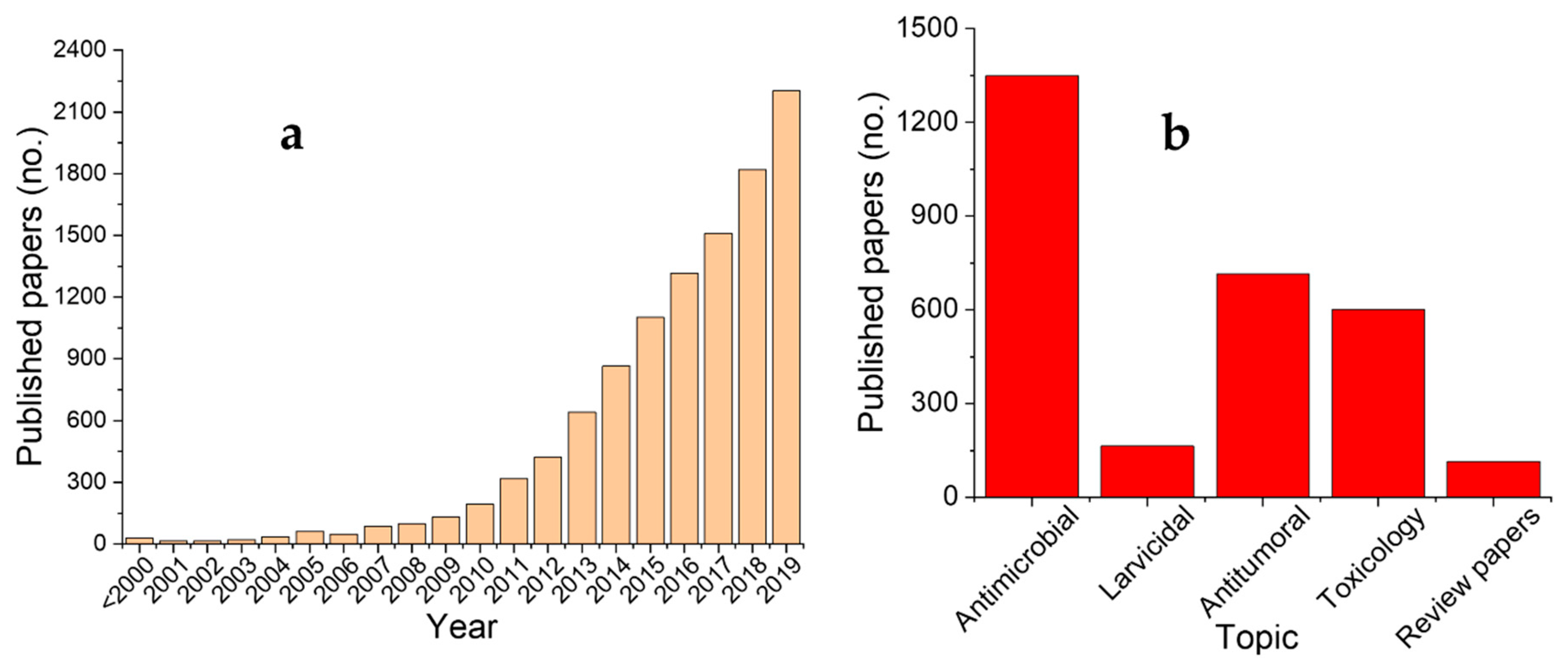
As previously presented, the number of articles about metallic nanoparticles phytosynthesis is increasing from year to year. This could be explained not only by the overall increase of the published scientific literature but also by a growing interest in this area. The field of phytosynthesized NPs, will continuously grow in the following years, as the use of different plant extracts and metallic salts precursors can offer a tremendous variety of differently shaped and sized nanoparticles. At the same time, the thorough understanding and a successful control of the phytosynthesis process in general towards homogenous nanoparticles could benefit from further studies; the continuous search for new alternatives to chemically or physically synthesized nanoparticles for various applications could find an adequate response in this area. Phytosynthesized NPs are close to industrial use for human-related applications. Phytosynthesized Au NPs (obtained by using aqueous extracts of Morinda lucida Benth. leaves) of a specific size and shape (spherical and 10 nm) have been proven to be able to penetrate Stratum Corneum by intercellular paths, opening the possibilities to use the NPs as transdermal transporter [22]. Considering their tremendous potential, the use of phytosynthesized NPs is expected in the near future to pass the barrier from laboratory studies to clinical trials. In this this context, it is worth mentioning that in the Cochrane Database of Systematic Reviews (section Clinical Trials), the metallic or metal oxide nanoparticles represent the subject of very few trials, while the phytosynthesized nanoparticles have not yet been evaluated. For example, some nanoparticles that were obtained by chemical reduction were evaluated in clinical trials, including Ag NPs’ antimicrobial activity and skin irritation potential (30 participants) [23], the antimicrobial potential of denture tissue conditioners, including Ag and ZnO NPs (42 participants) [24], Ag NPs for the treatment of pyorrhea (25 participants) [25], Ag NP-based sprays for reducing the pain that is associated with cesarean wounds (92 participants) [26], Ag NPs as antimicrobial coatings for venous catheters (472 participants) [27]. This would suggest the possibility, in the near future, of developing clinical trials, including phytosynthesized nanoparticles. Phytosynthesized nanoparticles represent a continuously increasing field of research, with numerous studies published each year. Together with the high interest in this area, the quality of the published works is also continuously increasing, switching from routine antioxidant or antimicrobial studies on trivial microbial lines to antibiotic-resistant strains and antitumoral studies. However, this growing interest is not reflected in the studies regarding the toxicological effects of NPs; this should be a subject of particular concern, as the increasing use of NPs in general (and the proposal of phytosynthesized NPs for future applications in particular) could lead to their accumulation in the environment. At the same time, the focus of the researchers should be also switched towards the phytosynthesis of other metallic NPs or metal oxide NPs, as well as the evaluation of their potential applications and toxicological effects.References
- Lekamge, S.; Miranda, A.F.; Abraham, A.; Li, V.; Shukla, R.; Bansal, V.; Nugegoda, D. The toxicity of silver nanoparticles (AgNPs) to three freshwater invertebrates with different life strategies: Hydra vulgaris, Daphnia carinata, and Paratya australiensis. Front. Environ. Sci. 2018, 6, 152.
- Stensberg, M.C.; Wei, Q.; McLamore, E.S.; Porterfield, D.M.; Wei, A.; Sepúlveda, M.S. Toxicological studies on silver nanoparticles: Challenges and opportunities in assessment, monitoring and imaging. Nanomedicine 2011, 6, 879–898.
- Fierascu, R.C.; Ortan, A.; Avramescu, S.M.; Fierascu, I. Phyto-nanocatalysts: Green synthesis, characterization and applications. Molecules 2019, 24, 3418.
- Sana, S.S.; Dogiparthi, L.K. Green synthesis of silver nanoparticles using Givotia moluccana leaf extract and evaluation of their antimicrobial activity. Mat. Lett. 2018, 226, 47–51.
- Abo-zeid, Y.; Urbanowicz, R.A.; Thomson, B.J.; Irving, W.L.; Tarr, A.W.; Garnett, M.C. Enhanced nanoparticle uptake into virus infected cells: Could nanoparticles be useful in antiviral therapy? Int. J. Pharm. 2018, 547, 572–581.
- Sutan, N.A.; Vilcoci, D.S.; Fierascu, I.; Neblea, A.M.; Sutan, C.; Ducu, C.; Soare, L.C.; Negrea, D.; Avramescu, S.M.; Fierascu, R.C. Phytosynthesis of gold and silver nanoparticles enhance in vitro antioxidant and mitostimulatory activity of Aconitum toxicum Reichenb. rhizomes alcoholic extracts. Mater. Sci. Eng. C Mater. Biol. Appl. 2018, 93, 746–758.
- Kim, D.H.; Park, J.C.; Jeon, G.E.; Kim, C.S.; Seo, J.H. Effect of the size and shape of silver nanoparticles on bacterial growth and metabolism by monitoring optical density and fluorescence intensity. Biotechnol. Bioproc. E 2017, 22, 210–217.
- Wang, L.; Hu, C.; Shao, L. The antimicrobial activity of nanoparticles: Present situation and prospects for the future. Int. J. Nanomed. 2017, 12, 1227–1249.
- Dong, Y.; Zhu, H.; Shen, Y.; Zhang, W.; Zhang, L. Antibacterial activity of silver nanoparticles of different particle size against Vibrio natriegens. PLoS ONE 2019, 14, 0222322.
- Cheon, J.Y.; Kim, S.J.; Rhee, Y.H.; Kwon, O.H.; Park, W.H. Shape-dependent antimicrobial activities of silver nanoparticles. Int. J. Nanomed. 2019, 14, 2773–2780.
- Subramanian, P.; Ravichandran, A.; Manoharan, V.; Muthukaruppan, R.; Somasundaram, S.; Pandi, B.; Krishnan, A.; Marimuthu, P.N.; Somasundaram, S.S.N.; You, S.G. Synthesis of Oldenlandia umbellata stabilized silver nanoparticles and their antioxidant effect, antibacterial activity, and bio-compatibility using human lung fibroblast cell line WI-38. Process. Biochem. 2019, 86, 196–204.
- Dakshayani, S.S.; Marulasiddeshwara, M.B.; Sharath Kumar, M.N.; Ramesh, G.; Raghavendra Kumar, P.; Devaraja, S.; Hosamani, R. Antimicrobial, anticoagulant and antiplatelet activities of green synthesized silver nanoparticles using Selaginella (Sanjeevini) plant extract. Int. J. Biol. Macromol. 2019, 131, 787–797.
- Al-Dhafri, K.; Ching, C.L. Phyto-synthesis of silver nanoparticles and its bioactivity response towards nosocomial bacterial pathogens. Biocatal. Agricult. Biotechnol. 2019, 18, 101075.
- Qais, F.A.; Shafiq, A.; Khan, H.M.; Husain, F.M.; Khan, R.A.; Alenazi, B.; Alsalme, A.; Ahmad, I. Antibacterial effect of silver nanoparticles synthesized using Murraya koenigii (L.) against multidrug-resistant pathogens. Bioinorg. Chem. Appl. 2019, 2019, 4649506.
- Tanase, C.; Berta, L.; Coman, N.A.; Roșca, I.; Man, A.; Toma, F.; Mocan, A.; Jakab-Farkas, L.; Biró, D.; Mare, A. Investigation of in vitro antioxidant and antibacterial potential of silver nanoparticles obtained by biosynthesis using beech bark extract. Antioxidants 2019, 8, 459.
- Gopinath, V.; Priyadarshini, S.; Ali, D.M.; Loke, M.F.; Thajuddin, N.; Alharbi, N.S.; Yadavalli, T.; Alagiri, M.; Vadivelu, J. Anti-Helicobacter pylori, cytotoxicity and catalytic activity of biosynthesized gold nanoparticles: Multifaceted application. Arab. J. Chem. 2019, 12, 33–40.
- Zhang, T.; Wang, L.; Chen, Q.; Chen, C. Cytotoxic potential of silver nanoparticles. Yonsei Med. J. 2014, 55, 283–291.
- Steckiewicz, K.P.; Barcinska, E.; Malankowska, A.; Zauszkiewicz–Pawlak, A.; Nowaczyk, G.; Zaleska-Medynska, A.; Inkielewicz-Stepniak, I. Impact of gold nanoparticles shape on their cytotoxicity against human osteoblast and osteosarcoma in in vitro model. Evaluation of the safety of use and anti-cancer potential. J. Mater. Sci. Mater. Med. 2019, 30, 22.
- El-Hawary, S.S.; El-Hefnawy, H.M.; Osman, S.M.; Mostafa, E.S.; Mokhtar, F.A.; El-Raey, M.A. Chemical profile of two Jasminum sambac l. (Ait) cultivars cultivated in Egypt–their mediated silver nanoparticles synthesis and selective cytotoxicity. Int. J. Appl. Pharm. 2019, 11, 154–164.
- Lee, Y.J.; Ahn, E.Y.; Park, Y. Shape-dependent cytotoxicity and cellular uptake of gold nanoparticles synthesized using green tea extract. Nanoscale Res. Lett. 2019, 14, 129.
- Fierascu, R.C.; Fierascu, I.; Lungulescu, E.M.; Nicula, N.; Somoghi, R.; Diţu, L.M.; Ungureanu, C.; Sutan, A.N.; Drăghiceanu, O.A.; Paunescu, A.; et al. Phytosynthesis and radiation-assisted methods for obtaining metal nanoparticles. J. Mater. Sci. 2020, 55, 1915–1932.
- Lin, Q.; Hong, X.; Zhang, D.; Jin, H. Biosynthesis of size-controlled gold nanoparticles using M. lucida leaf extract and their penetration studies on human skin for plastic surgery applications. J. Photochem. Photobiol. B Biol. 2019, 199, 111591.
- Abdellatif, A.A.H. Topical Silver Nanoparticles for Microbial Activity. 2019. Available online: https://clinicaltrials.gov/ct2/show/NCT03752424 (accessed on 28 December 2019).
- Aghajanzadeh, H. Antimicrobial Effects of Nanoparticles in Complete Prostheses. 2019. Available online: https://en.irct.ir/trial/38575 (accessed on 28 December 2019).
- Joshi, I. Use of Silver Nanoparticles for the Treatment of Pyorrhea. 2019. Available online: http://www.ctri.nic.in/Clinicaltrials/pmaindet2.php?trialid=33021 (accessed on 28 December 2019).
- Boroumand, Z.; Golmakani, N.; Mazloum, S.R.; Dadgar, S.; Golmohamadzadeh, S. The Effect of Spray Silver Nanoparticles (Nivasha) on Intensity of Cesarean Wound Pain; A Randomized Clinical Trial. 2018. Available online: https://clinicaltrials.gov/ct2/show/NCT01697748 (accessed on 28 December 2019).
- Antonelli, M. Comparison of Central Venous Catheters with Silver Nanoparticles versus Conventional Catheters (NanoAgCVC). 2011. Available online: https://clinicaltrials.gov/ct2/show/record/NCT00337714 (accessed on 28 December 2019).