Highly active anti-retroviral therapy (HAART) is prescribed for HIV infection and, to a certain extent, limits the infection’s spread. Nanopharmaceuticals offer excellent treatment options for HIV infections by improving the drug potency/efficacy, lowering the dose-related toxicities, and providing active targeting options to the remote HIV reservoirs, leading to the near-total eradication of the virus. Nanopharmaceuticals offer advantages over conventional drug delivery, such as an encapsulation of the drug in nanocarriers, despite its physiochemical properties, providing long-acting treatment options and reducing the dosing because of selective targeting and improvements to the bioavailability of the hydrophobic drugs and adherence of the patients.
- active targeting
- HIV reservoirs
- nanomedicine
- passive targeting
1. Human Immunodeficiency Virus (HIV)
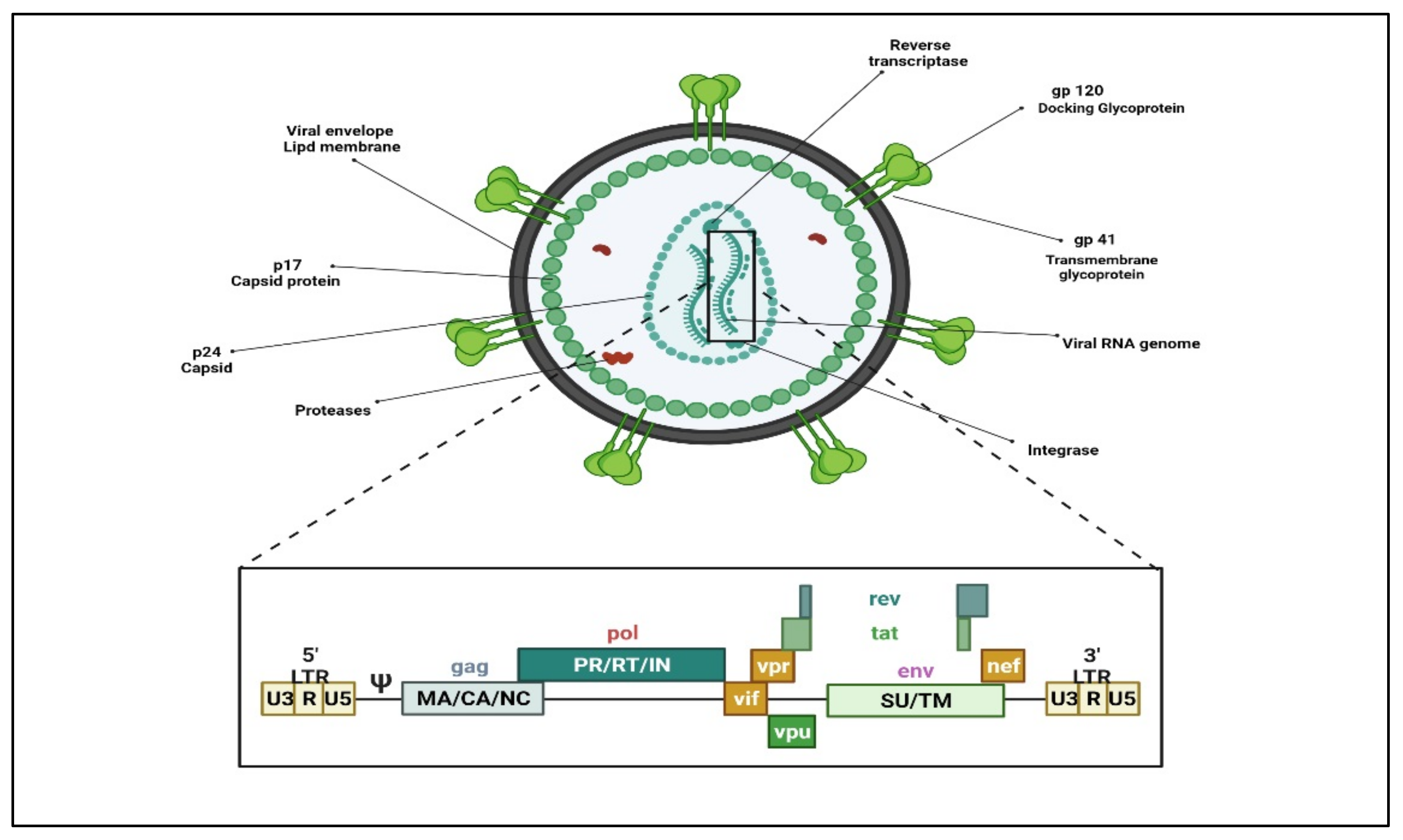
2. Nanopharmaceuticals; Novel Directions on HIV/AIDS Treatment Approaches
Novel ART approaches and nanopharmaceuticals have displayed promising results to a certain extent in HIV/AIDS therapy. Nanomedicine is a field of medicine that employs nanotechnology for the prevention and treatment of diseases utilizing NPs, such as biocompatible NPs [99][11] and nanorobots [100][12] for numerous applications comprising diagnosis [101][13], delivery [102][14], sensory [103][15], or the actuation purposes in a living organism [104][16]. Nanomedicine provides novel approaches to preventing viral infection, growth, and transmission. Many inorganic and metal NPs (i.e., gold, silver, and silica nanoparticles) have been intensively studied for application in imaging, bioassays, and therapeutics [105,106,107,108,109][17][18][19][20][21]. Despite significant advances in HIV/AIDS treatment, there are many lacunas in the HIV/AIDS treatments addressed by nanoart, which target different stages of the HIV lifecycle as per the drug/s mechanism of action [110][22] (Figure 52).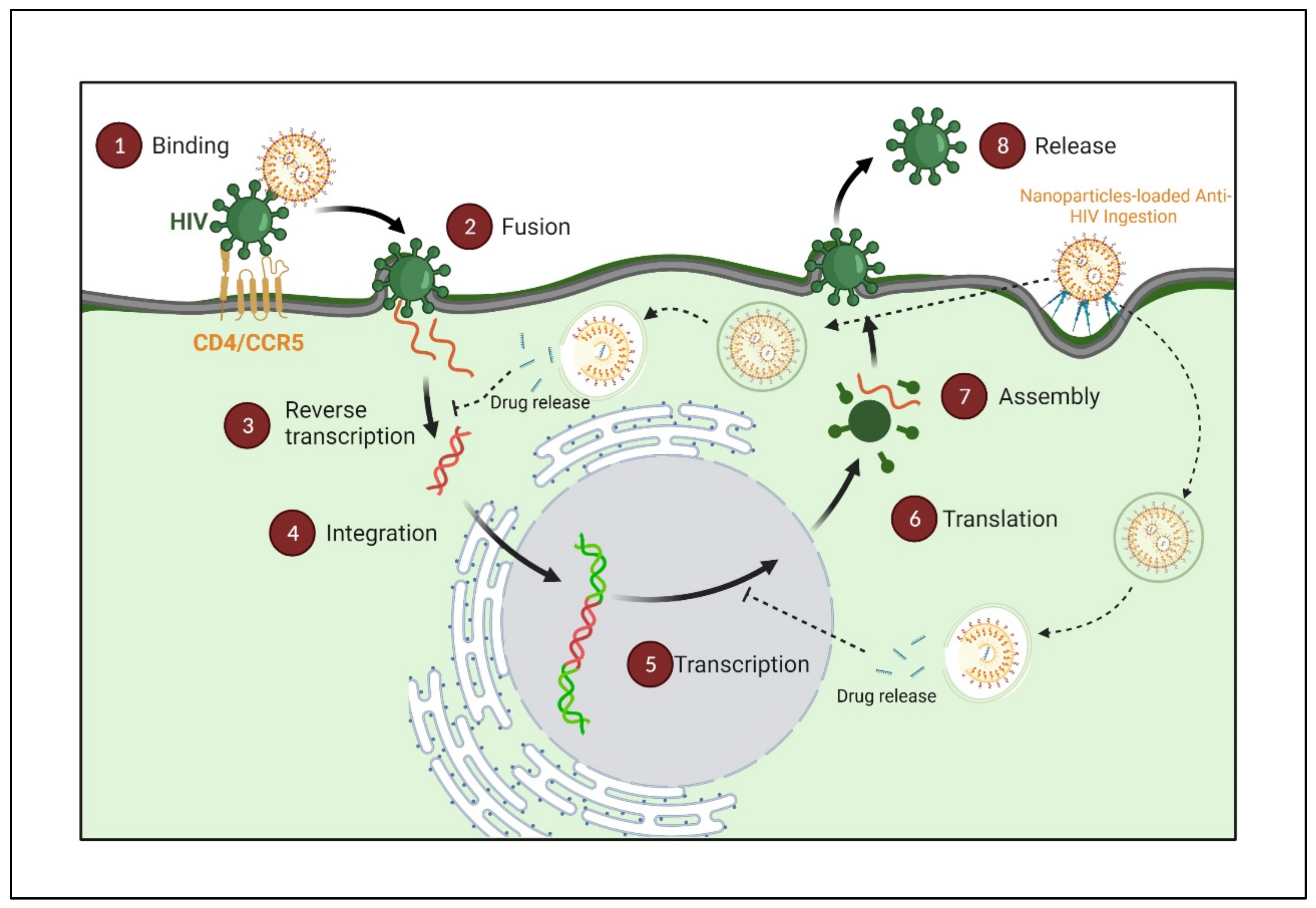
3. Nanoparticles Transport Approaches
Most nanoparticulate drug delivery systems need to cross the cell membrane and deliver the cargo (ART) to elicit an antiviral response. Hence it is imperative to familiarize oneself with the nanoparticle transport approaches. There are multiple ways for nanoparticles to cross the cell membranes, primarily through paracellular or transcellular pathways (Figure 63). The majority, however, reach the target cells via a substrate-specific process known as the endogenous transporter or carrier-mediated route determined by the concentration gradient of the substrates with the help of appropriate transporters [96,111,112][23][24][25].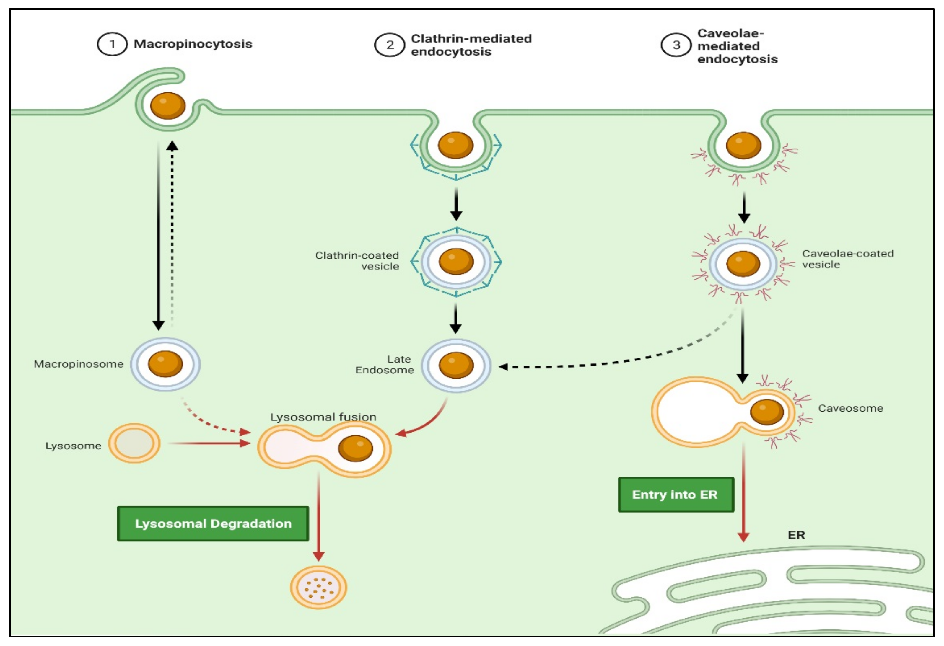
3.1. Active Transport
Active targeting involves the specific modification of drug/drug-loaded nanocarriers with site-specific ligands or “active” agents, which have a discriminating or specific affinity for distinguishing and interacting with a specific cell, tissue, or organ in the body based on its protein expression profile [113][26]. An active targeting approach significantly increases the possibility of the drug target cell’s interaction while sparing healthy cells. The active targeting of particulate carriers can be further classified as;Stimuli-Responsive Nanocarriers
Stimuli-sensitive nanocarriers are based on internal and external stimuli. The internal stimulus includes tumor microenvironment pH and temperature, while the external stimulus includes hyperthermia magnetic field or ultrasound energy [97,98,99,114,115,116][11][27][28][29][30][31].Antibody Targeted Nanocarriers
An antibody having a specific affinity towards an antigen found on the target cell’s surface could be anchored on the nanocarriers’ surface to increase their targeting efficiency. Earlier, this approach was widely explored in cancer therapy, but currently, it has become a key interest in HIV/AIDS therapy. HIV-infected cells express various molecules, such as gp120, the HLA-DR determinant of MHC-II, and CD receptors on their surface, which may be targeted by whole antibodies or fragments of an antibody. Immunoliposomes are widely explored nanocarriers amongst various nanocarriers. Cells, such as follicular dendritic cells, B cells, and macrophages can express the HLA-DR determinant of MHC-II and be theoretically targeted by anti HLA-DR monoclonal antibodies. Immunoliposomes anchored with anti-HLA-DR monoclonal antibodies revealed enhanced accumulation of Indinavir in the lymph nodes, with greater than a 126-fold area-under-the-curve than the free drugs in mice. Kumar et al. demonstrated the effective suppression of HIV infection in humanized mice by targeted siRNA delivery to T cells using an antibody (scFvCD7) specific to the CD7 receptor on the T cells [100,101,102][12][13][14].Receptor-Mediated Endocytosis (RME)
Target cells express various receptors on their surface to enable the internalization of drug-loaded cargoes into the cell and their degradation. The binding of ligand conjugated nanocarriers to receptors present on the cell surface generates a sequence of cellular actions that result in their internalization within the cell. Phagocytic processes are faster than RME, with the ligand playing an essential role in RME. The common RME mechanisms are micropinocytosis, clathrin-dependent endocytosis, caveolae-mediated endocytosis, and clathrin-independent endocytosis. Further, the uptake mechanism is often dependent on the nature of the ligand [96][23]. Macrophages are the primary differentiating cells of the mononuclear phagocyte system and are also responsible for disseminating the infection throughout the body, as mentioned elsewhere. Macrophages residing in the above-mentioned organs serve as a potential reservoir for HIV [97,98,113][26][27][28]. Targeting anti-HIV1 drugs to these macrophages residing in multiple HIV reservoirs would significantly benefit the therapy because many anti-HIV1 drugs administered via the conventional routes fail to penetrate these sites optimally.The d-Mannose Receptor Targeting
The d-mannose receptor (MR, CD206, or MRC1) is a transmembrane glycoprotein classified under the C-type lectin family and present on most macrophages’ surfaces. Its extracellular regions consist of an N-terminal cysteine-rich (CR) domain, which has an affinity to glycoproteins bearing sulfated sugars glycoproteins terminating in 4-SO4GalNAc, a fibronectin II (FNII) domain, and eight carbohydrate recognition domains (CRDs) that bind sugars, such as d-mannose and fucose, with high-affinity. Regardless of the continued development of drug delivery technologies, the effective targeting of drugs to macrophages to treat the underlying diseases remains proven [103,121][15][32]. Mannosylation is currently the best strategy to develop nanomedicines that target d-mannose receptors, which are highly expressed in cells of the immune system [109,133,134,135][21][33][34][35]. Based on the growing literature, nanocarrier mannosylation will increase uptake by macrophages to provide clinically relevant concentrations in target tissues or organs. Moreover, improved uptake is projected to require lower doses of the agents sufficient for therapeutic effects, thus providing reduced toxicity. Researchers have formulated mannosylated polymeric micelles for high efficient delivery of siRNA into macrophages [136][36]. Bhavin et al. have developed mannosylated PLGA nanoparticles that improve brain bioavailability [133,137][33][37]. Moreover, different novel drug delivery approaches in combination with mannosylation for the improvement in selective macrophage uptake, such as polymeric nanoparticle [133,138][33][38], polysaccharide-based vaccine [139][39], liposome [121][32], niosomes [140][40], NLC [134][34], dendrimer [135][35], solid lipid nanoparticles (SLN) [141][41], chitosan nanoparticles [62,142][42][43], and gelatin nanoparticles [128][44] have been evaluated.3.2. Passive Targeting
Passive targeting involves accumulating the drug-carrier system at a site due to physicochemical or pharmacological factors [1,42,46][1][45][46]. Nanoparticle accumulation is observed in the liver due to the large fenestrations. The nanocarriers are readily taken up by the monocyte phagocytic system (MPS) cells or reticuloendothelial system (RES) through phagocytosis. RES consists of fixed macrophage cells in organs, mainly the liver (Kupffer cells) and spleen, lung, kidney, bone marrow, circulating monocytes, macrophages, and polymorph nuclear leukocytes cells [143,144][47][48]. These RES cells cannot identify the particulate carriers themselves but recognize specialized opsonin proteins deposited on the particle surface, followed by MPS uptake [145][49].Endocytosis
Approaches involving passive targeting can result in the accumulation of higher concentrations of drugs at the target sites. This local gradient difference may allow the drug penetration by passive diffusion. Moreover, trafficking via non-receptor-mediated endocytosis (i.e., macropinocytosis) may enhance cellular drug uptake. Actively targeted drug trafficking can be possible via receptor-mediated endocytosis when the periphery of nanocarriers is tagged with ligand molecules matching the specific cell receptor (Figure 74) [112][25].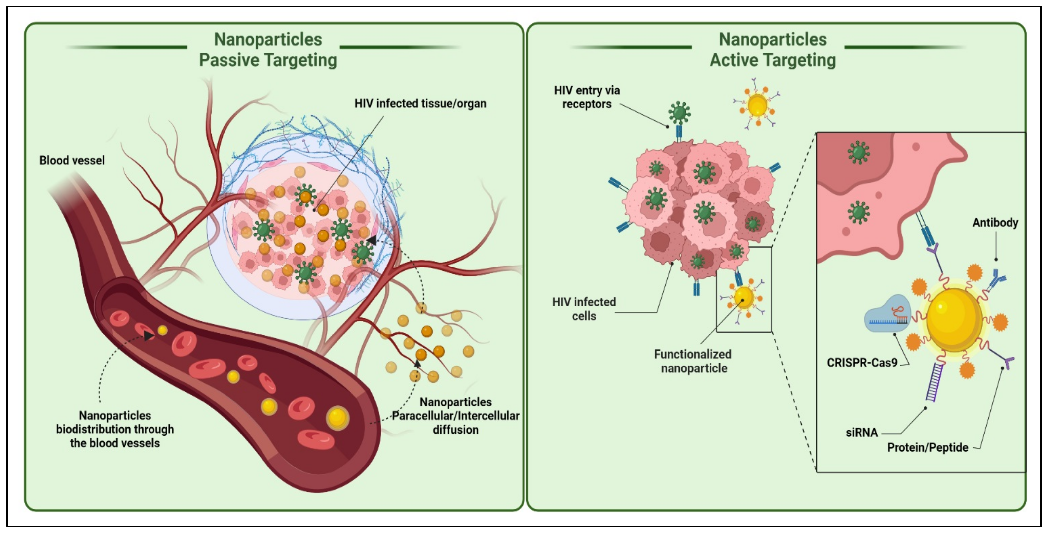
Phagocytosis
It is a process by which cells, generally macrophages, engulf solid particles. The first step of phagocytosis is opsonization, in which opsonin (antibody or complement molecules) cover solid particles [146][50]. Phagocytic cells express Fc and CR1 receptors that bind opsonin molecules and antibodies and complement C3b. Following ingestion, solid particles become trapped in phagocytic vesicles (phagosomes), which fuse with intracellular organelles containing digestive proteins and an acidic internal pH and mature into phagolysosomes that degrade the internalized nano drug delivery system. The nano drug delivery system is then eliminated by exocytosis after degradation or sequestered in residual cells’ bodies if it cannot be digested. Contacts between the nano drug delivery system and macrophages occur via the recognition of opsonin on the nano drug delivery system surface or through interactions with scavenger receptors on macrophages. This fact can target macrophages passively, lymph nodes, and the spleen to treat infections that affect RES (HIV/AIDS) [109,147,148][21][51][52]. Drug or drug carrier nanosystems can be passively targeted by manipulating their physicochemical factors, such as size, shape, surface charge, and surface hydrophobicity [46,130,149][46][53][54].4. Factors Impacting the Functionalities of Nanocarrier Targeted Delivery
4.1. Particle Size
Particle size affects the bioavailability and circulation time of the nanocarriers [134][34]. It also decides the mechanism through which it moves in the cell and its localization. The particle size of nanocarriers is suitable for passive targeting of various HIV reservoir sites. Nanoparticles of particle size >200 nm are opsonized, phagocytosed, and taken up by the macrophages of the RES organs, major HIV reservoir sites, while <200 nm escape phagocytosis and localize in remote reservoirs, such as bone marrow, brain, and gonads, in high concentrations [43][55]. HIV infection is an intracellular infection of macrophages localized primarily in the reticuloendothelial system (RES) organs, including the liver, spleen, lung, lymph node, genitals, lymphocytes, and brain [39][56]. Treating HIV infection with conventional therapies is unmanageable because of their major prevailing limitations, such as poor efficacy and drug resistance [150][57]. The drug did not reach the infected site in conventional therapies due to several HIV reservoir barriers [39,96][23][56]. Moreover, in the case of nanomedicine, it could passively or actively target these viral reservoir sites and eradicate the HIV infection [42,131,151][45][58][59]. Scientists have been studied the numerous nanomedicine for the improvement of anti-HIV therapies, such as liposome [54,152][60][61]; solid lipid nanoparticle [147,153][51][62]; polymeric nanoparticle [148,149][52][54]; and nanoemulsion [125[63][64],154], and nanostructured lipid carriers (NLCs) [143,155][47][65].4.2. Particle Shape
More recently, the effect of particle shape on cell uptake and biodistribution has been recognized. In one study, Mitragotri and Champion reported the surprising finding that particle size and particle shape also affect phagocytosis [151,156][59][66]. A macrophage internalized ellipse pointed end in a few minutes, while the same ellipse has not internalized via the flat region even over 12 h. Spherical nanoparticles were internalized through any point of attachment because of their symmetry. The asymmetric lipomer of doxycycline hydrochloride and amphotericin B revealed enhanced splenic uptake following intravenous administration [43,157][55][67].4.3. Surface Charge
Surface charge and functional groups present on the surface of particulate carrier influence interaction with the cells and further traverse across the negatively charged cell membrane. Positively charged nanoparticles demonstrated higher phagocytic uptake than neutral hydrophilic or negatively charged nanoformulations [158][68]. For instance, polystyrene nanoparticles with a primary amine group at the surface underwent significantly more phagocytosis than nanoparticles with sulfate, hydroxyl, and carboxyl groups. The extended blood circulation half-life of negatively charged particulate carriers could be due to the reduced adsorption of opsonin [159][69]. Further, negatively charged nanoparticles could effectively bound to cationic sites on the macrophages at the scavenger receptors, enabling their uptake by RES [160][70]. The cellular entry of antiretroviral agents that are negatively charged, such as phosphorylated nucleotide analogs and nucleic acids, get faster entry into the cells. The siRNA dendriplexes delivered to human astrocytes decreased the replication of HIV-1 due to higher intracellular concentration [161][71].4.4. Surface Hydrophobicity
The systemic circulation of particulate carriers is strongly influenced by surface hydrophobicity. Particles with hydrophobic surfaces are coated by the complement proteins, albumin, and immunoglobulins and further rapidly cleared by RES (reticuloendothelial system) from the circulation than those with a hydrophilic surface. Surface hydrophobicity thus impacts opsonization, phagocytosis, and the biodistribution of nanoparticles [162,163][72][73].5. Liposomes-Based Delivery Systems for Anti-HIV Therapeutics
Liposomes are small artificial spherical vesicles constituted by one or more phospholipid bilayers with the polar groups of phospholipids oriented to the inner and outer aqueous phase. Such a structure explains the high propensity of liposomes to be encapsulated with hydrophilic, hydrophobic, and amphiphilic drugs within the inner aqueous compartment, the lipid bilayers, and at their interfaces, respectively (Figure 85).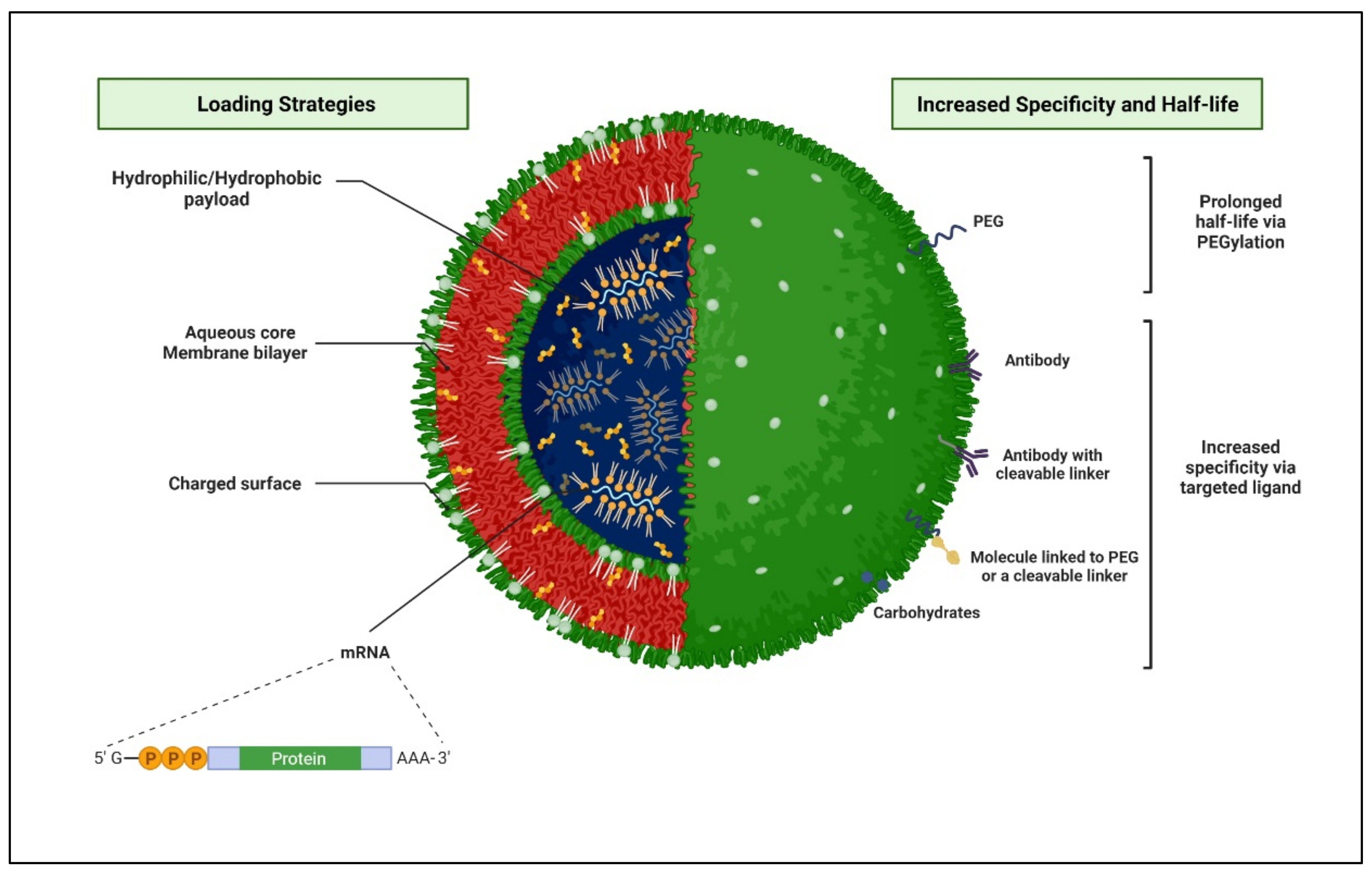
Liposomes-Based Delivery Systems of Ascorbic Acid to Increase the Bioavailability of ARTs
Vitamin C (ascorbic acid) is an essential water-soluble nutrient that functions as a cofactor for numerous enzymatic reactions. Vitamin C also serves as an antioxidant, anti-inflammatory, immunomodulatory, anti-viral, and anti-thrombotic agent and can potentially be used as a therapeutic or prophylactic agent [190][84]. Preliminary clinical evidence showed that massive doses of ascorbic acid (50–200 g per day) can suppress the symptoms of the HIV disease and can markedly reduce the tendency for secondary infections [191][85]. Oxidative stress influences viral replication, inflammatory responses, and immune cell proliferation, all of which contribute to the pathogenesis of HIV/AIDS. Hence, vitamin C can be used to reduce the damage caused by oxidative stress. Given the well-defined involvement of several lipids in the physiology of phagocytosis, the use of bioactive lipid nanoparticles to alter the phagosome maturation process has been proposed as a way to boost the efficacy of innate immunity mechanisms [192][86]. Many experimental findings suggested that a liposome-based delivery system of ascorbic acid may be potentially beneficial to reduce oxidative stress and prevent several HIV chronic conditions and immune system activation. This strategy might help the oral administration of the ARTs [193][87]. However, more research and trials are needed to determine the effects of vitamin C on HIV disease progression and prognosis.56. Nanotechnological Advantages for Effective Anti-HIV Therapy
Nanotechnology-based nanosize drug-loaded carrier design presents manifold advantages, including targeting different anatomical and cellular viral reservoirs, thereby completely eradicating the virus from the reservoir sites. Targeted drug delivery at the site of action improves the drug’s efficacy and reduces the off-target effect. Nanocarriers deliver drugs in a controlled manner, increasing residence time at the target sites, increasing bioavailability, and improving the quality of life of HIV patients [125,194][63][88]. More importantly, a salient feature of nanotechnology that can be exploited for anti-HIV therapy is altered in vivo biodistribution [1,195,196,197][1][89][90][91]. Although conventional drug distribution in the body is based on drug physicochemical properties, the drug properties are overshadowed to achieve carrier-mediated distribution when loaded in nanocarriers [198,199][92][93]. The physicochemical properties of the nanocarriers then become the rate-determining factor for the distribution in the body [200,201,202,203,204,205][94][95][96][97][98][99]. Therefore, altering the same could provide a promising approach to targeting HIV reservoir sites for effective anti-HIV therapy. Targeted drug delivery could be achieved via passive or active targeting [57,206,207][100][101][102].References
- Dalvi, B.R.; Siddiqui, E.A.; Syed, A.S.; Velhal, S.M.; Ahmad, A.; Bandivdekar, A.B.; Devarajan, P.V. Devarajan, P. Nevirapine Loaded Core Shell Gold Nanoparticles by Double Emulsion Solvent Evaporation: In Vitro and In Vivo Evaluation. Curr. Drug Deliv. 2016, 13, 1071–1083.
- Unaids.Org Global HIV & AIDS Statistics—2018 Fact Sheet|UNAIDS. Available online: http://www.unaids.org/en/resources/fact-sheet (accessed on 1 June 2022).
- Mahajan, K.; Rojekar, S.; Desai, D.; Kulkarni, S.; Vavia, P. Efavirenz Loaded Nanostructured Lipid Carriers for Efficient and Prolonged Viral Inhibition in HIV-Infected Macrophages. Pharm. Sci. 2020, 27, 418–432.
- Mahajan, K.; Rojekar, S.; Desai, D.; Kulkarni, S.; Bapat, G.; Zinjarde, S.; Vavia, P. Layer-by-Layer Assembled Nanostructured Lipid Carriers for CD-44 Receptor–Based Targeting in HIV-Infected Macrophages for Efficient HIV-1 Inhibition. AAPS PharmSciTech 2021, 22, 171.
- Rojekar, S.V.; Trimukhe, A.M.; Deshmukh, R.R.; Vavia, P.R. Novel pulsed oxygen plasma mediated surface hydrophılizatıon of ritonavır for the enhancement of wettability and solubility. J. Drug Deliv. Sci. Technol. 2021, 63, 102497.
- Rojekar, S.; Pai, R.; Abadi, L.F.; Mahajan, K.; Prajapati, M.K.; Kulkarni, S.; Vavia, P. Dual loaded nanostructured lipid carrier of nano-selenium and Etravirine as a potential anti-HIV therapy. Int. J. Pharm. 2021, 607, 120986.
- Rojekar, S.; Fotooh, L.; Pai, R.; Mahajan, K.; Kulkarni, S.; Vavia, P.R. Multi-organ Targeting of HIV-1 Viral Reservoirs with Etravirine Loaded Nanostructured Lipid Carrier: An In-vivo Proof of Concept. Eur. J. Pharm. Sci. 2021, 164, 105916.
- Brechtl, J.R.; Breitbart, W.; Galietta, M.; Krivo, S.; Rosenfeld, B. The Use of Highly Active Antiretroviral Therapy (HAART) in Patients with Advanced HIV Infection: Impact on Medical, Palliative Care, and Quality of Life Outcomes. J. Pain Symptom Manag. 2001, 21, 41–51.
- Simon, V.; Ho, D.D.; Karim, Q.A. HIV/AIDS epidemiology, pathogenesis, prevention, and treatment. Lancet 2006, 368, 489–504.
- Sharp, P.; Hahn, B.H. Origins of HIV and the AIDS Pandemic. Cold Spring Harb. Perspect. Med. 2011, 1, a006841.
- Nishioka, Y.; Yoshino, H. Lymphatic targeting with nanoparticulate system. Adv. Drug Deliv. Rev. 2001, 47, 55–64.
- Cavalu, S.; Banica., F.; Gruian, C.; Vanea, E.; Goller, G.; Simon, V. Microscopic and spectroscopic investigation of bioactive glasses for antibiotic controlled release. J. Mol. Struct. 2013, 1040, 47–52.
- Lee, E.S.; Na, K.; Bae, Y.H. Super pH-Sensitive Multifunctional Polymeric Micelle. Nano Lett. 2005, 5, 325–329.
- Kono, K. Thermosensitive polymer-modified liposomes. Adv. Drug Deliv. Rev. 2001, 53, 307–319.
- Kong, G.; Braun, R.D.; Dewhirst, M.W. Hyperthermia Enables Tumor-Specific Nanoparticle Delivery: Effect of Particle Size 1. Cancer Res. 2000, 60, 4440–4445.
- Chen, Q.; Krol, A.; Wright, A.; Nedham, D.; Dewhirst, M.W.; Yuan, F. Tumor microvascular permeability is a key determinant for antivascular effects of doxorubicin encapsulated in a temperature sensitive liposome. Int. J. Hyperth. 2008, 24, 475–482.
- Kim, E.; Yang, J.; Choi, J.; Suh, J.-S.; Huh, Y.-M.; Haam, S. Synthesis of gold nanorod-embedded polymeric nanoparticles by a nanoprecipitation method for use as photothermal agents. Nanotechnology 2009, 20, 365602.
- Kumar, P.; Ban, H.-S.; Kim, S.-S.; Wu, H.; Pearson, T.; Greiner, D.L.; Laouar, A.; Yao, J.; Haridas, V.; Habiro, K.; et al. T Cell-Specific siRNA Delivery Suppresses HIV-1 Infection in Humanized Mice. Cell 2008, 134, 577–586.
- Gagné, J.-F.; Désormeaux, A.; Perron, S.; Tremblay, M.J.; Bergeron, M.G. Targeted delivery of indinavir to HIV-1 primary reservoirs with immunoliposomes. Biochim. Et Biophys. Acta (BBA)—Biomembr. 2001, 1558, 198–210.
- Delcroix, M.; Riley, L.W. Cell-Penetrating Peptides for Antiviral Drug Development. Pharmaceuticals 2010, 3, 448–470.
- Azad, A.K.; Rajaram, M.V.S.; Schlesinger, L.S. Exploitation of the Macrophage Mannose Receptor (CD206) in Infectious Disease Diagnostics and Therapeutics. J. Cytol. Mol. Biol. 2014, 1, 1000003.
- Fraser, I.P.; Ezekowitz, R.A.B. Mannose Receptor and Phagocytosis. Adv. Cell. Mol. Biol. Membr. Organelles 1999, 5, 87–101.
- Shikuma, C.M.; Shiramizu, B. Mitochondrial toxicity associated with nucleoside reverse transcriptase inhibitor therapy. Curr. Infect. Dis. Rep. 2001, 3, 553–560.
- Bañó, M.; Morén, C.; Barroso, S.; Juárez, D.L.; Guitart-Mampel, M.; González-Casacuberta, I.; Canto-Santos, J.; Lozano, E.; León, A.; Pedrol, E.; et al. Mitochondrial Toxicogenomics for Antiretroviral Management: HIV Post-exposure Prophylaxis in Uninfected Patients. Front. Genet. 2020, 11, 497.
- Carr, A.; Miller, J.; Law, M.; Cooper, D.A. A syndrome of lipoatrophy, lactic acidaemia and liver dysfunction associated with HIV nucleoside analogue therapy: Contribution to protease inhibitor-related lipodystrophy syndrome. AIDS 2000, 14, F25–F32.
- Kim, J.; Song, S.Y.; Lee, S.G.; Choi, S.; Lee, Y.I.; Choi, J.Y.; Lee, J.H. Treatment of Human Immunodeficiency Virus-Associated Facial Lipoatrophy with Hyaluronic Acid Filler Mixed With Micronized Cross-Linked Acellular Dermal Matrix. J. Korean Med Sci. 2022, 37, e37.
- Baril, J.-G.; Junod, P.; LeBlanc, R.; Dion, H.; Therrien, R.; Laplante, F.; Falutz, J.; Côté, P.; Hébert, M.-N.; Lalonde, R.; et al. HIV-associated Lipodystrophy Syndrome: A Review of Clinical Aspects. Can. J. Infect. Dis. Med Microbiol. 2005, 16, 233–243.
- Guaraldi, G.; Stentarelli, C.; Zona, S.; Santoro, A. HIV-Associated Lipodystrophy: Impact of Antiretroviral Therapy. Drugs 2013, 73, 1431–1450.
- Kuo, H.-H.; Lichterfeld, M. Recent progress in understanding HIV reservoirs. Curr. Opin. HIV AIDS 2018, 13, 137–142.
- Liu, D.-Z.; Ander, B.P.; Xu, H.; Shen, Y.; Kaur, P.; Deng, W.; Sharp, F.R. Blood-brain barrier breakdown and repair by Src after thrombin-induced injury. Ann. Neurol. 2009, 67, 526–533.
- Teleanu, D.M.; Chircov, C.; Grumezescu, A.M.; Volceanov, A.; Teleanu, R.I. Blood-Brain Delivery Methods Using Nanotechnology. Pharmaceutics 2018, 10, 269.
- Banerjee, D.; Liu, A.P.; Voss, N.R.; Schmid, S.L.; Finn, M.G. Multivalent Display and Receptor-Mediated Endocytosis of Transferrin on Virus-Like Particles. ChemBioChem 2010, 11, 1273–1279.
- Jain, S.K.; Gupta, Y.; Jain, A.; Saxena, A.R.; Khare, P.; Jain, A. Mannosylated gelatin nanoparticles bearing an anti-HIV drug didanosine for site-specific delivery. Nanomed. Nanotechnol. Biol. Med. 2008, 4, 41–48.
- Dutta, T.; Jain, N.K. Targeting potential and anti-HIV activity of lamivudine loaded mannosylated poly (propyleneimine) dendrimer. Biochim. Biophys. Acta (BBA)—Gen. Subj. 2007, 1770, 681–686.
- Patel, B.K.; Parikh, R.H.; Patel, N. Targeted delivery of mannosylated-PLGA nanoparticles of antiretroviral drug to brain. Int. J. Nanomed. 2018, 13, 97–100.
- Kaur, A.; Jain, S.; Tiwary, A. Mannan-coated gelatin nanoparticles for sustained and targeted delivery of didanosine: In vitro and in vivo evaluation. Acta Pharm. 2008, 58, 61–74.
- Rao, K.S.; Reddy, M.K.; Horning, J.L.; Labhasetwar, V. TAT-conjugated nanoparticles for the CNS delivery of anti-HIV drugs. Biomaterials 2008, 29, 4429–4438.
- Kuo, Y.-C.; Wang, L.-J. Transferrin-grafted catanionic solid lipid nanoparticles for targeting delivery of saquinavir to the brain. J. Taiwan Inst. Chem. Eng. 2014, 45, 755–763.
- Lavigne, C.; Guedj, A.-S.; Kell, A.J.; Barnes, M.; Stals, S.; Gonçalves, D.; Girard, D. Preparation, characterization, and safety evaluation of poly (lactide-co-glycolide) nanoparticles for protein delivery into macrophages. Int. J. Nanomed. 2015, 10, 5965–5979.
- Shegokar, R. Preparation, Characterization and Cell Based Delivery of Stavudine Surface Modified Lipid Nanoparticles. J. Nanomed. Biother. Discov. 2012, 2.
- Yu, S.S.; Lau, C.M.; Barham, W.J.; Onishko, H.M.; Nelson, C.E.; Li, H.; Smith, C.A.; Yull, F.E.; Duvall, C.L.; Giorgio, T.D. Macrophage-Specific RNA Interference Targeting via “Click”, Mannosylated Polymeric Micelles. Mol. Pharm. 2013, 10, 975–987.
- Kedzierska, K. The Role of Monocytes and Macrophages in the Pathogenesis of HIV-1 Infection. Curr. Med. Chem. 2002, 9, 1893–1903.
- Jamari, J.; Ammarullah, M.I.; Santoso, G.; Sugiharto, S.; Supriyono, T.; Prakoso, A.T.; Basri, H.; van der Heide, E. Computational Contact Pressure Prediction of CoCrMo, SS 316L and Ti6Al4V Femoral Head against UHMWPE Acetabular Cup under Gait Cycle. J. Funct. Biomater. 2022, 13, 64.
- Garg, M.; Dutta, T.; Jain, N.K. Reduced hepatic toxicity, enhanced cellular uptake and altered pharmacokinetics of stavudine loaded galactosylated liposomes. Eur. J. Pharm. Biopharm. 2007, 67, 76–85.
- Cao, S.; Woodrow, K.A. Nanotechnology Approaches to Eradicating HIV Reservoirs. Eur. J. Pharm. Biopharm. 2019, 138, 48–63.
- Jindal, A.; Bachhav, S.; Devarajan, P.V. In situ hybrid nano drug delivery system (IHN-DDS) of antiretroviral drug for simultaneous targeting to multiple viral reservoirs: An in vivo proof of concept. Int. J. Pharm. 2017, 521, 196–203.
- Shibata, A.; McMullen, E.; Pham, A.; Belshan, M.; Sanford, B.; Zhou, Y.; Goede, M.; Date, A.; Destache, C.J. Polymeric Nanoparticles Containing Combination Antiretroviral Drugs for HIV Type 1 Treatment. AIDS Res. Hum. Retroviruses 2013, 29, 746–754.
- Liu, C.; Zhou, Q.; Li, Y.; Garner, L.V.; Watkins, S.P.; Carter, L.J.; Smoot, J.; Gregg, A.C.; Daniels, A.D.; Jervey, S.; et al. Research and Development on Therapeutic Agents and Vaccines for COVID-19 and Related Human Coronavirus Diseases. ACS Cent. Sci. 2020, 6, 315–331.
- Malik, T.; Chauhan, G.; Rath, G.; Kesarkar, R.N.; Chowdhary, A.S.; Goyal, A.K. Efaverinz and nano-gold-loaded mannosylated niosomes: A host cell-targeted topical HIV-1 prophylaxis via thermogel system. Artif. Cells Nanomed. Biotechnol. 2017, 46, 79–90.
- Vieira, A.C.; Magalhães, J.; Rocha, S.; Cardoso, M.S.; Santos, S.G.; Borges, M.; Pinheiro, M.; Reis, S. Targeted macrophages delivery of rifampicin-loaded lipid nanoparticles to improve tuberculosis treatment. Nanomedicine 2017, 12, 2721–2736.
- Makabi-Panzu, B.; Lessard, C.; Beauchamp, D.; Désormeaux, A.; Poulin, L.; Tremblay, M.; Bergeron, M.G. Uptake and Binding of Liposomal 2’,3’-Dideoxycytidine by RAW 264.7 Cells: A Three-Step Process. J. Acquir. Immune Defic. Syndr. Hum. Retrovirology 1995, 8, 227–235.
- Hillaireau, H.; Couvreur, P. Nanocarriers’ entry into the cell: Relevance to drug delivery. Cell. Mol. Life Sci. 2009, 66, 2873–2896.
- Kaur, C.D.; Nahar, M.; Jain, N.K. Lymphatic targeting of zidovudine using surface-engineered liposomes. J. Drug Target. 2008, 16, 798–805.
- Kulkarni, S.A.; Feng, S.-S. Effects of Particle Size and Surface Modification on Cellular Uptake and Biodistribution of Polymeric Nanoparticles for Drug Delivery. Pharm. Res. 2013, 30, 2512–2522.
- Klatzmann, D.; Barré-Sinoussi, F.; Nugeyre, M.T.; Danguet, C.; Vilmer, E.; Griscelli, C.; Brun-Veziret, F.; Rouzioux, C.; Gluckman, J.C.; Chermann, J.-C.; et al. Selective Tropism of Lymphadenopathy Associated Virus (LAV) for Helper-Inducer T Lymphocytes. Science 1984, 225, 59–63.
- Hajjar, A.M.; Lewis, P.F.; Endeshaw, Y.; Ndinya-Achola, J.; Kreiss, J.K.; Overbaugh, J. Efficient Isolation of Human Immunodeficiency Virus Type 1 RNA from Cervical Swabs. J. Clin. Microbiol. 1998, 36, 2349–2352.
- Jain, A.; Agarwal, A.; Majumder, S.; Lariya, N.; Khaya, A.; Agrawal, H.; Majumdar, S.; Agrawal, G.P. Mannosylated solid lipid nanoparticles as vectors for site-specific delivery of an anti-cancer drug. J. Control. Release 2010, 148, 359–367.
- Shahiwala, A.; Amiji, M.M. Nanotechnology-based delivery systems in HIV/AIDS therapy. Futur. HIV Ther. 2007, 1, 49–59.
- Edagwa, B.J.; Zhou, T.; McMillan, J.M.; Liu, X.-M.; E Gendelman, H. Development of HIV Reservoir Targeted Long Acting Nanoformulated Antiretroviral Therapies. Curr. Med. Chem. 2014, 21, 4186–4198.
- Eisele, E.; Siliciano, R.F. Redefining the Viral Reservoirs that Prevent HIV-1 Eradication. Immunity 2012, 37, 377–388.
- Trimukhe, A.; Rojekar, S.; Vavia, P.R.; Deshmukh, R. Pulsed plasma surface modified omeprazole microparticles for delayed release application. J. Drug Deliv. Sci. Technol. 2021, 66, 102905.
- Nie, S. Understanding and Overcoming Major Barriers in Cancer Nanomedicine. Opsonization Phagocytosis 2010, 5, 523–528.
- Campagne, M.V.L.; Wiesmann, C.; Brown, E.J. Macrophage complement receptors and pathogen clearance. Cell. Microbiol. 2007, 9, 2095–2102.
- Litzinger, D.C.; Buiting, A.M.; van Rooijen, N.; Huang, L. Effect of liposome size on the circulation time and intraorgan distribution of amphipathic poly(ethylene glycol)-containing liposomes. Biochim. Biophys. Acta (BBA)—Biomembr. 1994, 1190, 99–107.
- Saste, V.S.; Kale, S.S.; Sapate, M.K.; Baviskar, D.T. Modern Aspects for Antiretroviral Treatment. Int. J. Pharm. Sci. Rev. Res. 2011, 9, 18–24.
- Raina, H.; Kaur, S.; Jindal, A.B. Development of efavirenz loaded solid lipid nanoparticles: Risk assessment, quality-by-design (QbD) based optimisation and physicochemical characterisation. J. Drug Deliv. Sci. Technol. 2017, 39, 180–191.
- Alukda, D.; Sturgis, T.; Youan, B.C. Formulation of tenofovir-loaded functionalized solid lipid nanoparticles intended for HIV prevention. J. Pharm. Sci. 2011, 100, 3345–3356.
- Hari, B.V.; Narayanan, N.; Dhevendaran, K.; Ramyadevi, D. Engineered nanoparticles of Efavirenz using methacrylate co-polymer (Eudragit-E100) and its biological effects in-vivo. Mater. Sci. Eng. C 2016, 67, 522–532.
- Iannazzo, D.; Pistone, A.; Romeo, R.; Giofrè, S.V. Nanotechnology Approaches for Antiretroviral Drugs Delivery. J. AIDS HIV Infect. 2015, 1, 1–13.
- Kotta, S.; Khan, A.W.; Ansari, S.H.; Sharma, R.K.; Ali, J. Anti HIV nanoemulsion formulation: Optimization and in vitro–in vivo evaluation. Int. J. Pharm. 2014, 462, 129–134.
- Garg, B.; Beg, S.; Kumar, R.; Katare, O.; Singh, B. Nanostructured lipidic carriers of lopinavir for effective management of HIV-associated neurocognitive disorder. J. Drug Deliv. Sci. Technol. 2019, 53, 101220.
- Chakraborty, T.; Das, M.K.; Dutta, L.; Mukherjee, B.; Das, S.; Sarma, A. Successful Delivery of Zidovudine-Loaded Docosanol Nanostructured Lipid Carriers (Docosanol NLCs) into Rat Brain. In Surface Modification of Nanoparticles for Targeted Drug Delivery; Springer International Publishing: Berlin/Heidelberg, Germany, 2019; pp. 245–276.
- Dusserre, N.; Lessard, C.; Paquette, N.; Perron, S.; Poulin, L.; Tremblay, M.; Beauchamp, D.; Désormeaux, A.; Bergeron, M.G. Encapsulation of foscarnet in liposomes modifies drug intracellular accumulation, in vitro anti-HIV-1 activity, tissue distribution, and pharmacokinetics. AIDS 1995, 9, 833–842.
- Freitas, R.A. Pharmacytes: An Ideal Vehicle for Targeted Drug Delivery. J. Nanosci. Nanotechnol. 2006, 6, 2769–2775.
- Houacine, C.; Adams, D.; Singh, K. Impact of liquid lipid on development and stability of trimyristin nanostructured lipid carriers for oral delivery of resveratrol. J. Mol. Liq. 2020, 316, 113734.
- Désormeaux, A.; Harvie, P.; Perron, S.; Makabi-Panzu, B.; Beauchamp, D.; Tremblay, M.; Poulin, L.; Bergeron, M.G. Antiviral efficacy, intracellular uptake and pharmacokinetics of free and liposome-encapsulated 2’,3’-dideoxyinosine. AIDS 1994, 8, 1545–1554.
- Purvin, S.; Vuddanda, P.R.; Singh, S.K.; Jain, A. Pharmacokinetic and Tissue Distribution Study of Solid Lipid Nanoparticles of Zidovudine in Rats. J. Nanotechnol. 2014, 2014, 1–7.
- Pharmacokinetic and Tissue Distribution of Zidovudine in Rats Following Intravenous Administration of Zidovudine Myristate Loaded Liposomes. Available online: https://www.researchgate.net/publication/7448025_Pharmacokinetic_and_tissue_distribution_of_zidovudine_in_rats_following_intravenous_administration_of_zidovudine_myristate_loaded_liposomes (accessed on 22 March 2021).
- Destache, C.J.; Belgum, T.; Goede, M.; Shibata, A.; Belshan, M.A. Antiretroviral release from poly (DL-lactide-co-glycolide) nanoparticles in mice. J. Antimicrob. Chemother. 2010, 65, 2183–2187.
- Publications. Available online: https://universe.bits-pilani.ac.in/Hyderabad/punnaraoravi/Publications (accessed on 22 March 2021).
- Kawai, T.; Akira, S. Toll-like Receptor and RIG-1-like Receptor Signaling. Ann. N. Y. Acad. Sci. 2008, 1143, 1–20.
- Davidson, I.; Beardsell, H.; Smith, B.; Mandalia, S.; Bower, M.; Gazzard, B.; Nelson, M.; Stebbing, J. The frequency and reasons for antiretroviral switching with specific antiretroviral associations: The SWITCH study. Antivir. Res. 2010, 86, 227–229.
- Gaur, P.K.; Mishra, S.; Bajpai, M.; Mishra, A. Enhanced Oral Bioavailability of Efavirenz by Solid Lipid Nanoparticles: In Vitro Drug Release and Pharmacokinetics Studies. BioMed Res. Int. 2014, 2014, 1–9.
- Zardini, A.A.; Mohebbi, M.; Farhoosh, R.; Bolurian, S. Production and characterization of nanostructured lipid carriers and solid lipid nanoparticles containing lycopene for food fortification. J. Food Sci. Technol. 2017, 55, 287–298.
- Talegaonkar, S.; Bhattacharyya, A. Potential of Lipid Nanoparticles (SLNs and NLCs) in Enhancing Oral Bioavailability of Drugs with Poor Intestinal Permeability. AAPS PharmSciTech 2019, 20, 121.
- Desai, J.; Thakkar, H. Darunavir-Loaded Lipid Nanoparticles for Targeting to HIV Reservoirs. AAPS PharmSciTech 2017, 19, 648–660.
- Bhalekar, M.R.; Upadhaya, P.G.; Madgulkar, A.R.; Kshirsagar, S.J.; Dube, A.; Bartakke, U.S. In-vivo bioavailability and lymphatic uptake evaluation of lipid nanoparticulates of darunavir. Drug Deliv. 2015, 23, 2581–2586.
- Stavudine Entrapped Lipid Nanoparticles for Targeting Lymphatic HIV Reservoirs—PubMed. Available online: https://pubmed.ncbi.nlm.nih.gov/21612153/ (accessed on 30 August 2020).
- Kuo, Y.-C.; Chung, J.-F. Physicochemical properties of nevirapine-loaded solid lipid nanoparticles and nanostructured lipid carriers. Colloids Surf. B Biointerfaces 2011, 83, 299–306.
- Khan, A.A.; Mudassir, J.; Akhtar, S.; Murugaiyah, V.; Darwis, Y. Freeze-Dried Lopinavir-Loaded Nanostructured Lipid Carriers for Enhanced Cellular Uptake and Bioavailability: Statistical Optimization, in Vitro and in Vivo Evaluations. Pharmaceutics 2019, 11, 97.
- Endsley, A.N.; Ho, R.J. Enhanced Anti-HIV Efficacy of Indinavir After Inclusion in CD4-Targeted Lipid Nanoparticles. JAIDS J. Acquir. Immune Defic. Syndr. 2012, 61, 417–424.
- Makwana, V.; Jain, R.; Patel, K.; Nivsarkar, M.; Joshi, A. Solid lipid nanoparticles (SLN) of Efavirenz as lymph targeting drug delivery system: Elucidation of mechanism of uptake using chylomicron flow blocking approach. Int. J. Pharm. 2015, 495, 439–446.
- Park, K.S.; Bazzill, J.D.; Son, S.; Nam, J.; Shin, S.W.; Ochyl, L.J.; Stuckey, J.A.; Meagher, J.L.; Chang, L.; Song, J.; et al. Lipid-based vaccine nanoparticles for induction of humoral immune responses against HIV-1 and SARS-CoV-2. J. Control Release 2020, 330, 529–539.
- Saunders, K.O.; Pardi, N.; Parks, R.; Santra, S.; Mu, Z.; Sutherland, L.; Scearce, R.; Barr, M.; Eaton, A.; Hernandez, G.; et al. Lipid nanoparticle encapsulated nucleoside-modified mRNA vaccines elicit polyfunctional HIV-1 antibodies comparable to proteins in nonhuman primates. npj Vaccines 2021, 6, 50.
- Bae, M.; Kim, H. The Role of Vitamin C, Vitamin D, and Selenium in Immune System against COVID-19. Molecules 2020, 25, 5346.
- Cathcart, R.F. Vitamin C in the treatment of acquired immune deficiency syndrome (AIDS). Med. Hypotheses 1984, 14, 423–433.
- Cavalu, S.; Antoniac, I.V.; Mohan, A.; Bodog, F.; Doicin, C.; Mates, I.; Ulmeanu, M.; Murzac, R.; Semenescu, A. Nanoparticles and Nanostructured Surface Fabrication for Innovative Cranial and Maxillofacial Surgery. Materials 2020, 13, 5391.
- Berretta, M.; Quagliariello, V.; Maurea, N.; Di Francia, R.; Sharifi, S.; Facchini, G.; Rinaldi, L.; Piezzo, M.; Manuela, C.; Nunnari, G.; et al. Multiple Effects of Ascorbic Acid against Chronic Diseases: Updated Evidence from Preclinical and Clinical Studies. Antioxidants 2020, 9, 1182.
- Mallipeddi, R.; Rohan, L.C. Progress in antiretroviral drug delivery using nanotechnology. Int. J. Nanomed. 2010, 5, 533–547.
- Kumar, L.; Verma, S.; Prasad, D.N.; Bhardwaj, A.; Vaidya, B.; Jain, A.K. Nanotechnology: A magic bullet for HIV AIDS treatment. Artif. Cells Nanomed. Biotechnol. 2014, 43, 71–86.
- Date, A.A.; Destache, C.J. A review of nanotechnological approaches for the prophylaxis of HIV/AIDS. Biomaterials 2013, 34, 6202–6228.
- Gupta, U.; Jain, N.K. Non-polymeric nano-carriers in HIV/AIDS drug delivery and targeting. Adv. Drug Deliv. Rev. 2010, 62, 478–490.
