Separation and Analytical Techniques Used in Snake Venomics: Comparison
Please note this is a comparison between Version 2 by Peter Tang and Version 1 by christina Elie SAHYOUN.

The deleterious consequences of snake envenomation are due to the extreme protein complexity of snake venoms. Therefore, the identification of their components is crucial for understanding the clinical manifestations of envenomation pathophysiology and for the development of effective antivenoms. In addition, snake venoms are considered as libraries of bioactive molecules that can be used to develop innovative drugs. Numerous separation and analytical techniques are combined to study snake venom composition including chromatographic and electrophoretic techniques.

  • separation techniques
  • analytical techniques
  • snake venom
  • biomolecules
  • bioassay-guided fractionation
  • snake venomics

1. Introduction

Venomous animals are widely spread all over the world. Accordingly, animal–human encounters are frequent in endemic regions, possibly ending up with human envenomation. Animal venoms induce different types of symptoms, leading sometimes to death [1]. Snake venoms (SVs) are the highlights of research owing to their fascinating complexity and their deadly effects [2]. Snakebite envenoming is a global health issue affecting up to 2.7 million people per year. The death toll is estimated to reach 138,000 deaths per year (https://www.who.int/news-room/fact-sheets/detail/snakebite-envenoming (accessed on 23 January 2022)). Due to their highly diverse composition, SVs cause a variety of clinical manifestations ranging from local tissue necrosis to systemic cardiovascular and neurologic symptoms. Nowadays, the single efficient treatment for snake envenomation is antivenom. Yet, the development of effective antivenoms is hampered by the complexity and the limited understanding of the composition and biological activities of SVs, among others [3]. In addition to the appropriate development of antivenoms, a detailed characterization of SV components will help in the interpretation of clinical symptoms. It will also pave the way for the discovery of novel biomolecules with therapeutic interest [4]. To date, various drugs originating from SVs have been approved for clinical use. The most prominent drug CaptoprilTM, an angiotensin-converting enzyme inhibitor, is indicated for the treatment of hypertension. It was developed from a bradykinin-potentiating enzyme isolated from Bothrops jararaca venom [5]. AggrastatTM (Echistatin), another medication developed from Echis carinatus SV, is approved for the prevention of thrombotic complications. Reptilase isolated from different venoms of Bothrops species is approved for use during surgery for the prophylaxis and treatment of hemorrhage [6]. Accordingly, the exploration of SV components is of high significance to the medical field [7].
In fact, proteins and peptides constitute 90 to 95% of SV dry weight. They might contain more than 100 proteins mostly belonging to around 8–11 protein families [8]. Consequently, SVs are highly heterogeneous and require robust techniques to separate and identify protein isoforms. Recent advances in protein separation techniques provided means with higher resolution to evaluate these extremely heterogeneous mixtures. The separation of venom components is the first step in the process of venom decomplexation and analysis. It allows the repartition of molecules based on different biological, chemical, and physical properties [9].

2. Methods Used for Separation of Venom Complex Mixtures

2.1. Chromatographic Techniques

Chromatographic methods are mostly employed for the separation of SVs and are preferred over electrophoretic techniques since they provide better resolution [10]. Liquid chromatography (LC) involves the partition of molecules between two phases: a stationary and a mobile phase. Molecules with different biochemical and physical features will interact differently with the stationary phase and thus will be separated from other components of the mixture [11]. Different types of chromatography are currently used for the separation of SVs, namely (i) size exclusion chromatography, (ii) ion-exchange chromatography, (iii) affinity chromatography and (iv) reversed-phase chromatography. A single chromatographic step is usually insufficient to isolate a molecule or reveal composition of the complex SV mixture. Hence, multiple chromatographic steps can be coupled together, which is known as multidimensional chromatography. Additionally, LC might be coupled with electrophoretic techniques for an improved protein isolation.

2.2. Electrophoretic Techniques

The high versatility, feasibility, and practicality of chromatographic techniques outweighed those of electrophoretic techniques; nevertheless, the latter remain an important means for the separation of a variety of venoms (Table 1). For SVs, electrophoretic techniques are used rigorously for the separation and identification of protein content of whole venom and fractions. Even though electrophoretic methods have several limitations, their use is integral in the process of SV analysis. Two major types of electrophoretic methods are currently used: one-dimensional gel electrophoresis and two-dimensional gel electrophoresis.
Table 1.
The advantages and disadvantages of different separation techniques used for snake venom analysis.

Separation Technique

Advantages

Disadvantages

Size exclusion chromatography

-

Simple to design (isocratic gradient)

-

Arranges SV proteins into fractions of known MWs

-

No sample loss since solutes do not interact with stationary phase

-

Short analysis time

-

Low resolution compared to other techniques

-

Limited number of peaks can be resolved

-

Sample evidently requires further separation to reach purity

Ion-exchange chromatography

-

Resins have long life

-

Buffers are suitable and non-denaturing

-

High-resolution separation

-

Can only be used for charged molecules

-

Small changes in pH alter the binding of molecules

Affinity chromatography

-

High specificity

-

High resolution

-

Purification could be performed in a single step

-

Harsh buffers used could alter the structure and function of target proteins

-

Resin development is time-consuming and costly

RP-HPLC

-

Rapid, efficient, and accurate

-

Use of water-based solvents that are compatible with SV proteins

-

Most compatible with mass spectrometry

-

Costly organic solvents

-

Coelution of molecules with same polarity

1D gel electrophoresis

-

Gives an idea of the distribution of molecules in each fraction

-

Helps determine protein molecular weights

-

Requires small amount of sample

-

Procedure is time consuming

-

Large amounts of sample could not be separated at once

-

Suitable only for whole proteome analysis

2-D gel electrophoresis

-

Good for comparison of snake venoms

-

Whole proteome analysis could be performed in a single separation step

-

Inefficient separation

-

Poor reproducibility

-

Spot trains (hard to analyze each spot apart)

-

Low amount of proteins prevents identification my MS

3. Implementation of Separation Methods for SVs

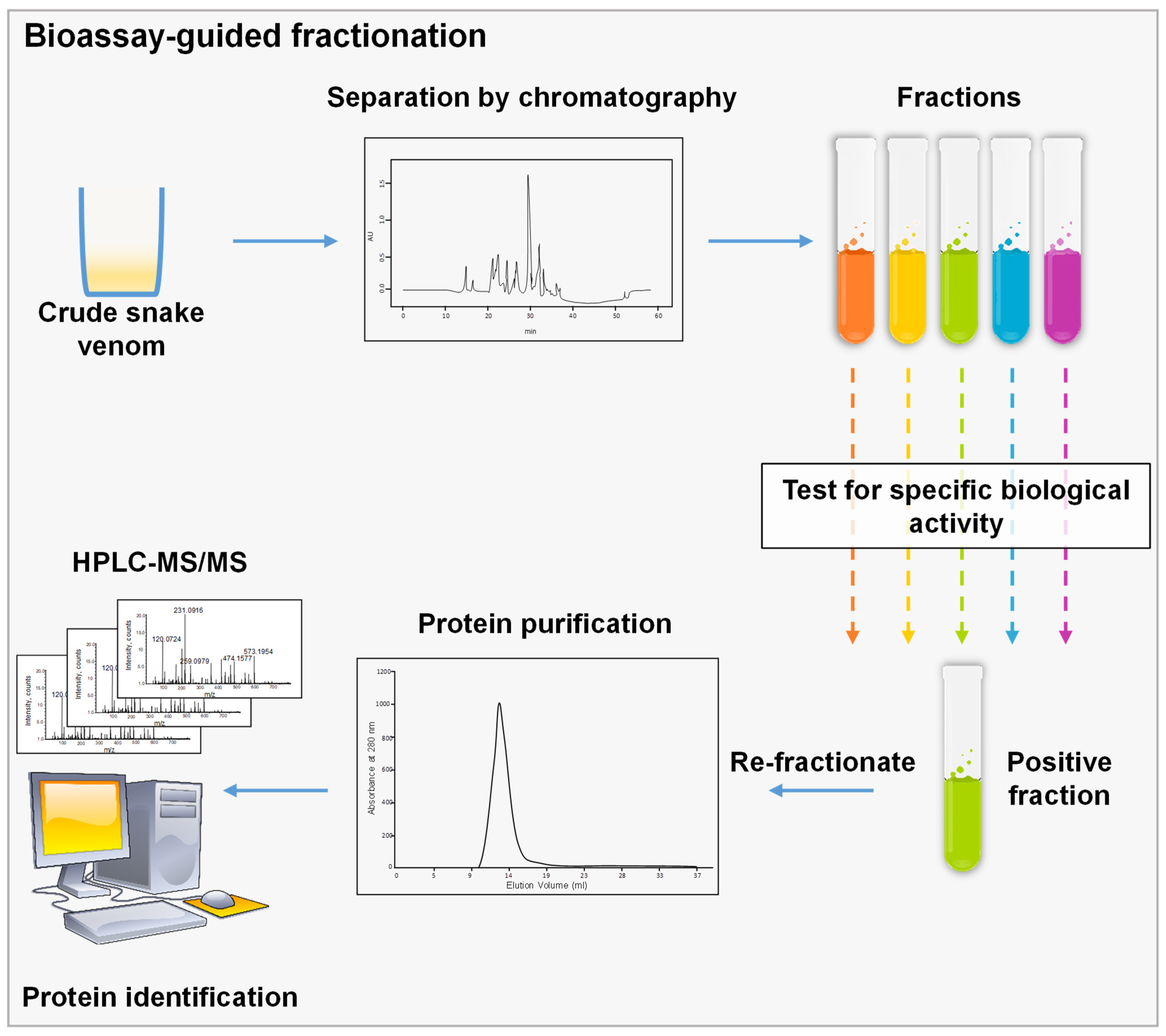
3.1. Bioassay-Guided Fractionation

It uses biological assays to perform a fractionation aiming to isolate a specific molecule. During the process, a complex mixture is separated typically by chromatographic techniques, and each fraction is tested separately for a specific biological feature. The fractions of interest are then re-fractionated until a pure protein is recovered (Figure 1) [58][12]. The choice of the assay to be used is directly related to the molecule to be isolated. For SVs, numerous bioassays are currently used for different specific components. In fact, SVs induce a wide spectrum of effects once injected in prey, since they target several organ systems: namely, the nervous and the cardiovascular systems [59][13]. Different bioassays are developed for the isolation of cardiotoxic and neurotoxic components. Since SV cardiotoxic components are known to have effects on the cardiac muscle or vascular smooth muscle [59][13], several bioassays might be used for the isolation of cardiotoxic components. Firstly, Langendorff model heart preparations could be used to screen for cardiotoxic molecules. For example, this model was used to screen for the cardiotoxic component of Vipera ammodytes venom. First, the venom was fractionated using SEC and generated four fractions with fraction C having the highest cardiotoxicity. The subfraction C1 induced an irreversible cardiac arrest of the isolated heart, while subfraction C2 induced an irreversible significant decrease in heart rate without inducing cardiac arrest. Consequently, both fractions were re-fractionated using an RP-HPLC C4 column, and Ammodytin L (AtnL) was identified as the major protein inducing cardiotoxicity [60][14]. Another robust biological model to analyze cardiotoxicity could be the zebrafish model. This model is used to monitor atrial and ventricular rates, blood flow and clot formation. Recently, wthe researchers took advantage of zebrafish embryos transparency to assess the direct cardiotoxicity of SV [61][15]. In addition, the zebrafish model was used to isolate the cardiotoxic molecule from Lachesis muta venom. First, crude venom was fractionated using SEC; then, the active fractions were fractionated using AEX, and fraction 8 with the highest cardiotoxicity was further purified using an RP-HPLC C18 column, generating a pure protein “mutacytin-1” [62][16]. Moreover, isolated rat mesenteric arteries might also be used to evaluate the vasorelaxant effect of fractions. This technique was used to isolate the vasorelaxant molecule in Bothrops leucurus venom. Crude venom was fractionated with CEX followed by RP-HPLC for the vasorelaxant fraction. After MS analysis, the molecule inducing vasorelaxation turned to be a PLA2 [63][17]. On the other hand, several neurotoxic components have been isolated and identified from SVs by the means of bioassay-guided fractionation. One of the most common neurotoxic assays used is the chick biventer cervicis nerve-muscle preparation. Neurotoxic components usually induce an inhibition of indirect twitches, leading to a decrease in muscle contraction in a concentration-dependent manner. This assay was used in tandem with SEC to isolate hostoxin-1 from Hoplocephalus stephensi [64][18], rufoxin from Rhamphiophis oxyrhynchus [65][19], and SPAN from Austrelaps species [66][20]. Another method to test neurotoxicity is the evaluation of neurotoxic effects on the sciatic nerve. The neurotoxic component of the venom of Daboia russelii was identified using this bioassay. The venom was fractionated by SEC and fraction 13 induced neurotoxic symptoms, such as respiratory distress, hind limb paralysis, lacrimation, convulsions, and profuse urination. This fraction was further purified using RP-HPLC, and the isolated protein was shown to inhibit indirectly stimulated twitches of sciatic nerve–muscle preparations [67][21]. Of note, SDS-PAGE is very useful in bioassay-guided fractionation, since it can indicate the protein content of each fraction eluted and the purity of the isolated protein [61][15]. Different combinations of separation techniques have been used for the isolation of novel SV molecules that upon biological characterization implicate the clinical outcome of the toxins or their potential use for drug development. 
Figure 1. Schematic representation of bioassay-guided fractionation, crude venom is separated using a chromatographic technique; then, fractions are assessed using a specified biological assay to choose the corresponding fraction. This step could be repeated several times until we reach a single peak or a pure molecule that could be identified by MS.

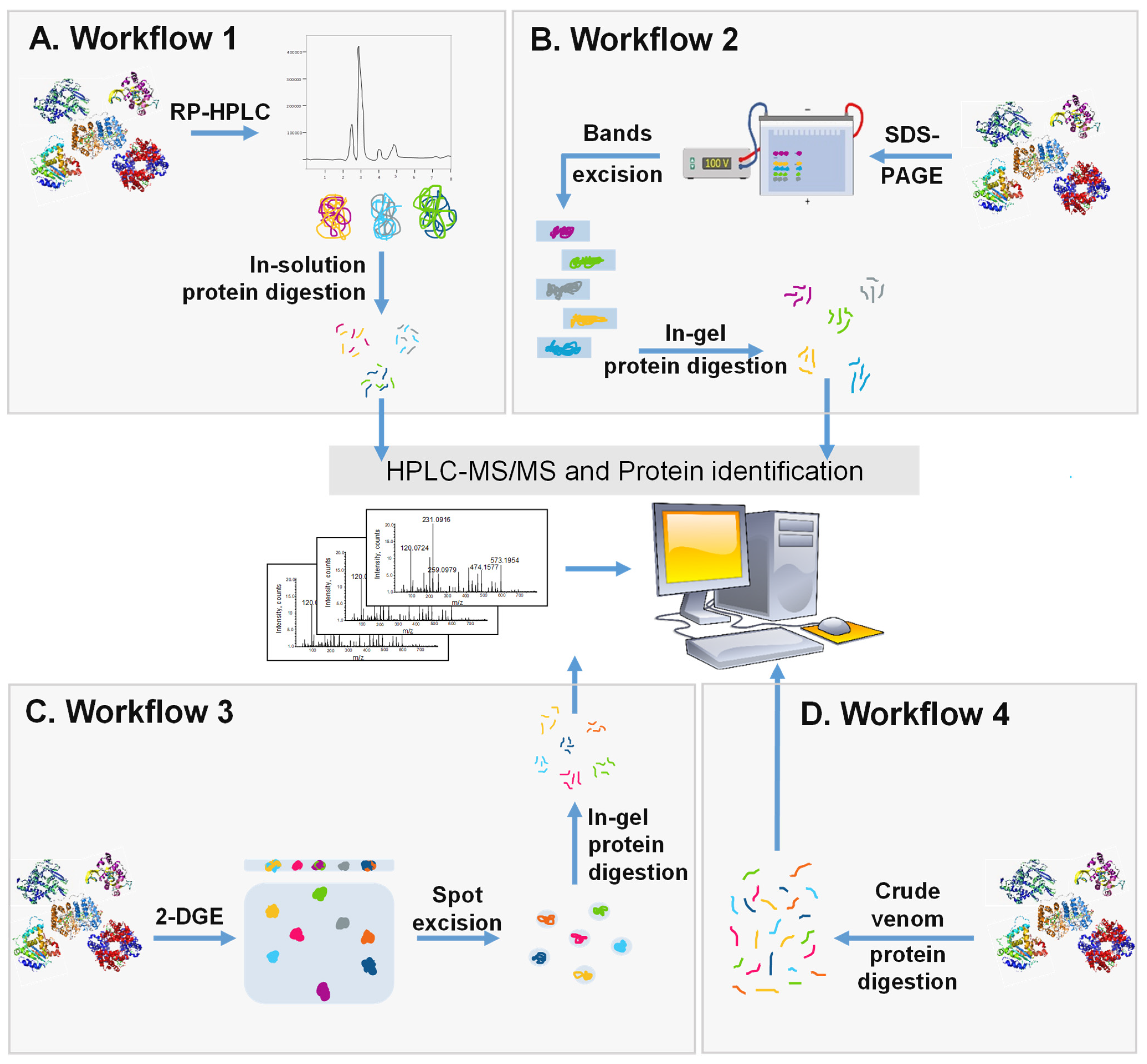
3.2. Whole Proteome Characterization and Identification

The currently available separation and analytical techniques allowed researchers to isolate and identify the most abundant constituents of the venoms. However, given the complexity of SVs, numerous molecules remain unexplored. More recently, the fundamental analysis of complex SV proteomes was made less challenging owing to the evolution of proteomics field. Accordingly, several proteomic techniques have been adapted, providing a rapid and relatively inexpensive method for the decomplexation of venom mixtures [96,97,98][22][23][24]. Both proteomic approaches, bottom–up (BU) and top–down (TD), might be used to analyze SVs composition. The latter has not been used in snake venomics until recently, since it is still in the course of progression [98,99][24][25]. On the other hand, bottom–up proteomics is more frequently used and typically involves the identification of trypsin-digested proteins by tandem mass spectrometry (MS/MS) after separation by both chromatographic and/or electrophoretic techniques. Multiple workflows are used for the BU proteomic analysis of SVs (Figure 2).
Figure 2. Schematic representation of the 4 major workflows used for whole venom separation and proteome identification. (A) Crude SV is separated with RP-HPLC followed by in-solution trypsin digestion and the identification of components by LC-MS/MS. (B) Crude SV is separated by SDS-PAGE and protein bands are cut, digested, and analyzed using LC-MS/MS. (C) Crude venom is separated using 2-DGE followed by protein spot excision, in-gel digestion, and then protein identification by LC-MS/MS. (D) Crude venom is directly digested into peptides and analyzed using LC-MS/MS.
The first workflow (Figure 2A) involves the separation of venom mixture with RP-HPLC using a stepwise or linear acetonitrile gradient. This step is followed by an in-solution trypsin digestion of fractions and LC-MS/MS. Prior to digestion, collected fractions can be tested using SDS-PAGE to evaluate the distribution of proteins in each fraction regarding the MW and number of protein bands. This technique was extensively used in the venomics field to reveal the proteomic composition of SVs such as Bungarus sindanus, Calliophis intestinalis, Deinagkistrodon acutus, Trimeresurus wiroti, Trimeresurus puniceus, and Hydrophis curtus [100,101,102,103,104,105][26][27][28][29][30][31]. This technique was also used in several studies to compare the variability between different SVs [106][32].
The second workflow (Figure 2B) involves the use of a electrophoretic separation. In this approach, crude venom is separated by SDS-PAGE, and protein bands are visualized using Coomassie Blue or silver stain. Later, the protein bands are cut, digested, and then analyzed by LC-MS/MS. An RP-HPLC step might be added before the gel separation to increase the resolution of the workflow. This approach was considerably used in venomics for distinct SVs. For example, it was used to characterize and compare venoms of Trimeresurus macrops and Trimeresurus hageni and led to the identification of 187 and 216 proteins, respectively [107][33].
In the third workflow (Figure 2C), 2-DGE is first performed and followed by the in-gel digestion of all protein spots that are then analyzed by LC-MS/MS. The strategy was shown to be effective for the separation and identification of several SV proteomes [108,109][34][35]. As with every technique, 2-DGE has its own advantages and disadvantages. In fact, it provides the advantage of high molecular weight proteins recovery; however, it is sometimes hard to distinguish protein spots due to inaccurate separation and spot trains [110][36]. In addition, if protein quantity is insufficient in protein spots, then further analysis might not be performed [111][37]. This technique has been used to analyze several snake proteomes such as Gloydius intermedius, Agkistrodon contortrix and Naja asehi [108,109,110][34][35][36]. A study comparing different workflows indicated that the in-gel digestion of proteins provides a lower resolution compared with direct in-solution digestion [112][38]. However, after HPLC, in-gel protein digestion provides higher resolution compared to in-solution protein digestion [113][39]. These findings show that the choice of the method should be related to the properties of the proteins present in the mixture and to the overall workflow adopted for the analysis.
The fourth workflow (Figure 2D) is known as shotgun analysis. This technique involves the tryptic digestion of crude venom proteins. Ultimately, the resulting peptides are separated by RP-HPLC using a C18 column followed by MS to identify proteins. Shotgun analysis has numerous advantages over other techniques, since it does not require a decomplexation process at the beginning that is time consuming and, in some cases, expensive. This strategy allows the recovery of low abundant proteins, since they may be lost during the separation step performed in other techniques [114][40]. This is reflected with a relatively high number of protein hits compared to other techniques. For example, the analysis of Vipera ammodytes ammodytes venom with the shotgun technique identified 99 proteins that is folds higher than other workflows used to analyze this SV [115][41]. This technique was also used for the analysis of Deinagkistrodon acutus venom and led to the identification of 84 proteins [116][42]. Despite the numerous advantages, some limitations have been reported such as the challenging identification of low molecular weight protein [115][41]. One way to overcome this limitation is to employ a combinatorial peptide ligand library and membrane filtration to recuperate low MW proteins [117][43]. This allows concentrating low-abundance proteins in the mixture, leading to a better protein coverage by shotgun analysis. The analysis of Naja ashei venom by Hus et al. indicated that the shotgun technique uncovers proteins that are not identified by other techniques such as 2-DGE, in which 19 proteins were identified compared to 39 proteins identified with shotgun. In this study, the authors indicate that a decomplexation step, although being time and labor consuming, provides higher coverage of proteins [110][36]. In another study, Bothrops leucurus venom yielded 137 proteins hits for shotgun analysis compared to 62 proteins identified using the first workflow [114][40].
Ultimately, even with the great advancement in the proteomic field, there is still no standard technique for SV analysis. Rather, a combination of several techniques could be used depending on the available resources and objectives of the study. This has been shown to provide a better coverage of the proteomes, since each technique might identify different sets of proteins. Proteomes of Naja mossambica and Naja naja nigricincta were analyzed using three different approaches, and the merged data identified 75 and 73 proteins, respectively, which was higher than the proteins identified by each approach alone [106][32]. Similarly, Naja naja nigricincta and Bungarus caeruleus venoms were analyzed by three different approaches simultaneously, using the second and fourth workflows, identifying a total of 81 and 46 proteins, respectively, for each SV [118][44].
To date, the literature available on snake venomics indicates undoubtedly that bottom–up proteomics is the strategy of choice for venom proteomes analysis. Yet, the use of such a strategy for the analysis of complex mixtures such as SVs is particularly challenging. This is due, in general, to the protein digestion step required prior to MS analysis. TD analysis provides an alternative to solve this problem, since intact proteins might be analyzed directly [119][45]. TD was not used in the field of venomics until a few years ago, and its use is limited, since it requires very sophisticated and costly instrumentation. As with any other type of analysis, TD requires the separation of complex protein mixtures prior to MS. Therefore, RP-HPLC constitutes the best method for separation, since it is compatible with the top–down downstream analysis [120][46]. However, other separation methods might be used as well [121][47]. A multidimensional separation seems to provide the best resolution, and it includes the use of gel-based separations followed by RP-HPLC [121][47]. TD proteomics was used successfully for the analysis of several SV proteomes including Dendroaspis jamesoni, Dendroaspis kaimosae, Dendroaspis viridis, and Ophiophagus hannah venoms [122,123][48][49]. In addition to being costly, TD analysis has other limitations, including its inconvenience for the identification of high molecular weight proteins that are an abundant constituent of viperid snakes [119,121][45][47]. Thus, to overcome the limitations of both strategies, an integration of TD and BU proteomics is currently employed and is designated as ‘middle–down proteomics’. This approach was used for the analysis of Echis carinatus sochureki, Protobothrops flavoviridis, and Vipera anatolica senliki venoms and proved to be the most effective compared to each method alone [124,125][50][51].

Finally, even with the scientific advancement, there is still no standard technique to be employed for the separation of SVs. Therefore, separation and analytical methods should be carefully chosen based on the objectives of the research and the available resources.

Finally, even with the scientific advancement, there is still no standard technique to be employed for the separation of SVs. Therefore, separation and analytical methods should be carefully chosen based on the objectives of the research and the available resources.

References

  1. Utkin, Y.N. Animal Venom Studies: Current Benefits and Future Developments. World J. Biol. Chem. 2015, 6, 28–33.
  2. Gutiérrez, J.M.; Calvete, J.J.; Habib, A.G.; Harrison, R.A.; Williams, D.J.; Warrell, D.A. Snakebite Envenoming. Nat. Rev. Dis. Primers 2017, 3, 17079.
  3. Yap, M.K.; Fung, S.Y.; Tan, K.Y.; Tan, N.H. Proteomic Characterization of Venom of the Medically Important Southeast Asian Naja Sumatrana (Equatorial Spitting Cobra). Acta Trop. 2014, 133, 15–25.
  4. Mohamed Abd El-Aziz, T.; Soares, A.G.; Stockand, J.D. Snake Venoms in Drug Discovery: Valuable Therapeutic Tools for Life Saving. Toxins 2019, 11, 564.
  5. Kini, R.M.; Koh, C.Y. Snake Venom Three-Finger Toxins and Their Potential in Drug Development Targeting Cardiovascular Diseases. Biochem. Pharmacol. 2020, 181, 114105.
  6. Blomback, B.; Blomback, M.; Nilsson, I.M. Coagulation Studies on Reptilase, an Extract of the Venom from Bothrops Jararaca. Thromb. Diath. Haemorrh. 1958, 1, 76–86.
  7. Frangieh, J.; Rima, M.; Fajloun, Z.; Henrion, D.; Sabatier, J.M.; Legros, C.; Mattei, C. Snake Venom Components: Tools and Cures to Target Cardiovascular Diseases. Molecules 2021, 26, 2223.
  8. Waheed, H.; Moin, S.F.; Choudhary, M.I. Snake Venom: From Deadly Toxins to Life-Saving Therapeutics. Curr. Med. Chem. 2017, 24, 1874–1891.
  9. Chan, Y.S.; Cheung, R.C.F.; Xia, L.; Wong, J.H.; Ng, T.B.; Chan, W.Y. Snake Venom Toxins: Toxicity and Medicinal Applications. Appl. Microbiol. Biotechnol. 2016, 100, 6165–6181.
  10. Tan, C.H.; Tan, K.Y.; Tan, N.H. A Protein Decomplexation Strategy in Snake Venom Proteomics. Methods Mol. Biol. 2019, 1871, 83–92.
  11. Bird, I.M. High Performance Liquid Chromatography: Principles and Clinical Applications. BMJ 1989, 299, 783–787.
  12. Kadi-Saci, A.; Laraba-Djebari, F. Purification and Characterization of a Thrombin-like Enzyme Isolated from Vipera Lebetina Venom: Its Interaction with Platelet Receptor. Blood Coagul. Fibrinolysis 2020, 31, 1–10.
  13. Averin, A.S.; Utkin, Y.N. Cardiovascular Effects of Snake Toxins: Cardiotoxicity and Cardioprotection. Acta Nat. 2021, 13, 4–14.
  14. Karabuva, S.; Lukšić, B.; Brizić, I.; Latinović, Z.; Leonardi, A.; Križaj, I. Ammodytin L Is the Main Cardiotoxic Component of the Vipera ammodytes ammodytes Venom. Toxicon 2017, 139, 94–100.
  15. Sahyoun, C.; Krezel, W.; Mattei, C.; Sabatier, J.-M.; Legros, C.; Fajloun, Z.; Rima, M. Neuro- and Cardiovascular Activities of Montivipera bornmuelleri Snake Venom. Biology 2022, 11, 888.
  16. Zanotty, Y.; Álvarez, M.; Perdomo, L.; Sánchez, E.E.; Giron, M.E.; Jimenez, J.C.; Suntravat, M.; Guerrero, B.; Ibarra, C.; Montero, Y. Mutacytin-1, a New C-Type Lectin-like Protein from the Venezuelan Cuaima (Lachesis muta muta Linnaeus, 1766)(Serpentes: Viperidae) Snake Venom Inducing Cardiotoxicity in Developing Zebrafish (Danio rerio) Embryos. Zebrafish 2019, 16, 379–387.
  17. Menezes, T.N.; Naumann, G.B.; Peixoto, P.; Rouver, W.N.; Gomes, H.L.; Campos, F.V.; Borges, M.H.; Dos Santos, R.L.; Bissoli, N.S.; Sanchez, E.F.; et al. Bothrops leucurus Venom Induces Acute Hypotension in Rats by Means of Its Phospholipase A2 (blD-PLA2). Toxicon 2020, 185, 5–14.
  18. Tan, L.C.; Kuruppu, S.; Smith, A.I.; Reeve, S.; Hodgson, W.C. Isolation and Pharmacological Characterisation of Hostoxin-1, a Postsynaptic Neurotoxin from the Venom of the Stephen’s Banded Snake (Hoplocephalus stephensi). Neuropharmacology 2006, 51, 782–788.
  19. Lumsden, N.G.; Banerjee, Y.; Kini, R.M.; Kuruppu, S.; Hodgson, W.C. Isolation and Characterization of Rufoxin, a Novel Protein Exhibiting Neurotoxicity from Venom of the Psammophiine, Rhamphiophis oxyrhynchus (Rufous Beaked Snake). Neuropharmacology 2007, 52, 1065–1070.
  20. Marcon, F.; Purtell, L.; Santos, J.; Hains, P.G.; Escoubas, P.; Graudins, A.; Nicholson, G.M. Characterization of Monomeric and Multimeric Snake Neurotoxins and Other Bioactive Proteins from the Venom of the Lethal Australian Common Copperhead (Austrelaps superbus). Biochem. Pharmacol. 2013, 85, 1555–1573.
  21. Venkatesh, M.; Prasad, N.; Sing, T.; Gowda, V. Purification, Characterization, and Chemical Modification of Neurotoxic Peptide from Daboia Russelii Snake Venom of India. J. Biochem. Mol. Toxicol. 2013, 27, 295–304.
  22. Lomonte, B.; Calvete, J.J. Strategies in “snake Venomics” Aiming at an Integrative View of Compositional, Functional, and Immunological Characteristics of Venoms. J. Venom. Anim. Toxins Incl. Trop. Dis. 2017, 23, 26.
  23. Abd El-Aziz, T.M.; Soares, A.G.; Stockand, J.D. Advances in Venomics: Modern Separation Techniques and Mass Spectrometry. J. Chromatogr. B Analyt. Technol. Biomed. Life Sci. 2020, 1160, 122352.
  24. Slagboom, J.; Kaal, C.; Arrahman, A.; Vonk, F.J.; Somsen, G.W.; Calvete, J.J.; Wüster, W.; Kool, J. Analytical Strategies in Venomics. Microchem. J. 2022, 175, 107187.
  25. Tasoulis, T.; Pukala, T.L.; Isbister, G.K. Investigating Toxin Diversity and Abundance in Snake Venom Proteomes. Front. Pharmacol. 2021, 12, 768015.
  26. Tan, K.Y.; Liew, J.L.; Tan, N.H.; Quah, E.S.H.; Ismail, A.K.; Tan, C.H. Unlocking the Secrets of Banded Coral Snake (Calliophis intestinalis, Malaysia): A Venom with Proteome Novelty, Low Toxicity and Distinct Antigenicity. J. Proteom. 2019, 192, 246–257.
  27. Oh, A.M.F.; Tan, C.H.; Tan, K.Y.; Quraishi, N.H.; Tan, N.H. Venom Proteome of Bungarus sindanus (Sind Krait) from Pakistan and in Vivo Cross-Neutralization of Toxicity Using an Indian Polyvalent Antivenom. J. Proteom. 2019, 193, 243–254.
  28. Chen, P.C.; Huang, M.N.; Chang, J.F.; Liu, C.C.; Chen, C.K.; Hsieh, C.H. Snake Venom Proteome and Immuno-Profiling of the Hundred-Pace Viper, Deinagkistrodon Acutus, in Taiwan. Acta Trop. 2019, 189, 137–144.
  29. Lee, L.P.; Tan, K.Y.; Tan, C.H. Snake Venom Proteomics and Antivenomics of Two Sundaic Lance-Headed Pit Vipers: Trimeresurus wiroti (Malaysia) and Trimeresurus puniceus (Indonesia). Comp. Biochem. Physiol. Part D Genom. Proteom. 2021, 40, 100875.
  30. Tan, C.H.; Tan, K.Y.; Ng, T.S.; Sim, S.M.; Tan, N.H. Venom Proteome of Spine-Bellied Sea Snake (Hydrophis curtus) from Penang, Malaysia: Toxicity Correlation, Immunoprofiling and Cross-Neutralization by Sea Snake Antivenom. Toxins 2018, 11, 3.
  31. Tan, K.Y.; Wong, K.Y.; Tan, N.H.; Tan, C.H. Quantitative Proteomics of Naja annulifera (Sub-Saharan Snouted Cobra) Venom and Neutralization Activities of Two Antivenoms in Africa. Int. J. Biol. Macromol. 2020, 158, 605–616.
  32. Hia, Y.L.; Tan, K.Y.; Tan, C.H. Comparative Venom Proteomics of Banded Krait (Bungarus fasciatus) from Five Geographical Locales: Correlation of Venom Lethality, Immunoreactivity and Antivenom Neutralization. Acta Trop. 2020, 207, 105460.
  33. Kumkate, S.; Chanhome, L.; Thiangtrongjit, T.; Noiphrom, J.; Laoungboa, P.; Khow, O.; Vasaruchapong, T.; Sitprija, S.; Chaiyabutr, N.; Reamtong, O. Venomics and Cellular Toxicity of Thai Pit Vipers (Trimeresurus macrops and T. hageni). Toxins 2020, 12, 54.
  34. Yang, Z.M.; Yang, Y.E.; Chen, Y.; Cao, J.; Zhang, C.; Liu, L.L.; Wang, Z.Z.; Wang, X.M.; Wang, Y.M.; Tsai, I.H. Transcriptome and Proteome of the Highly Neurotoxic Venom of Gloydius intermedius. Toxicon 2015, 107, 175–186.
  35. Bocian, A.; Urbanik, M.; Hus, K.; Łyskowski, A.; Petrilla, V.; Andrejčáková, Z.; Petrillová, M.; Legáth, J. Proteomic Analyses of Agkistrodon Contortrix Contortrix Venom Using 2D Electrophoresis and MS Techniques. Toxins 2016, 8, 372.
  36. Hus, K.K.; Buczkowicz, J.; Petrilla, V.; Petrillová, M.; Łyskowski, A.; Legáth, J.; Bocian, A. First Look at the Venom of Naja ashei. Molecules 2018, 23, 609.
  37. Leonardi, A.; Sajevic, T.; Pungerčar, J.; Križaj, I. Comprehensive Study of the Proteome and Transcriptome of the Venom of the Most Venomous European Viper: Discovery of a New Subclass of Ancestral Snake Venom Metalloproteinase Precursor-Derived Proteins. J. Proteome Res. 2019, 18, 2287–2309.
  38. Choksawangkarn, W.; Sriswasdi, S.; Kalpongnukul, N.; Wongkongkathep, P.; Saethang, T.; Chanhome, L.; Laoungbua, P.; Khow, O.; Sumontha, M.; Chaiyabutr, N.; et al. Combined Proteomic Strategies for In-Depth Venomic Analysis of the Beaked Sea Snake (Hydrophis schistosus) from Songkhla Lake, Thailand. J. Proteom. 2022, 259, 104559.
  39. Katali, O.; Shipingana, L.; Nyarangó, P.; Pääkkönen, M.; Haindongo, E.; Rennie, T.; James, P.; Eriksson, J.; Hunter, C.J. Protein Identification of Venoms of the African Spitting Cobras. Toxins 2020, 12, 520.
  40. Dias, Ê.; de Oliveira, L.A.; Sales Lauria, P.S.; Bordon, K.C.F.; Rodrigues Domênico, A.M.; da Silva Guerreiro, M.L.; Wiezel, G.A.; Cardoso, I.A.; Rossini, B.C.; Marino, C.L.; et al. Bothrops Leucurus Snake Venom Protein Profile, Isolation and Biological Characterization of Its Major Toxin PLA. Toxicon 2022, 213, 27–42.
  41. Gopcevic, K.; Karadzic, I.; Izrael-Zivkovic, L.; Medic, A.; Isakovic, A.; Popović, M.; Kekic, D.; Stanojkovic, T.; Hozic, A.; Cindric, M. Study of the Venom Proteome of Vipera ammodytes ammodytes (Linnaeus, 1758): A Qualitative Overview, Biochemical and Biological Profiling. Comp. Biochem. Physiol. Part D Genom. Proteom. 2021, 37, 100776.
  42. Nie, X.; He, Q.; Zhou, B.; Huang, D.; Chen, J.; Chen, Q.; Yang, S.; Yu, X. Exploring the Five-Paced Viper (Deinagkistrodon acutus) Venom Proteome by Integrating a Combinatorial Peptide Ligand Library Approach with Shotgun LC-MS/MS. J. Venom. Anim. Toxins Incl. Trop. Dis. 2021, 27, e20200196.
  43. Hus, K.K.; Marczak, Ł.; Petrilla, V.; Petrillová, M.; Legáth, J.; Bocian, A. Different Research Approaches in Unraveling the Venom Proteome of Naja Ashei. Biomolecules 2020, 10, 1282.
  44. Choudhury, M.; McCleary, R.J.R.; Kesherwani, M.; Kini, R.M.; Velmurugan, D. Comparison of Proteomic Profiles of the Venoms of Two of the “Big Four” Snakes of India, the Indian Cobra (Naja naja) and the Common Krait (Bungarus caeruleus), and Analyses of Their Toxins. Toxicon 2017, 135, 33–42.
  45. Ghezellou, P.; Garikapati, V.; Kazemi, S.M.; Strupat, K.; Ghassempour, A.; Spengler, B. A Perspective View of Top-down Proteomics in Snake Venom Research. Rapid Commun. Mass Spectrom. 2019, 33 (Suppl. 1), 20–27.
  46. Calvete, J.J.; Pla, D.; Els, J.; Carranza, S.; Damm, M.; Hempel, B.F.; John, E.B.O.; Petras, D.; Heiss, P.; Nalbantsoy, A.; et al. Combined Molecular and Elemental Mass Spectrometry Approaches for Absolute Quantification of Proteomes: Application to the Venomics Characterization of the Two Species of Desert Black Cobras, Walterinnesia aegyptia and Walterinnesia morgani. J. Proteome Res. 2021, 20, 5064–5078.
  47. Melani, R.D.; Skinner, O.S.; Fornelli, L.; Domont, G.B.; Compton, P.D.; Kelleher, N.L. Mapping Proteoforms and Protein Complexes From King Cobra Venom Using Both Denaturing and Native Top-down Proteomics. Mol. Cell. Proteom. 2016, 15, 2423–2434.
  48. Ainsworth, S.; Petras, D.; Engmark, M.; Süssmuth, R.D.; Whiteley, G.; Albulescu, L.O.; Kazandjian, T.D.; Wagstaff, S.C.; Rowley, P.; Wüster, W.; et al. The Medical Threat of Mamba Envenoming in Sub-Saharan Africa Revealed by Genus-Wide Analysis of Venom Composition, Toxicity and Antivenomics Profiling of Available Antivenoms. J. Proteom. 2018, 172, 173–189.
  49. Petras, D.; Heiss, P.; Harrison, R.A.; Süssmuth, R.D.; Calvete, J.J. Top-down Venomics of the East African Green Mamba, Dendroaspis angusticeps, and the Black Mamba, Dendroaspis polylepis, Highlight the Complexity of Their Toxin Arsenals. J. Proteom. 2016, 146, 148–164.
  50. Ghezellou, P.; Albuquerque, W.; Garikapati, V.; Casewell, N.R.; Kazemi, S.M.; Ghassempour, A.; Spengler, B. Integrating Top-Down and Bottom-Up Mass Spectrometric Strategies for Proteomic Profiling of Iranian Saw-Scaled Viper, Echis carinatus sochureki, Venom. J. Proteome Res. 2021, 20, 895–908.
  51. Hempel, B.F.; Damm, M.; Mrinalini; Göçmen, B.; Karış, M.; Nalbantsoy, A.; Kini, R.M.; Süssmuth, R.D. Extended Snake Venomics by Top-Down In-Source Decay: Investigating the Newly Discovered Anatolian Meadow Viper Subspecies. J. Proteome Res. 2020, 19, 1731–1749.
More
Video Production Service