Coronavirus Disease 19 Infection in Dental Practice: Comparison
Please note this is a comparison between Version 1 by Roberto Sorrentino and Version 2 by Vivi Li.

Dental professionals and patients are exposed to a high risk of COVID-19 infection, particularly in the prosthodontic practice, because of the bio-aerosol produced during teeth preparation with dental handpieces and the strict contact with oral fluids during impression making. 

  • coronavirus
  • COVID-19
  • SARS-CoV-2
  • prosthodontics
  • airborne
  • dental impression
  • pandemic
  • prevention

1. Introduction

In December 2019, severe acute respiratory syndrome coronavirus 2 (SARS-CoV-2), was first reported in Wuhan, Hubei province (China), and rapidly spread over 24 countries, leading the World Health Organization (WHO) to declare this severe pneumonia a global emergency on 30 January 2020 [1]. From 31 December 2019 to 17 June 2022, over 535,863,000 confirmed cases of coronavirus infectious disease-19 (COVID-19) have been reported, including approximately 6,315,000 deaths, and these numbers are increasing daily (Figure 1 and Figure 2) [2].
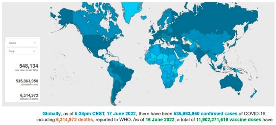
Figure 1. A screenshot of the interactive dashboard of COVID-19 global cases by the World Health Organization. This dashboard is continually updated and can be accessed at https://covid19.who.int/ (accessed on 17 June 2022).
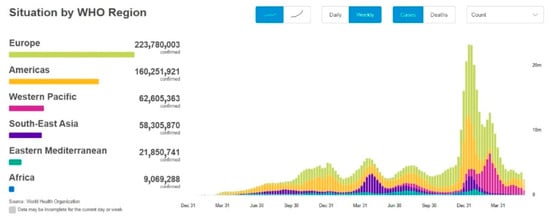
Figure 2. A screenshot of the COVID-19 situation by WHO Region. This dashboard is continually updated and can be accessed at https://covid19.who.int/ (accessed on 17 June 2022).
SARS-CoV-2, of the family Coronaviridae, is a single-stranded RNA beta coronavirus that is characterized by a diameter of 50–200 nm and probably originated from bats and pangolins [3]. It shares 85–92% nucleotide sequence homology with the pangolin coronavirus (CoV) genome and 96.2% nucleotide homology with bat CoV RaTG13, confirming the zoonotic origin of the virus [4]. The animal-to-human transmission event, the spillover phenomenon of SARS-CoV-2, can be plausibly imputed to the sale and killing of wildlife species at the Huanan Seafood Wholesale Market, where the initial cases of COVID-19 emerged [4]. The comprehension of the initial dynamics of the infection and the identification of the animal source of SARS-CoV-2 would help to prevent future new zoonosis by strengthening the control of food and hygiene within live animal markets.
Since the first COVID-19 infection, some variants of SARS-CoV-2 have been found [5]. These variants are adaptive mutations in the viral genome that can alter the virus’s pathogenic potential, and some of them were classified as variants of concern (VOCs) due to their public health implications [5][6][5,6]. In particular, VOCs have been associated with increased virulence or transmissibility, decreased neutralization by antibodies acquired through vaccination or natural infection, the ability to elude detection, and a reduction in therapeutic or vaccine efficiency [6]. Five SARS-CoV-2 VOCs have been recognized according to the WHO: Alpha (B.1.1.7, first report in the United Kingdom); Beta (B.1.351, first report in South Africa); Gamma (P.1, first report in Brazil); Delta (B.1.617.2, first report in India); and Omicron (B.1.1.529, first report in South Africa) [6]. Among the listed VOCs, the Omicron variant is the most severely altered, paving the path for increased transmissibility and partial resistance to COVID-19 vaccine-induced immunity (Figure 3 and Figure 4) [5][7][8][5,7,8].
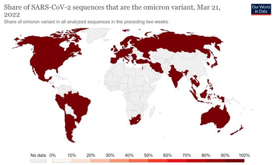
Figure 3. Share of SARS-CoV-2 sequences that are the omicron variant. This dashboard is continually updated and can be accessed at https://ourworldindata.org/grapher/covid-cases-omicron?country=GBR~FRA~BEL~DEU~ITA~ESP~USA~ZAF~BWA~AUS (accessed on 17 June 2022).
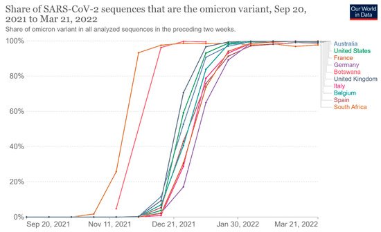
Figure 4. Share of omicron variant in all analyzed sequences. This dashboard is continually updated and can be accessed at https://ourworldindata.org/grapher/covid-cases-omicron?tab=chart&country=GBR~FRA~BEL~DEU~ITA~ESP~USA~ZAF~BWA~AUS (accessed on 17 June 2022).
The time from the infection to the onset of symptoms can vary from 2 to 14 days, and the median incubation period reported by the WHO was 5–6 days [9][10][9,10].
Older age and other risk factors, such as a history of underlying diseases and/or co-infections, play an important role in determining the severity of symptoms, leading to a higher risk to develop severe illness and death [11].
The clinical initial manifestations of COVID-19 can be aspecific. First of all, a large number of patients show symptoms of common cold, such as dry cough, sore throat, low-grade fever, or myalgia [11]. Less frequent manifestations of the infection may be nausea, diarrhea, dysgeusia, and persistent olfactory dysfunction (hyposmia) due to mucosal edema and nasal inflammation [12][13][12,13]. Some findings underlined the potential transmission of SARS-CoV-2 mediated by asymptomatic subjects, who may represent potential reservoirs for the spreading and re-emergence of the infection since the viral load in such patients was comparable to that of symptomatic patients [14].
Even if the mechanism of SARS-CoV-2 infection is not yet completely known, the high transmissibility of the virus can be partially explained by the higher affinity of the virus for cells located in the lower airways, where the virus binds using the host receptor for the conversion enzyme of angiotensin 2 (ACE2) and replicates, causing pneumonia [15].
The radiologic signs of bilateral pneumonia are abnormal ground-glass opacities found in chest X-ray and computed tomographic (CT) scans. The worst clinical picture patients can present is multiorgan dysfunction, acute renal failure, and acute respiratory distress syndrome (ARDS) [16].
People older than 60 years old and patients with chronic comorbidities (especially diabetes, high blood pressure, and cardiovascular diseases) were found to be more prone to develop severe clinical disease and fatal outcomes, which occurred only in critical cases. Fortunately, children often seem to show mild symptoms of COVID-19 [15].
SARS-CoV-2 has a human-to-human typical transmission by means of respiratory droplets and indirect contact, whereas the conjunctival and mother-to-fetus modes still need to be confirmed [17].
Some evidence suggested that infected individuals can spread infection through bodily secretions, such as the saliva and nasal fluid produced by talking, sneezing, and coughing. The high presence of the virus in saliva may be explained by the binding of SARS-CoV-2 with ACE2 receptors, which are highly concentrated in salivary glands [18][19][20][18,19,20].
Moreover, the contact of contaminated hands with the mucous membranes of the mouth and/or nose may lead to the onset of COVID-19 infection. The fecal–oral mode also may be another important route for nosocomial spread. Consequently, the disinfection of objects, handwashing, and social distancing (beyond six feet) are strongly recommended in order to control the community outbreak of the disease [17][18][19][20][17,18,19,20].
In particular, dental offices could be easily contaminated since the use of high-speed handpieces or ultrasonic instruments could cause the aerosolization of patients’ secretions, such as saliva or blood. Thus, dental professionals are exposed to a high risk of contracting SARS-CoV-2 because social distancing is unachievable during dental procedures [21].
On the other hand, the increased susceptibility of patients in dental offices is another important concern: elderly age, diabetes, chemotherapy, pregnancy, or conditions of an impaired immune defense system may easily lead to a worsening clinical picture or fatal outcome in the case of SARS-CoV-2 infection [11][22][11,22].
Among the branches of dentistry, prosthodontics is the part of restorative dentistry concerned with the design, manufacture, and fitting of artificial replacements for missing teeth and the associated soft tissues. Many aspects and devices used during prosthetic procedures may offer the opportunity for cross-contamination, requiring careful attention and rigorous protocols for the prevention of infection spreading.
To date, there are scarce data in the scientific literature about the management of COVID-19 infection during prosthodontics procedures in order to prevent COVID-19 cross-infections.

2. Dental Impression

Dental impression and wax or silicone interocclusal records could be contaminated with patients’ saliva and, just as frequently, blood (Figure 56).
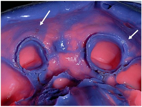
Figure 56.
 Debris of saliva and blood on a conventional elastomeric dental impression (arrows indicated).
Some investigations found low bacterial contamination (median number of 1.3 × 10² cfu/20 mm3) on a high percentage of dental impressions (more than 70%), whereas higher contamination was found (103–3.4 × 10⁴ cfu/20 mm3) on the resultant percentage of samples. Bacteria can be opportunistic or non-opportunistic species, and low-pathogenic species may promote the onset of latent infections and/or reactivate ones [23][24][25,26]. Regarding the viral contaminations of dental impressions, it is widely known that patients’ body fluids (i.e., saliva and blood) might be potential reservoirs and sources of the disease, hence the urge to pursue meticulous protocols of decontamination and disinfection of the impressions before sending them to dental laboratories in order to reduce gypsum cast contamination with patient-derived microbes. Regarding conventional dental impressions, a prior accurate debridement of saliva and blood is recommended by brushing and/or rinsing under running water to allow complete contact between the dental impressions and disinfectant materials [25][26][27,28]. Afterward, conventional dental impressions can be disinfected by immersion or spraying techniques. Disinfection by spraying is considered a suitable method for alginate and polyethers, which are more prone to dimensional distortion after 10 min of immersion, even if the risk of disinfectant inhalation cannot be excluded. On the other hand, the immersion technique, suitable for impression materials less prone to expansion or swelling (i.e., polyvinylsiloxane), grants a full coverage of the disinfectant [25][26][27,28]. Glutaraldehyde and chlorine compounds are generally preferable to chemically disinfect dental impressions, with a time of contact established by providers’ instructions and specific guidelines [27][29]. Concerning SARS-CoV-2, some investigations found out that coronavirus contamination can be significantly reduced with 62–71% ethanol or 0.1% sodium hypochlorite solutions within 1 min of exposure; in addition, other biocidal agents (i.e., 70%–75% 2-propanol and 0.23% povidone-iodine) can effectively reduce the viral infectivity [28][29][30,31]. Furthermore, the use of automatic mixers is strongly recommended in order to avoid the microbial contamination derived from handling some impression materials (i.e., addition silicones) without gloves. Regarding the disinfection procedures, the two following physical events are worthy of notice. The first is the negative effect of the disinfection treatment on the dimensional stability of the impression material and the accuracy of its surface detail. The second event is the deactivation effect of the impression material on the disinfecting solution that might decrease the efficiency of the disinfection process [30][32]. Such events must be kept in check and avoided when possible. For this purpose, it is advisable to follow the instructions provided by the manufacturers of the disinfectants. Customized trays should be properly disinfected after intraoral try-in and before the use of brushes for impression material adhesives [23][25]. Regarding digital technologies and optical impressions, a significant advantage is the possibility to autoclave the latest-generation scanners’ tips, and proper disinfectant commercial products can be used for other scanner items and scanning devices (i.e., implant-supported scan-bodies) to reduce the risk of cross-contamination, according to manufacturers’ suggestions [23][31][25,33]. Moreover, intraoral scanner systems provide the advantage of avoiding the transfer of a conventional impression and therefore the risk of contagion to the laboratory via the physical impression or the gypsum cast. Besides, digital technologies are also useful during the present pandemic for prosthetic planning and the manufacturing of removable or fixed prostheses. Indeed, compared to conventional procedures, the digital workflow allows the fabrication of reliable prostheses in fewer appointments and with quicker chairside times [32][34].

3. Prosthodontics Aids

In order to reduce the probability of contagion, maximum attention has to be paid to the disinfection and sterilization of the instruments and aids that can be used during prosthetic clinical practice. For this purpose, it is useful to dwell on the following indications reported in the literature. The disinfection of some aids, such as occlusal rims or casts, could be achieved by spraying with 5.25% sodium hypochlorite solution and allowing to set for 10 min [33][34][35][35,36,37]. Moist heat sterilization is performed using a steam autoclave at 121 °C for 15–20 min at 15 lb pressure/square inch, and it is suggested for handpieces, stainless steel instruments, tissue retraction pluggers, dapen dishes, and glass slabs in combination with ethylene oxide at a concentration of 450–800 mg/L [36][37][38,39]. Moreover, ethylene oxide might be used for the disinfection of mouth and face mirrors, carbon steel hand instruments, three-way syringes, saliva ejectors, and evacuators [38][40]. Regarding impression trays, metallic ones should be heat-sterilized via autoclave, chemical vapor, or dry heat and ethylene oxide sterilization. Custom acrylic resin should be disinfected with tuberculocidal hospital disinfectant for reuse during the same patient’s next visit, while it is suggested to avoid using plastic trays because they are more difficult to sterilize efficiently [39][41]. Acrylic dentures must be soaked in glutaraldehyde solution for 12 h after being rinsed with running water and stored in an ultrasonic cleaner. Subsequently, they must be cleaned with running water, scraped with chlorhexidine, and then exposed to chlorine dioxide for 3 min. Then, ethylene oxide is used to sterilize [38][40]. Regarding dentures made of metal, it is suggested to spray with 2% glutaraldehyde solution and place the prostheses in a plastic bag for 10 min [30][32].

4. Metal–Ceramic Materials

Immersion in glutaraldehydes for the duration indicated by the disinfectant manufacturer can be used to disinfect fixed metal/porcelain prostheses. In addition, fixed prostheses may be disinfected by immersing them in diluted hypochlorite for a brief period of time without causing harm. The higher the noble metal concentration, the lower the risk of harmful effects on the metal core [40][42]. Care should be taken to limit the amount of time in which metals are exposed to potentially corrosive chemicals. Iodophors might be utilized as well, but there is no evidence to back this up. Fixed metal prostheses can be disinfected with ethylene oxide or even autoclaving if needed. Conversely, unglazed porcelain should not be exposed to any disinfectant because porcelain firing/glazing might be sufficient [40][42]. Before delivering to the patient, any aid that was treated with a disinfectant must be properly cleaned.

5. All-Ceramic Materials

All-ceramic materials require surface treatments aimed at favoring the adhesion of the material to the dental surfaces. This surface treatment is called “etching” and mainly concerns the glass ceramics, such as feldspathic or leucite-based ceramics, lithium silicates, and disilicates, in order to cement these materials to the prepared tooth adhesively, increasing the resistance to fracture of the final restoration [41][42][43][43,44,45]. Regarding zirconia ceramics, they are characterized by the absence of glass in their composition, and the surface treatment takes place through a tribochemical silica coating or low-pressure air abrasion (Al2O3, 50 µm at 2 bars) for both conventional and adhesive cementations [44][45][46][46,47,48]. The surface treatment procedures of all-ceramic materials require some rinsing and drying steps, both in the laboratory and in the dental office. Unfortunately, the rinsing and drying phases lead to nebulization and the formation of bio-aerosols. Bio-aerosols are liquid/solid particles generated by different sources and are responsible for the transmission of airborne microorganisms, using droplet nuclei (1–5 μm) or droplets (>5 μm). High-speed handpieces, ultrasonic scalers, air turbines, air polishing, and air–water syringes are all sources of bio-aerosols during dental practice. These particles can stay suspended in the air for a variable time and fall, contaminating all the surfaces of dental offices [47][49]. Tooth preparation procedures or chairside prosthesis modifications require the use of dental handpieces, and the production of aerosols is unavoidable, resulting in a high risk of indirect infections for patients and dental professionals, which is difficult to contain [48][50]. SARS-CoV-2 spread may occur via respiratory droplets and contact transmission. Hence, it is advisable to meticulously manage the patients to minimize the risk of COVID-19 infections.
Video Production Service