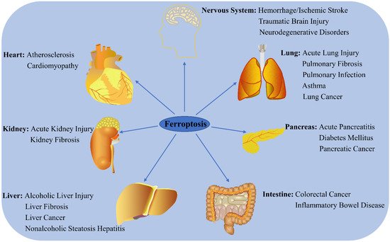
1. Role of Ferroptosis in Chronic Diseases
Ferroptosis has recently become a hotspot of research focus in the field of disease prognosis and therapy, with numerous studies reporting that it regulates the occurrence and progress of multiple disorders (Figure 12). Here, the latest progress in ferroptosis research and its association with various diseases are summarized.
Figure 12. Role of ferroptosis in chronic diseases. Ferroptosis participates in the regulation of many system disorders, including nervous system diseases, cardiovascular diseases, liver diseases, kidney diseases, lung diseases, pancreatic diseases, and intestinal diseases.
1.1. Cancers
1.1.1. Lung Cancer
Lung cancer is one of the most common causes of cancer-related deaths worldwide. Some key regulators, including KRAS, TP53, Nrf2, YAP, NFS1, STYK1, LSH, RNF113A, and non-coding RNA, are reported to participate in ferroptosis regulation
[1][67]. KRAS mutant lung cancer cells are vulnerable to the ferroptosis induced by SLC7A11 inhibition
[2][68]. P53 also inhibits SLC7A11 expression and cystine uptake, consequently inducing ferroptosis
[3][43]. The activation of Nrf2 negatively regulates ferroptosis by up-regulating various target genes, such as HO-1. Acetaminophen (APAP) sensitizes non-small-cell lung cancer (NSCLC) to erastin-mediated ferroptosis by negatively regulating the Nrf2/HO-1 signaling pathway
[4][69]. Curcumin triggers ferroptosis in NSCLC by activating autophagy
[5][70]. DHA induces lung cancer cell ferroptosis via the inactivation of the PRIM2/SLC7A11 axis
[6][71]. Orlistat promotes lung cancer cell ferroptosis by reducing GPX4 expression and inducing lipid peroxidation
[7][72]. A pure compound extracted from danshen, dihydroisotanshinone I, suppresses the growth of lung cancer cells by triggering both ferroptosis and apoptosis
[8][73]. The artemisinin derivatives artesunate and DHA induce ROS-dependent apoptosis/ferroptosis in NSCLC cells
[9][74]. MiR-27a-3p promotes NSCLC through SLC7A11-mediated-ferroptosis
[10][75], while MiR-302a-3p induces the ferroptosis of NSCLC cells by targeting FPN
[11][76].
1.1.2. Liver Cancer
Liver cancer is the sixth most common malignancy and the third primary cause of cancer-related deaths worldwide
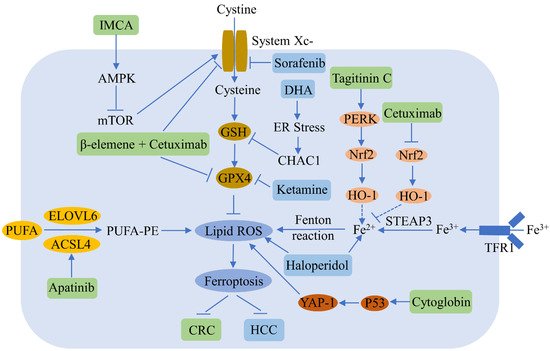
[12][77]. Ferroptosis plays an important role in the regulation of hepatocellular carcinoma (HCC) (
Figure 23). Sorafenib, which is a multikinase inhibitor used widely in the treatment of advanced HCC, induces ferroptosis by inhibiting SLC7A11; however, activation of the p62-Keap1-Nrf2 pathway suppresses ferroptosis in HCC
[13][78]. Erastin and sorafenib induce ferroptosis by inhibiting Nrf2 expression and activity in HCC
[14][79]. Sorafenib also decreases retinoblastoma protein levels, thereby increasing ferroptosis
[15][80], while the inhibition of metallothionein enhances sorafenib-induced ferroptosis in HCC
[16][81]. Haloperidol strongly promotes erastin- and sorafenib-induced ferroptosis by increasing Fe
2+ levels and lipid peroxidation in HCC
[17][82]. Ketamine induces ferroptosis via the inhibition of lncRNA PVT1 and GPX4 in liver cancer cells
[18][83]. DHA induces ferroptosis by activating unfolded protein response and upregulating CHAC1 expression in primary liver cancer cells
[19][84]. MiR-214-3p promotes erastin-mediated ferroptosis by decreasing ATF4 expression in hepatoma cells
[20][85]. Artesunate increases the sensitivity of HCC to sorafenib by inducing ferroptosis
[21][86], while O-GlcNAcylation sensitizes liver cancer to RSL3-induced ferroptosis through the YAP/TFRC pathway
[22][87].
Figure 23. Ferroptosis regulators and pathways in hepatocellular carcinoma (HCC) and colorectal cancer (CRC). Ferroptosis can be a negative regulator of HCC and CRC. Various compounds can inhibit HCC and CRC growth by inducing ferroptosis. The blue and green patterns correspond to regulators for HCC and CRC, respectively. Sorafenib induces HCC ferroptosis by inhibiting SLC7A11. Dihydroartemisinin (DHA) triggers HCC ferroptosis by activating unfolded protein response and upregulating CHAC1. Ketamine increases HCC ferroptosis by inhibiting GPX4. Haloperidol promotes ferroptosis via the increase of Fe2+ levels and lipid peroxidation in HCC. IMCA induces CRC ferroptosis by inhibiting SLC7A11. β-elemene and cetuximab combined treatment results in CRC ferroptosis by downregulating GPX4 and SLC7A11. Apatinib enhances CRC ferroptosis by up-regulating ELOVL6/ACSL4 signaling. Tagitinine C induces CRC ferroptosis via the up-regulation of the PERK-Nrf2-HO-1 signaling pathway. Cetuximab increases CRC ferroptosis by downregulating the Nrf2/HO-1 pathway. Cytoglobin increases CRC ferroptosis through the upregulation of p53 and YAP1.
1.1.3. Colorectal Cancer
Recent studies have reported that ferroptosis participates in the regulation of colorectal cancer (CRC), a common malignancy of the digestive system (
Figure 23). RSL3 is known to induce CRC ferroptosis by inactivating GPX4 and producing ROS
[23][88]. Tagitinine C, a natural product, acts synergistically with erastin to induce ferroptosis in CRC cells via the PERK-Nrf2-HO-1 signaling pathway
[24][48]. The benzopyran derivative IMCA induces ferroptosis by downregulating SLC7A11 and the AMPK/mTOR pathway in CRC
[25][89]. Inhibiting the KIF20A/NUAK1/Nrf2/GPX4 signaling pathway triggers ferroptosis and sensitizes CRC to oxaliplatin
[26][90]. Cetuximab increases the ferroptosis of KRAS mutant CRC, induced by RSL3, by inhibiting the Nrf2/HO-1 signaling pathway
[27][50]. A combined treatment of β-elemene and cetuximab induces KRAS mutant CRC cell ferroptosis via the downregulation of GPX4 and SLC7A11
[28][91], and the inhibition of SLC7A11 induces the ferroptosis of CRC stem cells
[29][92]. Apatinib enhances ferroptosis in CRC cells by upregulating ELOVL6/ACSL4 signaling
[30][93]. Cytoglobin sensitizes the CRC cells to RSL3 and erastin through the upregulation of p53 and YAP1
[31][94]. Andrographis enhances the effect of 5-fluorouracil (5FU) against CRC by inducing ferroptosis and inhibiting β-catenin/Wnt-signaling pathways
[32][95].
1.1.4. Breast Cancer
Breast cancer is the most common cancer among women. Sulfasalazine has been found to trigger breast cancer cell ferroptosis via the repression of GPX4 and SLC7A11 expressions and an increase in TFR1 and DMT1 expressions, particularly in cells with low estrogen receptor (ER) expression
[33][96]. The combined treatment of siramesine and lapatinib triggers breast cancer cell ferroptosis by increasing TF expression and reducing FPN expression
[34][97]. Metformin triggers ferroptosis via a reduction in the UFMylation of SLC7A11 in breast cancer
[35][98]. Curcumin promotes the ferroptosis of breast cancer cells by increasing lipid ROS levels, lipid peroxidation end-product MDA accumulation, and intracellular free iron levels
[36][99]. Targeted exosome-encapsulated erastin promotes the ferroptosis of triple-negative breast cancer (TNBC) cells
[37][100]. The GSK3β/Nrf2 signaling pathway strengthens the ferroptosis of breast cancer induced by erastin
[38][101]. Inhibiting GPX4 increases gefitinib-induced ferroptosis in TNBC cells
[39][102], while simvastatin induces ferroptosis in TNBC cells
[40][103]. Metformin promotes ferroptosis via the up-regulation of miR-324-3p and down-regulation of GPX4 in breast cancer
[41][104]. Lidocaine induces the ferroptosis of ovarian and breast cancers by increasing miR-382-5p and decreasing SLC7A11
[42][105], while ketamine induces ferroptosis by targeting the KAT5/GPX4 axis in breast cancer cells
[43][106].
1.1.5. Ovarian Cancer
Ovarian cancers are prevalent female malignancies, seriously affecting women’s health and life quality in the world. Lidocaine promotes ferroptosis in ovarian and breast cancer cells by enhancing miR-382-5p and down-regulating SLC7A11 expression
[42][105]. Stearoyl-CoA desaturase 1 (SCD1) suppresses ovarian cancer cell ferroptosis
[44][107], while the inhibition of pharmaceutical SCD1 promotes ferroptosis in vitro and in vivo, and the combined treatment of SCD1 inhibitors and ferroptosis inducers significantly inhibits ovarian tumor growth
[45][108]. SNAI2 knockdown promotes ferroptosis in ovarian cancer
[46][109]. Sodium molybdate induces the ferroptosis of ovarian cancer cells via labile iron elevation and GSH depletion
[47][110]. Ferroptosis inducers increase the sensitivity of BRCA-proficient ovarian cancer cells to PARP inhibitor by repressing SLC7A11
[48][25]. Human serum incubated-superparamagnetic iron oxides promote ferroptosis via p53 overexpression in ovarian cancer cells
[49][111]. Superparamagnetic iron oxide nanoparticles increase oxidative stress, reduce autophagy, and activate ferroptosis in ovarian cancer stem cells
[49][111]. GALNT14 induces ferroptosis in ovarian cancer via the EGFR/mTOR pathway
[50][112]. MAP30 protein from
Momordica charantia and cisplatin synergistically induce ferroptosis in ovarian cancer
[51][113].
1.1.6. Pancreatic Cancer
Pancreatic cancer, one of the most fatal of all cancers, is seen mainly in males and the older population (40–85 years)
[52][114]. The inhibition of cytosolic aspartate aminotransaminase promotes pancreatic cancer cell ferroptosis by repressing mitochondrial metabolism and promoting a catabolic state
[53][115]. The natural compound artesunate induces ferroptosis in pancreatic cancer cells
[54][116]. Cysteine depletion promotes the ferroptosis of pancreatic tumor cells in mice
[55][117]. Ruscogenin, a saponin found in the root of
Ophiopogon japonicus, increases intracellular ferrous iron and ROS, thereby inducing ferroptosis in pancreatic cancer cells
[56][118]. The abrogation of ADP ribosylation factor 6 promotes RSL3-induced ferroptosis in pancreatic cancer cells
[57][119]. DHA acts synergistically with cisplatin to trigger ferroptosis in pancreatic ductal adenocarcinoma (PDAC) by regulating iron metabolism
[58][120]. Combining artesunate with GRP78 inhibition facilitates ferroptosis in KRAS mutant PDAC
[59][121]. Chrysin enhances pancreatic cancer sensitivity to gemcitabine by inducing autophagy-dependent ferroptosis via the targeting of human carbonyl reductase 1
[60][122]. Ponicidin suppresses pancreatic cancer growth by inducing ferroptosis
[61][123], while piperlongumine induces ferroptosis in human pancreatic cancer cells by suppressing the gamma-glutamyl cycle and regulating the metabolism of PUFAs
[62][124].
1.2. Nervous System Diseases
1.2.1. Stroke
Stroke is one of the leading causes of death and disability in the world. Selenium activates homeostatic transcription to protect neuron cells from ferroptosis and treat stroke
[63][125]. ACSL4 enhances ischemic stroke by inducing neuronal ferroptosis-related brain injury and neuroinflammation
[64][126], while the inhibition of ACSL4 improves neurological functioning after stroke via the suppression of ferroptosis
[65][127]. Reducing NCOA4 inhibits the ferritinophagy-mediated ferroptosis of neurons, and thus can be used to treat ischemic stroke
[66][63]. The hemin-induced hemorrhagic stroke model is one of classic neuronal ferroptosis
[67][128]. Intracerebral hemorrhage (ICH) is a type of severe stroke, the pathology of which is closely related to ferroptosis. Curcumin nanoparticles inhibit ferroptosis and enhance ICH treatment
[68][129], and baicalin can also suppress ferroptosis in ICH
[69][130]. Tau-mediated iron export inhibits ferroptosis after ischemic stroke
[70][131]. Supplementing lactoferrin reduces the ferroptosis of nerve cells after diabetic ICH
[71][132], while crocin reduces ICH-induced neuronal ferroptosis by increasing Nrf2 expression and nuclear translocation
[72][133]. Dauricine represses nerve cell ferroptosis and brain injury after ICH by up-regulating GPX4 expression
[73][134], while pyridoxal isonicotinoyl hydrazone, a lipophilic iron-chelating agent, alleviates hemorrhage stroke by decreasing ferroptosis and inflammation
[74][135].
1.2.2. Traumatic Brain Injury
Traumatic brain injuries (TBIs) occur worldwide and result in serious economic burden. Inhibiting ferroptosis reduces tissue damage and ameliorates long-term motor and cognitive function after TBI in mice
[75][136]. Biomarkers of ferroptosis are increased after TBI; however, baicalein decreases neuronal ferroptosis and improves post-TBI outcome
[76][137]. Ferritin H deletion in nerve cells abolishes the neuroprotection of melatonin against TBI-mediated ferroptosis
[77][138]. Prokineticin-2 prevented neuronal cells from ferroptosis in a TBI model
[78][139]. Polydatin alleviates TBI via the inhibition of ferroptosis
[79][140]. MiR-212-5p attenuates neuronal ferroptosis after TBI by targeting PTGS2
[80][141]. Ruxolitinib was found to protect against neuronal ferroptosis in a mouse TBI model
[81][142]. Ferristatin II, an iron uptake inhibitor, prevents neuronal ferroptosis after TBI
[82][143], while tetrandrine ameliorates TBI by regulating autophagy to reduce ferroptosis
[83][144].
1.2.3. Alzheimer’s Disease
Alzheimer’s disease (AD) is the most ordinary type of dementia, featuring β-amyloid and Tau protein accumulation and aggregation
[84][145]. FPN loss induces ferroptosis and promotes memory impairment in AD
[85][146]. NADPH oxidase 4 promotes astrocytes ferroptosis via oxidative-stress-induced lipid peroxidation in AD
[86][147]. Mitochondrial aldehyde dehydrogenase reduces AD-induced cardiac anomalies by inhibiting ACSL4-mediated ferroptosis
[87][148]. Tetrahydroxy stilbene glycoside was shown to improve AD in APP/PS1 mice via the inhibition of GPX-related ferroptosis
[88][149], while eriodictyol improves cognitive disorder in APP/PS1 mice by suppressing ferroptosis via Nrf2 activation mediated by a vitamin D receptor
[89][150]. In addition, forsythoside A alleviates AD by reducing Nrf2/GPX4 activation-induced ferroptosis
[90][151].
1.2.4. Parkinson’s Disease
Parkinson’s disease (PD) is a common neurodegenerative disorder characterized by motor impairment. FTH1 was reported to suppress ferroptosis by impairing ferritinophagy in a 6-OHDA-induced PD model
[91][152]. Activating the p62-Keap1-Nrf2 pathway inhibits the ferroptosis triggered by 6-OHDA in dopaminergic cells
[92][153]. Thioredoxin-1 reduces ferroptosis in PD by up-regulating GPX4 and GSH
[93][154]. Super-enhancer-driven sorting nexin 5 expression facilitated the ferroptosis of dopaminergic neurons in PD models
[94][155]. MiR-335 enhanced ferroptosis by degrading FTH1 in in vitro and in vivo models of PD
[95][156]. Moxibustion presents a neuroprotective function via anti-ferroptosis in PD
[96][97][157,158]. Ferritinophagy-mediated ferroptosis involves in paraquat-induced neurotoxicity in PD
[98][159].
1.2.5. Huntington’s Disease
Huntington’s disease (HD) is an autosomal dominant and fatal neurodegenerative disease resulting from abnormal cytosine-adenine-guanine repetition in the huntingtin gene
[99][160], for which there is currently no effective treatment. A few ferroptotic characteristics have been observed in HD animal models and patients, such as iron accumulation, oxidative stress, GSH depletion, and reduced GPX activity
[100][161], which suggests the participation of ferroptosis in the regulation of HD pathogenesis. Several ferroptosis regulators have also been found to take effect in HD models. For instance, Fer-1 suppressed oxidative lipid damage and ferroptosis in HD cellular models
[101][162], while the intraventricular delivery of the iron chelator DFO led to motor phenotype improvement in R6/2 HD mice
[102][163].
1.3. Metabolic Diseases
1.3.1. Cardiovascular Diseases
Cardiovascular diseases are those involving the heart and blood vessels, such as atherosclerosis, peripheral vascular diseases, and cerebrovascular diseases. Ferritin plays a key role in inhibiting cardiac ferroptosis and succedent heart failure, while cardiac ferritin H loss promotes cardiomyopathy by increasing SLC7A11-mediated ferroptosis
[103][23]. Mitochondria-dependent ferroptosis plays an important role in doxorubicin-induced cardiomyopathy
[104][164]. ENPP2, a lipid kinase participating in lipid metabolism, reduces erastin-induced ferroptosis in cardiomyocytes by regulating GPX4, ACSL4, and Nrf2 expression and increasing AKT signaling
[105][165]. The inhibition of ferroptosis alleviates atherosclerosis by reducing lipid peroxidation and endothelial dysfunction
[106][9]. Rapamycin plays a key role in reducing excess iron and ferroptosis in cardiomyocytes
[107][166]. MSC exosomes derived from human umbilical cord blood inhibit ferroptosis and attenuate myocardial injury, possibly by inhibiting the expression of DMT1 by miR-23a-3p
[108][167]. GPX4 down-regulation during myocardial infarction results in ferroptosis in cardiomyocytes
[109][32]. TRIM21 ablation alleviates cardiotoxicity of the chemotherapeutic agent doxorubicin by suppressing ferroptosis
[110][168].
1.3.2. Diabetes Mellitus
Diabetes mellitus is classified as either type 1 diabetes mellitus (T1DM) or type 2 diabetes mellitus (T2DM), the latter of which affects approximately 90% to 95% of all diabetes mellitus patients. Quercetin potentially alleviates T2DM by decreasing pancreatic iron deposition and pancreatic β cell ferroptosis
[111][169]. SLC40A1 mediates ferroptosis and cognitive dysfunction in T1DM
[112][170]. SIRT3 deficiency suppresses autophagy-mediated ferroptosis by suppressing AMPK-mTOR pathway activation and enhancing GPX4 levels, thus providing a potential therapeutic approach for gestational diabetes mellitus
[113][34]. Melatonin reduces ferroptosis levels by activating the Nrf2/HO-1 signaling pathway in T2DM osteoporosis
[114][55]. Ferroptosis participates in the development of diabetic nephropathy (DN); however, Nrf2 upregulation inhibits ferroptosis and delays the progression of DN
[115][171]. Ferroptosis might enhance diabetic renal tubular injury via the HIF-1α/HO-1 pathway
[116][172]. HMOX1 upregulation enhances ferroptosis in diabetic atherosclerosis
[117][173]. Autophagy inhibition leads to the ferroptosis of cardiomyocytes and worsens diabetic cardiomyopathy development in mice by targeting Nrf2
[118][174]. DFO treatment alleviates poststroke cognitive impairment in diabetes by inhibiting ferroptosis
[119][175]. Caveolin-1 reduces diabetes-related cognitive disorder by regulating neuronal ferroptosis-mediated mitochondrial homeostasis
[120][176].
1.3.3. Liver Diseases
Increasing evidence indicates that the suppression of ferroptosis may slow the development of several liver diseases, including alcoholic liver injury, nonalcoholic steatosis hepatitis (NASH), and fibrosis
[121][177]. Intestinal SIRT1 deficiency attenuates alcoholic liver injury via the mitigation of hepatic ferroptosis in mice
[122][178]. Ginkgolide B, a primary ingredient of
Ginkgo biloba extracts, alleviates nonalcoholic fatty liver disease in obese mice by inhibiting ferroptosis, possibly through the Nrf2 signaling pathway
[123][179]. Ferroptosis inhibitors reduce methionine/choline-deficient diet-induced NASH by inhibiting liver lipotoxicity
[124][180]. (+)-Clausenamide, an active alkaloid isolated from the leaves of
Clausena lansium (Lour.) Skeels, alleviates drug-induced liver injury by suppressing hepatocyte ferroptosis. The radical oxidation of n-6 PUFAs promotes ferroptosis and APAP-induced acute liver failure
[125][181]. Ferroptosis plays a dual role in liver fibrosis. Some evidence suggests the pathogenic role of ferroptosis in iron-overload-induced liver fibrosis, and inhibiting ferroptosis potently prevents liver fibrosis. Hepatic TF plays a role in maintaining liver function and preventing ferroptosis-induced liver fibrosis
[126][182]. Artesunate relieves liver fibrosis via downregulation of the ferroptosis signaling pathway
[127][183]. Fibroblast growth factor 21 reduces iron-overload-induced liver injury and fibrosis by suppressing ferroptosis
[128][184]. Activation of hepatic stellate cells (HSCs); that is, transdifferentiation into matrix-producing myofibroblasts, is considered the central driver of hepatic fibrosis
[129][185]. Recent studies demonstrate the potential of inducing ferroptosis in HSCs as a therapeutic strategy designed to alleviate the development of liver fibrosis. Artemether relieves carbon-tetrachloride-induced liver fibrosis and inhibits HSCs activation through p53-dependent ferroptosis induction
[130][186]. Sorafenib suppresses liver fibrosis by inducing HSCs ferroptosis via the inactivation of the HIF-1α/SLC7A11 pathway
[131][187]. Moreover, certain regulators of ferroptosis in HSCs, including p53
[130][186], ELAV-like protein 1 (ELAVL1)
[132][188], and zinc finger protein 36 (ZFP36)
[133][189], have been reported as potential targets for the treatment of liver fibrosis.
1.3.4. Kidney Diseases
Ferroptosis has recently been associated with diverse kidney diseases, including acute kidney injury (AKI) and polycystic kidney disease. Quercetin reduces AKI by suppressing ferroptosis
[134][190], while nuciferine also reduces folic-acid-induced AKI via the suppression of ferroptosis
[135][191], and the inactivation of ferroptosis regulator GPX4 results in AKI in mice
[136][37]. Inhibiting ferroptosis attenuates AKI in rats with severe acute pancreatitis
[137][192], while the silencing of miR-182-5p and miR-378a-3p reduces ischemia/reperfusion-induced renal injury in rats by suppressing ferroptosis
[138][193]. The inhibition of ferroptosis by XJB-5-131 reduces renal tubular cell injury in kidney diseases
[139][194]. Targeted inhibition of the circadian clock components Rev-erb-α/β suppresses ferroptosis to reduce folic acid-induced AKI
[140][195]. Dimethyl fumarate inhibits ferroptosis to attenuate AKI by targeting Nrf2
[141][196]. Fenofibrate inhibits ferroptosis by upregulating Nrf2, thereby resisting DN progression
[115][171]. Activating the vitamin D receptor attenuates cisplatin-induced AKI by repressing ferroptosis partially through GPX4 trans-regulation
[142][197]. Legumain enhances tubular ferroptosis via the activation of chaperone-mediated GPX4 autophagy in AKI
[143][27]. Tocilizumab mimotopes reduce kidney injury and fibrosis by blocking IL-6 signaling and ferroptosis
[144][198]. ACSL4 deficiency alleviates ferroptosis-mediated AKI
[145][199].
1.3.5. Lung Diseases
Recent studies have suggested that ferroptosis plays an important role in a number of lung diseases, including acute lung injury (ALI), chronic obstructive pulmonary disease, pulmonary fibrosis, pulmonary infection, and asthma. Fer-1 relieves LPS-induced ALI by suppressing ferroptosis
[146][200]. Nrf2 inhibits ferroptosis and alleviates ALI by increasing SLC7A11 and HO-1 expression
[147][24]. The inhibitor of apoptosis-stimulating protein of p53 reduces IIR-ALI via the suppression of ferroptosis
[148][201]. Panaxydol attenuates LPS-induced ALI by up-regulating the Keap1-Nrf2/HO-1 pathway to inhibit ferroptosis
[149][56]. Wedelolactone mitigates acute pancreatitis associated with lung injury by increasing GPX4 to suppress pyroptosis and ferroptosis
[150][202]. Nrf2 alleviates seawater-drowning-induced ALI by suppressing ferroptosis
[151][203]. Nrf2 and STAT3 reduce IIR-ALI by decreasing SLC7A11-mediated ferroptosis
[152][26]. Melatonin reduces PM2.5-induced lung injury through the suppression of lung epithelial cell ferroptosis via Nrf2 activation
[153][204]. Sevoflurane reduces LPS-induced ALI by repressing ferroptosis via the upregulation of HO-1 expression
[154][205]. Obacunone reduces LPS-induced ALI by suppressing ferroptosis via the increase of Nrf2-dependent antioxidant responses
[155][206]. Hydrogen sulfide reduces ferroptosis and activates autophagy by downregulating mTOR signaling in sepsis-induced ALI
[156][207]. The inhibition of ACSL4 mitigates ferroptosis in ischemia/reperfusion-induced lung injury by decreasing lipid peroxidation and increasing the GSH and GPX4 levels
[157][208].
1.4. Inflammatory Bowel Diseases
Inflammatory bowel disease is a chronic relapsing disease mainly affecting the intestinal tract, and includes ulcerative colitis (UC) and Crohn’s disease (CD). Ferroptosis participates in intestinal epithelial cell death in UC
[158][209]. The suppression of ferroptosis ameliorates DSS-induced UC by suppressing the Nrf2/HO-1 signaling pathway
[159][49]. Curculigoside protects against ferroptosis in UC by inducing GPX4
[160][210]. Fer-1 reduces TNBS-induced colitis by inhibiting ferroptosis
[161][211]. Furin inhibits the DSS-induced ferroptosis of epithelial cells and reduces experimental colitis via Nrf2-GPX4 signaling pathway activation
[162][212]. Astragalus polysaccharide was shown to inhibit ferroptosis in a murine model of experimental colitis and human Caco-2 cells by blocking the Nrf2/HO-1 pathway
[163][213]. Dietary lipids are a trigger of GPX4-restricted enteritis resembling CD
[164][214].
2. Regulatory Effects of Food-Borne Active Ingredients on Ferroptosis
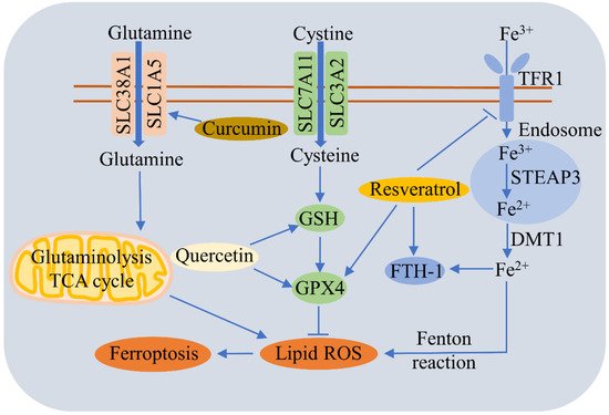
A large number of studies have reported that food-borne active ingredients, such as polyphenols, can regulate ferroptosis (Figure 34).
Figure 34. Regulatory effects of food-borne active ingredients on ferroptosis. This schematic diagram shows the regulation pathways of ferroptosis by polyphenols such as quercetin, curcumin, and resveratrol. Curcumin triggers ferroptosis by promoting SLC1A5-mediated glutamine uptake. Quercetin alleviates ferroptosis by upregulating GSH and GPX4. Resveratrol reduces ferroptosis by decreasing TfR1 expression and increasing the expressions of FTH1 and GPX4.
Quercetin (QCT), a natural flavonoid found commonly in fruits and vegetables, alleviates T2DM by inhibiting the ferroptosis of pancreatic β cells via the upregulation of GSH and GPX4, and by increasing mitochondria membrane-associated protein VDAC2, which functions as an antioxidant
[111][169]. QCT also reduces AKI by repressing ferroptosis via a reduction in MDA and lipid ROS levels and an increase in GSH
[134][190]. QCT can suppress the erastin-induced ferroptosis of bone-marrow-derived mesenchymal stem cells, probably via the antioxidant pathway
[165][215]. QCT triggers p53-mediated cancer cell ferroptosis by promoting lysosome-dependent ferritin degradation and ROS generation
[166][216]. Dihydroquercetin reverses cigarette-smoke-induced ferroptosis in the pathogenesis of chronic obstructive pulmonary disease by up-regulating the Nrf2-dependent pathway
[167][217].
Gallic acid (GA) is a natural polyhydroxy phenolic compound seen in various foods, such as edible mushrooms, fruits, and vegetables. GA triggers cancer cell death via the activation of apoptotic, ferroptotic, and necroptotic pathways
[168][218]. Preirradiation therapy followed by GA treatment inhibits the survival of cancer cells more effectively than GA treatment alone via the apoptosis and ferroptosis cell death mechanisms
[169][219].
Curcumin, a polyphenol compound extracted from the turmeric plant, enhances the treatment effect of NSCLC by activating autophagy-dependent ferroptosis
[5][70]. Curcumin decreases rhabdomyolysis-related renal damage by reducing ferroptosis, and mechanistic studies have shown that curcumin downregulates the TLR4/NF-κB axis and activates HO-1
[170][220]. Since curcumin nanoparticles suppress ferroptosis, they can be used to strengthen the treatment of ICH
[68][129]. Curcumin also induces the ferroptosis of breast cancer cells by upregulating SLC1A5
[36][99].
Epigallocatechin gallate (EGCG), a major polyphenol in green tea, protects against radiation-induced intestinal injury by scavenging ROS and repressing apoptosis and ferroptosis via the Nrf2 signal pathway
[171][221]. EGCG pretreatment reduces doxorubicin cardiotoxicity-induced ferroptosis by increasing AMPKα2 and promoting adaptive autophagy
[172][222]. Apigenin is a flavonoid found in green leafy herbs and vegetables, including celery, parsley, spinach, chamomile, green pepper, and eggplant, as well as oranges and red wine
[173][223]. Apigenin is able to alleviate myeloperoxidase-mediated oxidative stress and repress ferroptosis in neuronal cells
[174][224].
Resveratrol is a polyphenol that exists commonly in various vegetables and fruits, such as grapes. Resveratrol alleviates ferroptosis-induced myocardial ischemia/reperfusion injury, reduces TfR1 expression, and increases the expressions of FTH1 and GPX4
[175][225]. Resveratrol nanoparticles can repress erastin-induced ferroptosis in HT22 mouse hippocampal cells
[176][226], and also inhibits acrolein-induced ferroptosis and insulin secretion disorder via the ER-stress-related PERK pathway in mouse pancreatic β cells
[177][227]. Nobiletin, a critical active flavonoid in citrus fruits, was found to reduce ferroptosis-related renal injury, inflammation, and fibrosis in a unilateral ureteral obstruction mouse model
[178][228]. Nobiletin triggers the ferroptosis of human skin melanoma cells via the GSK3β-mediated Keap1/Nrf2/HO-1 signaling pathway
[179][229].