Analytical techniques play a fundamental role in heritage science (HS). Among them, Particle Induced X-ray Emission (PIXE) and X-ray Fluorescence (XRF) techniques are widely used in many laboratories for elemental composition analysis. Although they are well-established, a strong effort is put on their upgrade, making them suitable for more and more applications.
1. Technological Advance: The Micro-Particle Induced X-ray Emission (PIXE) XE Technique at The Laboratory of Nuclear Techniques for The Environment and Cultural Heritage of The Italian National Institute of Nuclear Physics ( the INFN-LABEC)
The capability to produce ion beams with size lower than 100 μm, known as microbeams, has been a natural development of PIXE technique. This is of great importance for samples with microstructures such as biological tissues, geological materials, microelectronic devices, or small specimens such as cultured individual cells, microcrystals, and single aerosol particles.
In the scanning system of the microbeam facility at the INFN-LABEC, the beam was swept over the target in horizontal and vertical directions by a magnetic field, perpendicular to the beam direction, generated by ferrite-cored coils positioned immediately before the lens. The magnetic deflector coils allowed, in principle, a maximum scanned area of several mm2 for 3 MeV protons but was, however, limited by the exit window aperture (2 × 2 mm2, typically).
Among the different applications, this beamline was used to analyse lapis lazuli
[1][2][43,44], a blue semi-precious stone used for more than 7000 years for carved jewels, decorative objects, as well for pigments. This study, besides increasing the knowledge on this blue rock, could shed light on many unresolved questions, especially regarding the trade routes exploited in ancient times.
Further information about the micro-beam line at INFN-LABEC and additional results can be found in
[3][4][47,48].
2. X-Ray Fluorescence (XRF)F: From Point Analysis to Elemental Maps with Portable Equipment
The main limit of the PIXE and the other IBA techniques is the lack of portability, a feature that is a severe limitation when a work of art cannot be moved to a laboratory. The XRF technique preserves non-invasive, non-destructive, and multi-elemental characteristics; moreover, it exploits portable instrumentation. For this reason, it is one of the most widely used techniques for material analysis in the field of cultural heritage. Its main drawback is a much lower sensitivity than PIXE to light elements
[5][49] and the limited possibility of a quantitative analysis in
heritage science (HS
) [6][26].
At INFN-LABEC, a portable XRF spectrometer was designed and assembled, exploiting the experience acquired over the years with X-ray spectroscopy using ion beams. In this device, different tubes (Mo, Ti and W anodes available) were used for maximising the efficiency of the production of X-rays over a wide range of energies, as it depends on the main emission lines of the anode material.
Among the many applications, see for example
[7][8][50,51], this instrument was also successfully employed for the discrimination between polishing methods of Japanese swords, or “katanas”. For the study, a set of katanas, both damaged and well-conserved, from the Stibbert Museum of Florence, were analysed, exploiting the XRF spectrometer. The measurements were conducted on a set of 13swords, confirming the goodness of this method to distinguish between the two groups of katanas. Further information is available in the publication
[9][52].
A natural development of the XRF technique is to allow scanning without disclaiming the portability of the device. With this aim, within the INFN-CHNet group, a MA-XRF scanner was designed with a special focus on portability and lightness. The technical characteristics and analytical capabilities (detection efficiency,
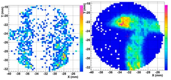
spatial resolution, and so onetc.) of this equipment are thoroughly described in [53]. Comparing the maps with the measurements conducted in Sections 3 and 5, the advantage of a scanning system is evident: it is straightforward to match the visible pattern with the elemental distribution and therefore to identify the material that was probably used for painting layers.
3. Comparison between PIXE and XRF Techniques at the INFN-LABEC
As a practical example, here, the same area of the parchment irradiated at the PIXE beamline described i
n Section 3 is reported and with the XRF spectrometer presented
in Section 5. The scan was conducted with the motor system shown in
Figure 12.
Figure 1. Scanning system at the end of the PIXE beamline.
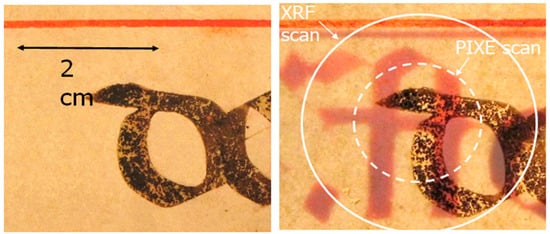
Figure 26.
The area irradiated by PIXE and XRF. The backlit image shows the presence of the backside.
As can be seen from the backlit picture (Figure 26), lyrics are present on the backside of the bifolio.
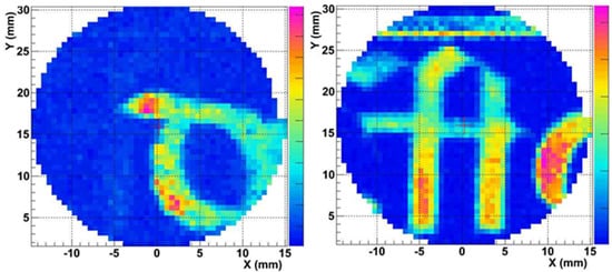
Results of the irradiation are presented in Figure 37 and Figure 48. As can be seen from Figure 37, with XRF it is possible to detect the ink from the two sides of the parchment, both iron from the front side (Figure 37, left) and mercury (Figure 37, right).
Figure 37.
Maps of Fe (
left
) and mercury (
right
) of the area scanned in
Figure 2 by XRF technique.
6 by XRF technique.
Figure 48.
Maps of Fe (
left
) and mercury (
right
) of the area scanned in
Figure 2 by PIXE technique.
6 by PIXE technique.
On the contrary, with the PIXE technique, only the ink on the front side was clearly detected, whereas the letter on the other side is not clearly visible from the map of mercury.
This result is a consequence of the higher penetration depth of XRF in comparison with PIXE. This can be considered an example of the advantage of matching the two techniques; indeed, the detection of the materials on the verso of the folio together with those on the recto may complicate the reading. On the other hand, two materials can be detected with only one measurement (easily distinguishable thanks to the mapping methods) and, moreover, the eventual interaction with these materials may be observed.