The SARS-CoV-2 pandemic represents the focus of the biomedical research worldwide. The identification of the molecular events related to the SARS-CoV-2 infection, as well as the characterization of its clinical features, could put an end to this dramatic health emergency. In this scenario, the interpretation of histopathological data in light of the clinical imaging characteristics of SARS-CoV-2 infection can provide the scientific rationale to develop diagnostic and therapeutic protocols that are capable of improving the management of infected patients. Specifically, morphological and molecular analysis of SARS-CoV-2 infected tissues could highlight new useful prognostic and predictive biomarkers for in vivo investigations.
- SARS-CoV-2
- imaging diagnostic
- Anatomic Pathology
- Radiology
- Pandemic
- Therapy
- ACE2
- biomarkers
1. Introduction
In December 2019, physicians reported numerous patients showing pneumonia of unknown origin in the Chinese region of Wuhan
In December 2019, physicians reported numerous patients showing pneumonia of unknown origin in the Chinese region of Wuhan
[1]
. Thanks to genomic investigations of the pathogen related to these diseases, Chinese health authorities demonstrated that the pneumonia outbreak was correlated to the infection of a new coronavirus, whose genetic sequence is homologous to that of the coronavirus causing severe acute respiratory syndrome coronavirus-2 (SARS-CoV-2)
[2]
. Following the spreading of the infection in all the world, The World Health Organization (WHO) on 11 March 2020 declared the novel SARS-CoV-2 outbreak a global pandemic.
Despite the SARS-CoV-2 infection appearing very complex, the initial common clinical manifestation of SARS-CoV-2-related disease, which facilitated patient’s detection, was pneumonia
. Several studies described the molecular mechanisms involved in the infection of pulmonary epithelium by SARS-CoV-2 as well as the immune-mediated response (
)
[2]
, but still, little is known about infections in non-pulmonary sites. While the latest literature provides insight into clinical manifestations of SARS-CoV-2 disease, histopathology and autopsy findings currently remain scarce. Similarly, imaging diagnostic analysis, such as computerized tomography (CT), have well described pulmonary abnormalities, but still not the involvement of other organs.
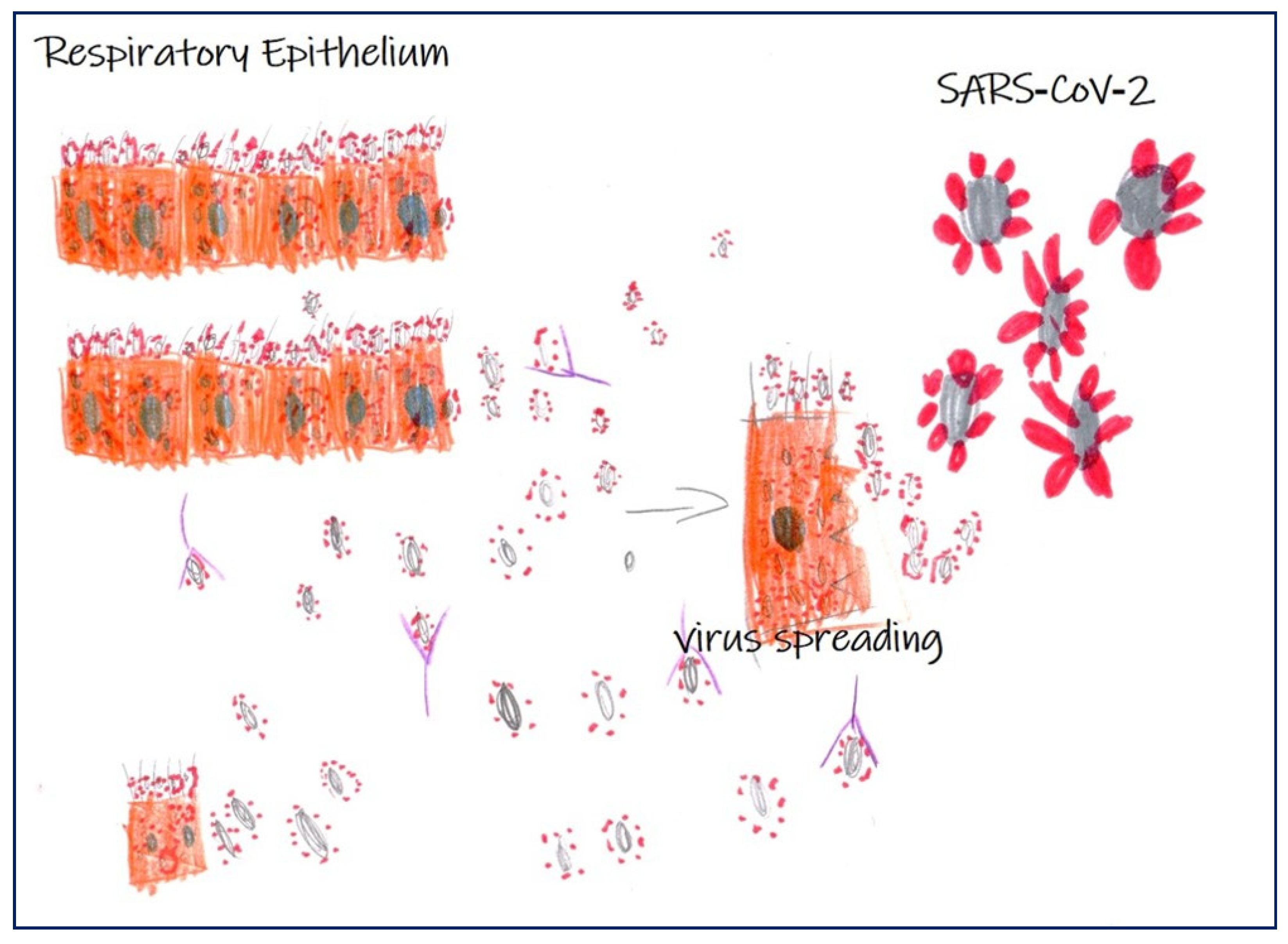
Figure 1.
Scheme of severe acute respiratory syndrome coronavirus-2 (SARS-CoV-2) infection in respiratory epithelium. Image shows infection of SARS-CoV-2 in respiratory epithelium, virus spreading, and the antibody response (Martina Gioia Simeca, 5-year-old).
2. Understanding SARS-CoV-2 Infection
SARS-CoV-2 infections are clinically characterized by two phases correlated with a different immune response
SARS-CoV-2 infections are clinically characterized by two phases correlated with a different immune response
[3]
. During the incubation stages, no severe clinical manifestations are generally observed in healthy population; these subjects are characterized by an appropriate genetic background for specific adaptive immune response, which frequently proves to be competent to eliminate the virus, precluding disease progression to severe stages
[3]
. However, if the immune response in positive patients does not eliminate the virus, subjects go through the most severe stages of disease, which are characterized by a damaging inflammatory response mainly involving lungs and resulting in severe diffuse alveolar damage (DAD). At this stage of disease, some other organs with high expression may be involved. The worse outcome of SARS-CoV-2 includes older age (e.g., >65 years), concomitant cardiovascular disease, hypertension, diabetes, obesity, kidney disease, cancer, and other immunodeficiency conditions
[3]
.
Indeed, recent clinical reports also describe gastrointestinal symptoms, heart injury, vasculitis, kidney dysfunction, and thrombocytopenia in SARS-CoV-2 positive patients
.
A better understanding of the histological changes observed during SARS-CoV-2 infection may increase our knowledge of the pathogenesis of the disease. The identification of specific histological characteristics of the SARS-CoV-2-related diseases, including biomarkers expression, along with specific morphological and molecular alterations detected by imaging methods, will help to formulate an earlier diagnosis and therefore to establish the most appropriate therapeutic protocols in order to prevent the frequent complications caused by this virus.
In this context, interdisciplinarity is fundamental. Specifically, a great contribution can be provided by the association and interpretation of data derived from medical disciplines based on the study of images, such as radiology, nuclear medicine, and pathology. Additionally, the histopathological characterization of tissues of SARS-CoV-2 patients, by both biopsy and autopsy, has made it possible to elucidate some mechanisms related to the SARS-CoV-2 infection. The first image of the SARS-CoV-2 that showed the structure of the virus in the world was caught using a transmission electron microscope
[1]
. Both the histological and ultrastructural analysis of lung, kidney, heart, and vascular compartments are elucidating the tissue alteration induced by SARS-CoV-2 infection. Furthermore, the use of ancillary techniques, such as immunohistochemical or in situ hybridization analyses, can provide molecular information related not only to the presence of the virus, but also to the virus-related cellular adaptations or, even more importantly, the inflammatory infiltrate associated to SARS-CoV-2 infection. These cellular and molecular biomarkers may also constitute a substrate for developing: (a) new diagnostic protocols based on radiotracers for PET or SPECT investigations, (b) predictive and prognostic assays, (c) new drugs, and/or (d) re-evaluation of already approved drugs for others diseases or viral infections.
Therefore, here, we highlighted the most recent histopathological and imaging data concerning the SARS-CoV-2 infection in lungs and others human organs such as the kidney, heart, and vascular system. In addition, we evaluated the possible match among data of radiology, nuclear medicine, and pathology departments in order to support the intense scientific work against the SARS-CoV-2 pandemic. In this regard, the development of artificial intelligence algorithms that are capable of correlating these clinical data with the new scientific discoveries concerning the SARS-CoV-2 might be the keystone to get out of the pandemic.
References
- Zhu, N.; Zhang, D.; Wang, W.; Li, X.; Yang, B.; Song, J.; Zhao, X.; Huang, B.; Shi, W.; Lu, R.; et al. China Novel Coronavirus Investigating and Research Team. A novel coronavirus from patients with pneumonia in China. N. Engl. J. Med. 2020, 382, 727–733.
- Yan, F.; Gao, Y.; Pang, X.; Xu, X.; Zhu, N.; Chan, H.; Hu, G.; Wu, M.; Yuan, Y.; Li, H.; et al. Evolution of the novel coronavirus from the ongoing Wuhan outbreak and modeling of its spike protein for risk of human transmission. J. Exp. Bot. 2020, 63, 457–460.
- Park, S.E. Epidemiology, virology, and clinical features of severe acute respiratory syndrome -coronavirus-2 (SARS-CoV-2; Coronavirus Disease-19). Clin. Exp. Pediatr. 2020, 63, 119–124.
- Li, M.Y.; Li, L.; Zhang, Y.; Wang, X.S. Expression of the SARS-CoV-2 cell receptor gene ACE2 in a wide variety of human tissues. Infect. Dis Poverty 2020, 9, 45.
- Zhao, X.Y.; Xu, X.X.; Yin, H.S.; Hu, Q.M.; Xiong, T.; Tang, Y.Y.; Yang, A.Y.; Yu, B.P.; Huang, Z.P. Clinical characteristics of patients with 2019 coronavirus disease in a non-Wuhan area of Hubei Province, China: A retrospective study. BMC Infect. Dis. 2020, 20, 311.

