Freezing of Gait: Comparison
Please note this is a comparison between Version 1 by Roger Clotet and Version 4 by Jamir Pitton Rissardo.

Parkinson Disease (PD) primarily affects older adults. It is the second-most common neurodegenerative disease after Alzheimer’s disease. Freezing of Gait (FoG) is a symptom present in approximately 80% of advanced-stage PD’s patients. FoG episodes alter the continuity of gait, and may be the cause of falls that can lead to injuries and even death. The recent advances in the development of hardware and software systems for the monitoring, stimulus, or rehabilitation of patients with FoG has been of great interest to researchers because detection and minimization of the duration of FoG events is an important factor in improving the quality of life. 

  • Parkinson Disease
  • freezing of gait

1. Introduction

Parkinson Disease (PD) is the second-most common neurodegenerative disease, after Alzheimer’s disease, among the elderly. At present, there are 10 million people patients with PD worldwide [1][2][1,2]. Statistics show that the prevalence of PD is higher in Europe, North America, and South America in comparison with Africa and Asia, with an incidence of 13.4 per 100,000 people per year [2][3][4][2,3,4]. The causes of PD remain unknown, but some studies have attributed it to environmental exposure factors and genetic factors [5]. Moreover, sex and ethnicity have been shown to be influencing factors, with the male:female ratio of patients with PD being approximately 3:2 [3]. Age remains the main risk factor for the development of PD, and the prevalence and incidence of the disease increases exponentially after 60 years of age [3][6][3,6]. This trend has important implications for public health, since greater longevity, which is the trend in most countries, is expected to increase the number of people with PD by 50% in 2030 [3][7][3,7].
Parkinson disease is associated with non-motor and motor symptoms. Non-motor symptoms include dementia, depression, psychotic characteristics, autonomic dysfunction, oculomotor abnormalities, and olfactory and visual impairments. Though not as visible as these motor symptoms, non-motor symptoms are also experienced by many individuals with PD as a part of their disease. Patients with PD may experience nonmotor symptoms related to the disease itself or to the medications used to treat it.[8] The moto include tr symptoms include tremor, stiffness, bradykinesia, postural instability, festination, decreased blink frequency, blepharospasm, and Freezing of Gait (FoG) [9][8]. Medical evaluation of non-motor symptoms is usually performed by a neuropsychologist, while motor symptoms are diagnosed by a neurologist on the basis of medical history, a review of signs and symptoms, and physical and neurological examinations.
The FoG symptom is related on bradykinesia, rigidity, tremor, and postural instability, together with perceptive malfunction and frontal executive dysfunction [9][10][8,9]. It presents as a reduction in the forward progression of the feet, despite the person’s intention to walk [11][12][13][10,11,12]. This symptom occurs in 21–27% of patients in the early stages of PD [14][15][13,14], and this percentage continues to increase during PD evolution, with the symptom appearing in 80% of patients more than 17 years from the initial diagnosis. FoG may be the cause of falls that can lead to injuries and even death [16][15].
FoG has its origin in the brain, specifically in the mesencephalic locomotive region (MLR), where the performance of the pedunculopontinal nucleus (PPN) diminishes their connections with basal ganglia. Similarly, the region of the brain stem is related to the condition of freezing, which has been validated with Functional Magnetic Resonance Imaging (FMRI) and ElectroEncephaloGraphy (EEG) data. FoG is no longer considered a strictly motor symptom but is a part of cognitive impairments that originate from areas of the brain that allow the body to be able to walk without hindrance [17][18][16,17].
FoG does not respond to existing drugs, and neurorehabilitation exercises tend to be repetitive and tiring. Several invasive methods, such as deep brain stimulation (DBS) [19][18] or vagus nerve invasive stimulation (VNS) [20][19], have been developed for the treatment of FoG, but they are expensive, do not guarantee elimination of freezing, and may increase other symptoms. Patients with an episode of FoG can resume walking after receiving external stimulation, and non-invasive methods such as visual, vibratory, and tactile stimulation devices can provide such stimulation at a relatively low cost and without health risks.

2. Brain Activity during a FoG Episode

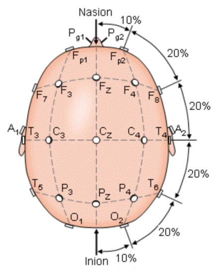
To understand the parameters to be measured by those systems, it was necessary to analyze the brain motor activity involved in PD during episodes FoG. Brain activity occurs when the brain generates electrical impulses known as action potentials, which travel through neurons. Electrical impulses contain information that travels from neuron to neuron making use of hundreds of thousands of them to get transported and perform a specific function, any alteration provokes a change in their contiguous connections [21][20]. When the brain generates an impulse to move a muscle, the impulse passes through the basal ganglia that help to smooth muscle movements and coordinate changes in posture, such as the gait. A statistical parametric mapping analysis applied to healthy subjects during the gait revealed that the following areas were activated in their brain activity: supplementary motor, medial primary sensorimotor, striatum, cerebellar vermis, and visual cortex. These results indicate that the cerebral cortexes that control: motor functions, visual cortex, basal ganglia, and cerebellum, may be involved in the bipedal locomotor activities in humans [22][21]. When a person has PD, there is a degeneration in the cells of the basal ganglia that causes a decrease production of dopamine and reduces connectivity between nerve cells and muscles [23][22]. Encephalography was used to understand the bioelectrical connections of those who suffer from PD and present FoG. The Figure 1 shows the electrode arranged system in a 10–20 scheme. This scheme was used to analyze brain activity utilizing electrodes on the hair scalp in FOG patients. The presence of FOG episodes generates different levels of energy in the brain waves of the parietal zone (P4), suggesting that this zone has been deeply affected by the disease. Measurements of P4 and the central zone (Cz) are the features that most contributed to the analysis for detecting FoG transition in PD patients [24][25][26][23,24,25].
Figure 1. The international 10–20 system seen above the head. A = Ear lobe, C = central, Pg = nasopharyngeal, P = parietal, F = frontal, Fp = frontal polar, O = occipital [24][23].
When the fronto-parietal zone is affected, there is a decrease in executive functions (including cognitive skills), aggravating the problems in people with FoG, who also have failures in their visuospatial network. Some authors such as Amboni et al. compared the progression of cognitive impairment in 26 Parkinsonian patients with FoG (FoG+) and without FoG (FoG-) over a follow-up period of 2 years, finding that FoG+ patients had a faster progression of cognitive impairment, while in FoG- patients, cognitive alteration remained unchanged during this period [27][26]. Another technique used is magnetic resonance imaging, where it was concluded that FoG+ patients present predominantly frontal-executive dysfunction compared to FoG- patients [28][27]. At the same time, the right hemisphere of the brain looked more affected in FoG+ patients, which would make sense due to the great influence of this hemisphere on visuospatial abilities [29][28]. At a neuronal level, the important role of the pedunculopontine nucleus (PPN) located in the MLR, likely play a crucial role in the appearance of axial symptoms in PD. The aim was to activate the remaining PPN cholinergic neurons to improve axial symptoms including FoG and balance deficits in PD patients. Its importance resides in that it is the main center of the mesencephalic locomotor region and controls the initiation, maintenance, and modulation of posture and gait [30][29]. The presence of FoG was associated with altered functional connectivity between the PPN and the corticopontine-cerebellar pathways (in the bilateral cerebellum and in the pons) and visual temporal areas compared to healthy subjects, additionally marked abnormalities in white matter extending to motor, sensory and cognitive regions [31][30]. A significant number of PD patients increasingly rely on visual and auditory signals to control the locomotion [32][31], which creates a problem because visual impairments have been correlated with gait disturbance, deficits in visual attention, memory, and visuospatial abilities [33][34][32,33]. Lenka et al. managed to establish that a reduction in inter-hemispheric connectivity between bilateral parietal operculum, somatosensory cortex, and primary auditory areas are correlated with FoG [35][34]. The hearing deficiency was tested with the Rey Auditory-Verbal Learning Test (RAVLT) [36][35]. This shows a decline in vision and hearing in those who have episodes of FoG.

3. Motor Characteristics during FoG

Although FoG originates in the brain, it manifests as an irregularity in gait. As PD progresses, this irregularity becomes more frequent and disabling for the patient, which results in longer FoG episodes. These episodes can present as complete akinesia when the patient is not medicated (off state, FoG-). When the patient is medicated (on state, FoG+), the FoG episodes are shorter and rarely become akinetic. FoG+ patients present deficiencies during motor initiation that are not present in FoG- patients or healthy people. Additionally, FoG+ patients are slower to initiate motor activity, and in particular, to respond to the signal from the brain to initiate walking. Moreover, these patients require more time to react to stop walking in comparison with FoG- patients and healthy people [37][38][36,37]. In muscular-system analyses, FoG is characterized by co-contraction of agonist and antagonist muscles [11][39][10,38]. The patients show changes in the pressure of the foot and the behavior of the distance between the steps, as these become shorter compared to those in a slow walk. The frequency of this type of walk is between 4 and 5 Hz [40][41][39,40]. Patients with FoG respond to classic auditory, visual, and tactile stimulations [42][41]. However, these stimulations cannot effectively induce the muscular system to react to bradykinesia [43][42], which results in alterations in symmetry, rhythm, and bilateral coordination in the patients’ walk with FoG [44][45][43,44]. On the other hand, dysfunction in cognitive networks and interictal gait changes may contribute to Parkinson’s disease patients presenting with episodes of FoG. An analysis of patients performing two activities at the same time is presented in one study [46][45], and the FoG+ patients showed a shorter stride length and slower speed, except during postural balance, in that study. In contrast, during the turn, both groups (FoG+ and FoG-) showed a slower turning speed in the tests involving double activities compared to the findings for the single-task condition. Analyses of the FoG to date have focused on the lower extremities. However, some studies have revealed determining characteristics in the upper limbs, specifically in the wrists, which show FoG earlier than the legs and feet [37][47][36,46]. These studies revealed that FoG can be detected using wrist motion and machine learning models with an FoG hit rate of 0.9 and specificity between 0.66–0.8. Additionally, the standard deviation, acceleration, and rotation were analyzed, in addition to the power between the frequency ranges of 0 to 1 Hz, 5 to 6 Hz, and in the intervals of 0 to 4 Hz, 5 to 8 Hz, and 9 to 12 Hz [47][48][46,47]. Some studies have focused on analyzing single-leg posture using portable inertial sensors and performing statistical calculations. The results of these studies indicate that the acceleration peak of the medial-lateral trunk in FoG+ and FoG- patients was significantly lower than that of healthy patients (p < 0.05), and the equilibrium was longer in FoG- patients in comparison with that in FoG+ patients [49][48]. Various studies have revealed significant differences in the duration of the balanced phase of the feet during walking in FoG+ and FoG- patients and healthy subjects. This reduction in the duration of equilibrium is generally associated with an increased risk of falls [50][51][52][53][54][49,50,51,52,53]. In one study [55][54], the two main reasons for falls in PD patients were identified as FoG and impaired balance. This argument is supported by other investigations [56][57][58][55,56,57], which validated the relationship between FoG and falls. These falls are consequences of FoG and appear from the early stages of the disease [59][60][61][58,59,60]. Additionally, falls could be disabling and can deteriorate the quality of life of patients [62][63][64][65][61,62,63,64]. These falls can occur in multiple directions and are associated with motor symptoms but also with non-motor symptoms, such as mood and cognitive disorders [66][65]. When the FoG occurs, the center of gravity continues to advance due to the effect of inertia, but the feet stop moving body while the body continues forward, which causes the majority of falls. Therefore, to initiate the walk, FoG+ patients require wider movements than FoG- patients [18][17].