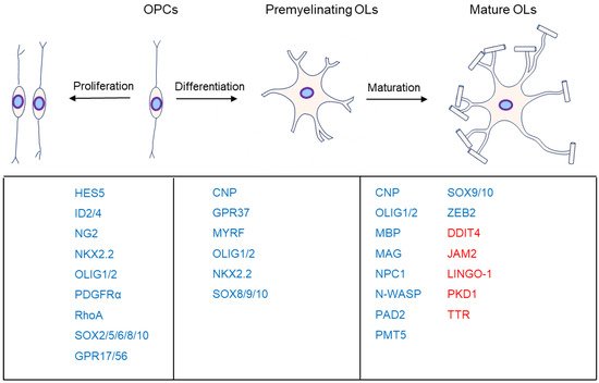
2. OL Differentiation
OLs generated from OPCs are fundamental to myelin formation, and all myelination processes are composed of OPC proliferation, OL differentiation, and maturation. Although a supra-threshold axon diameter (>0.3 μm) is necessary for axonal myelination during optic nerve development, OL differentiation is independent of dynamic signals from the myelinated neuron
[6][74]. Supra-threshold axon diameter is only an insufficient necessary factor.
Notably, the first territories to be occupied by OPCs are not necessarily the first regions to be myelinated
[7][75], suggesting a potential regulation of other environmental factors. However, dynamic neuronal signals such as transcriptional changes or neuronal activity may also mediate physiological processes, such as OL differentiation and maturation
[8][76]. The optic nerves are almost myelinated, but most brain regions are partially myelinated, even though the axon caliber is far more than the supra-threshold. Consequently, potential inhibitory factors or repulsive signals likely prevent the onset of myelination
[9][77]. Conversely, it is also possible that attractive factors or signals exist to initiate the onset of myelination
[6][74].
2.1. AKT-mTOR
AKT (AKT serine/threonine kinase 1), as an essential effector of PI3K (phosphatidylinositol-4,5-bisphosphate 3-kinase), promotes myelination in the CNS
[10][11][78,79] through the mTOR (mechanistic target of rapamycin) pathway (
Figure 23). Akt stimulates axonal wrapping and raises myelin thickness by the mTOR pathway. Moreover, maintaining AKT activation induces hypermyelination or demyelination
[12][80]. Accordingly, sustaining the PI3K signaling pathway in OPCs results in gradual hypermyelination, leading to leukodystrophy
[13][81]. Conversely, PTEN (phosphatase and tensin homolog)
[14][82], as an inhibitor of the PI3K-Akt-mTOR signaling pathway, negatively regulates myelination, and DLG1 (discs large MAGUK scaffold protein 1, an interactor of PTEN) has a similar effect during myelination
[13][15][81,83]. CNP (2,3-cyclic nucleotide 3-phosphodiesterase) controlling constitutive AKT activation boosts MBP staining and increases the corpus callosum size, with pronounced hypermyelination in small-caliber axons
[12][80].
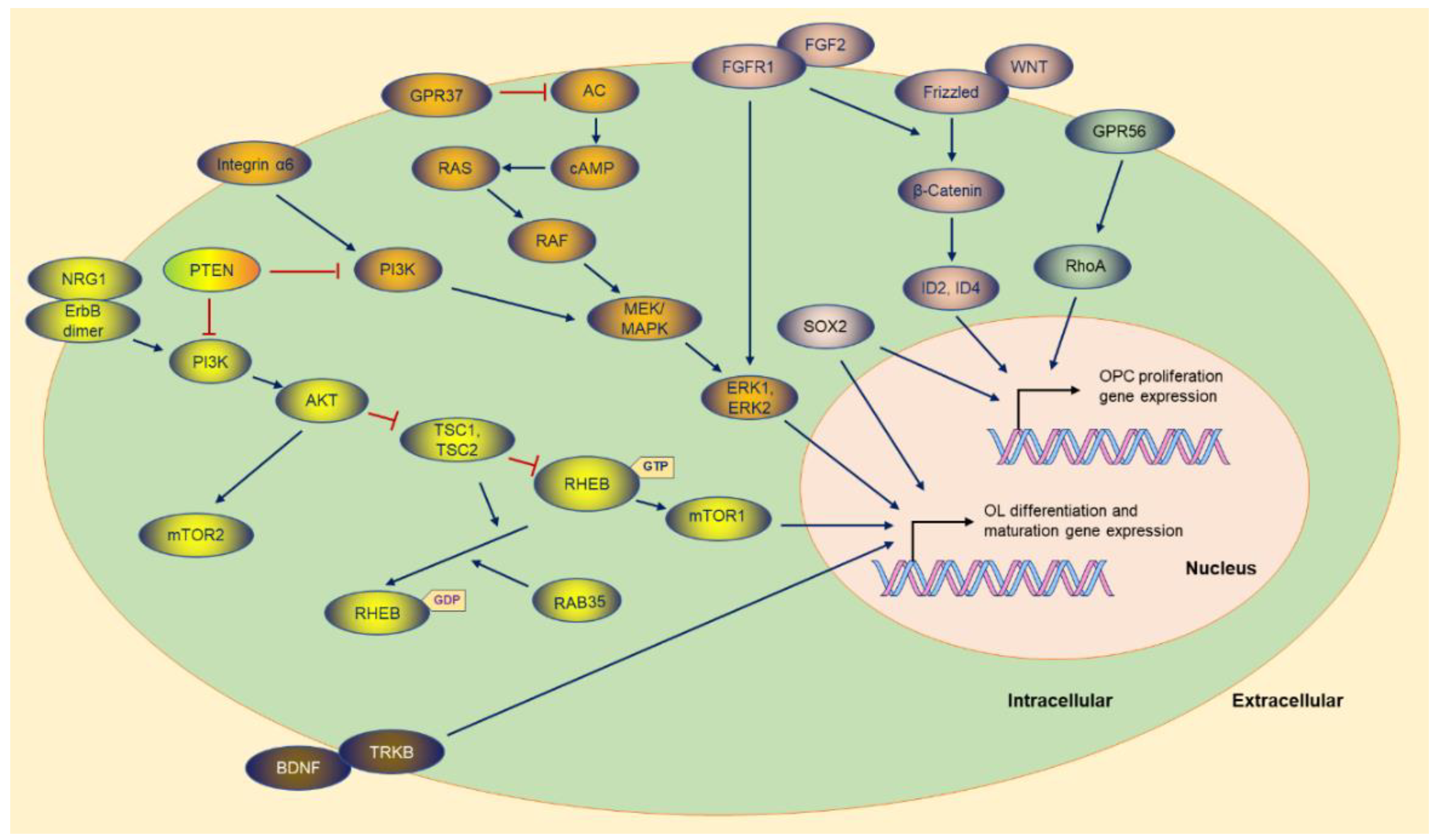 Figure 2. The
 m
olecules and the potential signaling pathway regulate the OPC proliferation and OL differentiation gene expression. Most of the molecules are exhibited, and the majority of the upstream molecules originate from the cytoplasmic membrane. T
he sign of blue arrows represents promoting, and the sign of red lines represents inhibiting. AC: Adenylate cyclase.
mTOR, as the downstream of the PI3K-AKT signaling pathway, is the core element of two complexes (mTORC1 and mTORC2) with different functions in the complex
[14][16][82,84]. Ablation of RAPTOR (regulatory-associated protein of MTOR complex 1), as an element of the mTORC1, leads to hypomyelination. Conversely, RICTOR (RPTOR independent companion of MTOR complex 2) deficiency does not affect myelination as an essential element of the mTORC2
[14][82].
Tuberous sclerosis protein 1 (TSC1) and TSC2 negatively regulate mTORC1 signaling
[14][17][18][82,85,86], and downregulation of mTORC1 is essential for OPC differentiation and the subsequent myelination initiation. TSC2, as a member of GAP (GTPase-activating protein), constrains mTORC1 by stimulating the GTPase activity of RHEB (Ras homolog, mTORC1 binding), while RHEB binding to GTP causes the activation of mTORC1
[18][86]. Although TSC1 is not a GAP (RAS p21 protein activator 1), it stabilizes TSCs to maintain GTPase activity
[14][82]. Thus, TSC1 or TSC2 ablation is supposed to strengthen RHEB-GTP stability, and consequently, abnormal activation of mTORC1 results in arrested differentiation in the early stage
[16][19][84,87]. In some situations, mTORC1 hyperactivity causes distinctly delayed myelination and remyelination after injury
[19][87]. Surprisingly, TSC1 deletion is detrimental to OL myelination
[20][21][88,89].
mTORC1 activity also is downregulated by RAB35 (RAB35, member RAS oncogene family), a Ras-related GTPase controlling myelin growth through MTMR2 (myotubularin-related protein 2) and MTMR13
[22][90]. Accordingly, disruption of
Rab35 causes hypermyelination via elevation of PI3P signaling and mTORC1 hyperactivation
[22][90].
Some factors can switch the PI3K pathway to the MAPK pathway, such as Integrin α6 in OLs, a receptor for laminins in the neuronal axon. When the two types of molecules contact each other, myelin-forming OLs activate the switch in survival signaling dependence
[23][91], which reverses neuregulin’s inhibition in OL differentiation, subsequently promoting the myelination process
[23][24][91,92].
2.2. ERK1/2
The ERK1/2 advocates myelin wrapping during myelination and remyelination, and maintained OL ERK1/2 activation causes hypermyelination in the CNS
[25][1] (
Figure 23). ERK1/2 contributes to remyelination after the demyelination induced by lysophosphatidylcholine (LPC) injection into the corpus callosum
[26][93]. MEK1 (MAPK kinase 1) is the upstream activator of ERK1/2 and displays declined expression in experimental autoimmune encephalomyelitis (EAE) induction
[25][1]. Accordingly, maintained ERK1/2 activation upgrades myelin thickness after the LPC injection
[27][28][94,95]. MEK inhibitors, such as PD0325901, AZD6244, AZD8330, CI-1040, and U0126
[29][30][31][96,97,98], significantly rectify OPC differentiation in a time- and dose-dependent manner, suggesting that regulation of the MAPK–ERK signaling pathway is sufficient to accelerate OL generation, facilitating myelin sheath formation
[29][96].
2.3. GPR37
GPR37 is highly expressed in OLs and significantly intensified during OL differentiation and myelination (
Figure 23). GPR37, also known as PAELR (Parkin-associated endothelin B-like receptor), has been identified as a substrate of parkin (an E3 ubiquitin ligase)
[32][99]. Although the OPC number was not affected in
Gpr37 null mice, GPR37 is regarded as an inhibitor of OL differentiation and myelination. Yang HJ et al. found that GPR37 restricts OL differentiation and hypermyelination by suppressing the cAMP-dependent Raf-MAPK-ERK1/2 cascade
[33][35].
Gpr37 knockout leads to dramatically diminished MAG expression in the mouse brain. The mutant mice exhibit strikingly enlarged myelin loss during the cuprizone demyelination model without impacting the number of OPCs and OLs
[34][100]. GPR37 suppresses activation of the cAMP-dependent Raf-MAPK-ERK1/2 cascade via inhibiting AC (adenylate cyclase)
[33][35].
2.4. SOX10-MYRF
MYRF (myelin regulatory factor) is a membrane-bound transcriptional factor on the endoplasmic reticulum (ER). It forms homo-trimers in the ER
[35][101] and then undergoes self-cleavage via the intrinsic peptidase
[36][37][38][102,103,104]. TMEM98 (transmembrane protein 98) can block MYRF self-cleavage in vitro and in vivo
[39][40][105,106]. Following the self-proteolysis, the homo-trimer of MYRF N-terminal fragments is translocated to the nucleus and binds the motif to activate the transcription of myelin genes
[36][37][41][42][43][44][45][46][102,103,107,108,109,110,111,112].
MYRF is required to initiate and maintain myelination
[5][47][8,113] (
Figure 23).
Myrf knockout mice sustain the premyelinating stage, leading to myelination failure and postnatal death
[48][49][10,114], a similar phenotype to that in
Olig1 null mice
[50][115] and
Sox10 mutant Zebrafish
[51][116]. Loss of
Myrf in OPCs does not alter OPC proliferation and recruitment after demyelination but impairs remyelination because of diminishing OL differentiation
[41][52][107,117]. In addition, OLs deriving from OPCs with
Myrf deletion produce few myelin proteins in response to demyelination
[41][107]. Not surprisingly, conditioned knockout
Myrf in mature OLs leads to a dramatic downregulation of myelin gene expression and impairment of myelin sheaths
[47][52][113,117].
Myrf expression is enormously intensified during the initiation of OL differentiation, and SOX10 acts as a
Myrf gene enhancer
[53][118]. SOX10 binds the first intron of
Myrf, which is also the region regulated by OLIG2
[54][119]. Once induced by SOX10, MYRF redirects SOX10 to myelin gene expression
[46][112]. Therefore, the feedback and forward regulatory loops compose an essential molecular circuit for OL differentiation and maturation. Surprisingly, despite the importance of MYRF in OL differentiation and maturation, most of the genetic mutations of
Myrf do not cause apparent myelin-related human diseases
[55][56][57][58][59][120,121,122,123,124].
2.5. NRG1
NRG1 (Neuregulin-1) promotes OPC migration, proliferation, and differentiation into OLs
[60][61][62][125,126,127], and NRG1/ErbB (Erb-b2 receptor tyrosine kinase) signaling (
Figure 23) is required for OL survival and maturation
[63][64][65][128,129,130].
NRG1 has more than 30 splice isoforms, sharing an EGF-like function domain to activate the receptors, ErbB2/ErbB3 heterodimer, or ErbB4 homodimer
[66][67][68][131,132,133]. Immature NRG1 is a transmembrane protein that releases soluble N-terminal moieties containing the EGF-like domain after proteolytic processing
[68][133]. Soluble NRG1 is a mitogen for OLs, provides an axonal signal for OL survival and increases myelination
[69][134]. Inhibiting its receptors reversed the positive effects of the administration of soluble NRG1
[60][125]. BACE1 (Beta-site APP-cleaving enzyme 1) cleaves NRG1 type Ⅰ and type Ⅲ.
Bace1 deficiency leads to hypomyelination and impairs remyelination
[64][129].
NRG1, acting through ErbB, is an important regulators in OPC proliferation and OL differentiation during development
[70][135]. NRG1-ErbB modulates myelin-related gene expression depending on the PI3K-AKT-mTOR pathway
[67][71][72][132,136,137]. Recently, Ding Z et al. found that NRG1 can convert astrocytes into OL lineage cells via the PI3K-AKT-mTOR signaling activation and eventually improves remyelination
[73][138]. Increasing AKT activity of the OLs in
Bace1 null mice is sufficient to normalize myelination without inducing hypermyelination
[74][139].
Nrg1 type Ⅲ null mice died at birth owing to the deficiency of functional neuromuscular junction, which also hampers the analysis of NRG1 in the CNS
[75][76][140,141]. Although OLs normally differentiate when cocultured with
Nrg1 type Ⅲ null dorsal root ganglia, the myelination of dorsal root ganglia is impaired in the
Nrg1 type Ⅲ deficient mice
[77][142]. Intriguingly, heterozygous
Nrg1 type Ⅲ mutant mice exhibit hypomyelination in the brain, but myelination in the spinal cord and the optic nerve is normal
[77][142]. However, others have not found CNS hypomyelination in heterozygous
Nrg1 type Ⅲ mutant or
Nrg1 null mice
[78][143]. Surprisingly, mice overexpressing
Nrg1 type Ⅲ have hypomyelinated axons in the CNS
[78][143].
Nrg1 type Ⅲ overexpression in the spinal cord improves motor function and increases motor neuron survival in mice with amyotrophic lateral sclerosis
[79][144].
2.6. BDNF
CNS myelinating cells develop from slowly dividing adult progenitor cell OPCs through a premyelinating OL stage before maturing into myelinating OLs.
Bdnf (brain-derived neurotrophic factor) heterozygous mice display hindered myelination in the CNS
[80][145] (
Figure 23). BDNF functions via two different classes of transmembrane receptors: TRKB (tropomyosin-related kinase receptor B) and p75NTR (p75 neurotrophin receptor). Maintaining BDNF enhances myelination, but it cannot be attenuated in the
p75Ntr knockout mice
[80][145]. Therefore, p75NTR is not necessary for promoting myelination. Conversely, BDNF cannot rescue the impeding myelination induced by the impairment of TrkB signaling
[80][145].
2.7. GlcNAc
Recently, Michael et al.
[81][146] found that GlcNAc (N-acetylglucosamine) is essential in triggering OL differentiation. N-glycan branching and GlcNAc support OPC differentiation into OLs via suppressing PDGF-α. Furthermore, primary myelination in newborn pups is strengthened when the lactating mice are supplemented with oral GlcNAc. Conversely, by blocking N-glycan branching, primary myelination is impeded. Intriguingly, oral GlcNAc protects neuronal axons from damage in the cuprizone-induced demyelination mouse model via enhancing myelin reparation
[81][146].
2.8. OLIG2
OLIG2 is regarded as a marker of OL family cells, although it is also expressed during development in motoneurons, subgroups of astrocyte precursors, and Purkinje cell precursors
[82][83][84][85][86][147,148,149,150,151]. OLIG2 acts as a binding upstream enhancer to induce the expression of target genes such as
Nkx2.2 (NK2 homeobox 2) and
Sox10. Conditional knockout of
Olig2 in embryonic neural stem cells confines OL differentiation without the alteration of OL specification, resulting in hypomyelination
[87][152].
OLIG2 is critical to OPC specification and OL differentiation. When cortical OPCs are conditional knockout
Olig2, the OPCs are transformed into astrocytes
[88][153]. OPCs are completely absent from most regions of the CNS in
Olig2 knockout mice
[89][90][91][92][154,155,156,157]. However, OPCs are still present in dramatically reduced numbers in the forebrain and hindbrain of
Olig2 knockout mice
[89][93][154,158]. Conditioned knockout of
Olig2 in OPCs leads to hypomyelination because of the limitation of OL differentiation, while conditional knockout of
Olig2 in immature OLs accelerates OL myelination by facilitating maturation
[93][158].
Though
Olig2 is closely related to
Olig1 [94][159],
Olig1 provides little compensation for
Olig2 loss
[82][147]. OLIG2 forms a homodimer or a heterodimer to induce OPC specification
[83][148]. Moreover, OLIG2 exhibits versatile functions in OPCs and OLs via post-translational modification, which was reviewed thoroughly by H Li and WD Richardson
[86][151].
2.9. PDE
PDEs (phosphodiesterases) have been implicated in OL maturation and myelination in the CNS (
Figure 23). Inhibitors of PDEs, such as PDE1 inhibitor vinpocetine
[95][160] and PDE5 inhibitor sildenafil
[96][161], not only spoil inflammation but also exert a negative impact on the CNS OL differentiation processes, including diminishing myelin gene expression and enhancing myelination negative transcriptional regulators (such as ID2 and ID4)
[96][161].
3. OL Maturation
3.1. DDIT4
DDIT4 (DNA damage inducible transcript 4, also known as REDD1/Dig2/RTP801) is a negative regulator of myelination
[15][83]. DDIT4 maximum expression is in line with the peak activity of AKT and DLG1. Moreover,
Ddit4 deficiency provokes hypermyelination by enhancing mTOR activation and enlarged myelin thickness. Intriguingly,
Ddit4-deficient mice do not present myelin out-folding (depending on PIP3) or macula (depending on AKT)
[15][83].
3.2. JAM2
Neuronal JAM2 (junction adhesion molecule 2) is sufficient and necessary in the somatodendritic membrane to inhibit OL myelination in the neuronal cell body
[9][97][77,162], while Galectin-4 is an inhibitor in the axons
[98][163]. Galectin-4 is specifically sorted into segmental domains along the axon membrane, and OLs do not deposit myelin on Galectin-4 covered surfaces, leading to long unmyelinated axon segments
[98][163]. After the JAM2 extracellular portion is fused to the immunoglobulin Fc region, the formed JAM2-Fc is added into cultured OLs, with higher JAM2-Fc binding to MBP+ myelinating OLs than to OPCs, suggesting that the JAM2 receptor is upregulated on the surface during OL differentiation
[9][97][77,162]. Furthermore, soluble JAM2-Fc arrests myelin formation in cultured OLs from wild-type mice
[9][97][77,162]. Intriguingly, recent studies have found that JAM2 is also associated with primary familial brain calcification, an uncommon degenerative neurological disease due to abnormal calcium phosphate deposits in the brain
[99][100][164,165]. In
Jam2 null mice, the neuronal soma is sheathed, and contactin-associated protein, which typically localizes only with the paranodal structures of the Nodes of Ranvier, clusters on the neuronal somatic member
[9][77].
3.3. PKD1
PKD1 (protein kinase D1), a serine/threonine kinase belonging to the calcium/calmodulin-dependent kinase family, is implicated in OL maturation, except for its role in tumor progression
[101][102][166,167].
Pkd1 homozygous deficiency is lethal to the mutant mice, and
Pkd1 heterozygous mutant mice exhibit quickly inducible epilepsy and hypermyelination, supporting the finding of the epilepsy patient
[103][168]. In addition, PKD1 can elevate functional synapse formation by enhancing N-cadherin’s stability in an activity-dependent manner
[104][169].
3.4. TTR
TTR (Transthyretin) binds and transfers thyroid hormones in cerebrospinal fluid and blood.
Ttr mutation is related to familial amyloid polyneuropathy, a neurodegenerative disorder with TTR deposition in the peripheral nervous system
[105][106][170,171]. However,
Ttr null mice exhibit hypermyelination, magnified OL density in the corpus callosum, and anterior commissure during postnatal development
[107][172]. Moreover,
Ttr deficiency magnifies OPC migration and proliferation with debased apoptosis
[107][172]. Intriguingly, TTR is expressed in OPCs, and boosting the pAKT level in OLs may be the mechanism of hypermyelination
[107][172]. During remyelination in the adult mouse corpus callosum,
Ttr null mice exhibit an expedited remyelination rate, preferentially remyelinating small axons
[108][173]. Moreover,
Ttr null mice display thicker myelin than wild-type mice
[108][173].
3.5. LINGO-1
LINGO-1 (leucine-rich repeat and Ig-like domain-containing Nogo receptor interacting protein 1), a transmembrane protein, is expressed explicitly in OLs and neurons, serving as a potent negative modulation of axonal myelination and regeneration in the CNS
[3][109][6,174]. Downregulating LINGO-1 functions
[3][6], such as
Lingo-1 RNAi
[110][175], sh-RNA
[109][111][174,176], anti-LINGO-1 antibody
[112][113][114][115][116][177,178,179,180,181], dominant-negative LINGO-1, or soluble LINGO-1-Fc
[117][118][182,183], improves OL differentiation and myelination, accompanied by prolonged process length and augment branching, and downregulated RhoA
[116][119][181,184] activity is the potential mechanism
[3][6]. Conversely,
Lingo-1 overexpression results in RhoA activation, negatively regulating OL differentiation and myelination
[3][6]. Neuronal LINGO-1 is a critical component of the Nogo receptor complex, restricting axonal growth via RhoA. The Nogo receptor is absent in OLs, and consequently, LINGO-1 prevents OL myelination through intercellular interactions with self-association in the trans
[118][183] or cytoplasmic gelsolin signaling pathway
[114][179].
3.6. N-WASP
N-WASP (Neural Wiskott–Aldrich syndrome protein) is essential for myelin wrapping in Schwan cell and OL myelination
[120][121][122][123][185,186,187,188]. Conditional knockout
N-Wasp continues to ensheathe the onset myelinating axons but fails to extend circumferentially to elaborate myelin, and the affected mice demonstrate apparent motor deficits without progress
[122][187]. In
N-Wasp-deficient nerves, most cells arrest at the premyelinating stage and subsequently fail to myelinate, with occasional misfolding myelin forming unusually short internodes and thin myelin sheaths
[121][186]. Strikingly,
N-Wasp deficiency leads to hypomyelination and induces remarkably focal hypermyelination, representing long myelin out-folds enclosing neuronal cell bodies and unmyelinated axons
[120][185].
3.7. PRMT5
PRMT5 (protein arginine methyltransferase 5) is a histone arginine methyltransferase catalyzing histone H4R3 methylation. Although suppression or knockout of
Prmt5 does not affect OPC proliferation, it attenuates OPC survival and differentiation leading to hypomyelination
[124][189]. Its potential mechanism is heightened nuclear acetylation of H4K5 following histone H4R3 methylation reduction, which can be rescued via bridling histone acetyltransferases
[124][189].
3.8. ZEB2
ZEB2 (zinc finger E-box binding homeobox 2, also known as Zfhx1b and Sip1), a transcription factor, contributes to many essential neurodevelopmental processes
[125][126][127][128][190,191,192,193]. ZEB2 heterozygous mutation in humans leads to Mowat–Wilson syndrome
[125][128][190,193].
Zeb2 knockout mice have severe impairment of myelination, failing to express myelin genes, and ZEB2 may be one of the direct transcriptional targets of
Olig2 [129][130][194,195]. Moreover, ZEB2 controls the onset of Schwann cell differentiation by recruiting HDAC1/2 and nucleosome remodeling and deacetylase complex co-repressor complexes in mice
[127][192].
3.9. PAD2
Citrullination, a modification converting peptidyl-arginine residues to peptidyl-citrulline, has been associated with the etiology of several diseases, including inflammation in the CNS
[131][196]. Citrullination by PAD2 (peptidyl-arginine deiminase 2)
[132][197] contributes to OL differentiation and myelination by modifying myelin and chromatin-related proteins
[133][198]. In
Pad2 transgenic mice, homozygous mice exhibit thinner myelin and more severe focal demyelination than heterozygous mice
[134][199]. Overexpression of
Pad2 increases levels of TNF-α (tumor necrosis factor-α), TNF-α induces predominantly cytosolic PAD4 translocation into the nucleus, and high citrullination of histones by PAD4 causes irreversible changes to OLs, which may contribute to apoptosis
[134][135][199,200]. Moreover, citrullination of MBP by PAD2 leads to reduced interaction of the arginine residues with negatively charged lipids, forming incompact sheaths lacking an attraction between the MBP and lipids, and consequently, the myelin becomes unstable or remains immature
[136][201].
3.10. NPC1
CNS hypomyelination is one of the pathological characteristics of Niemann–Pick Type C disease (NPC), a rare childhood-onset neurodegenerative disorder due to mutations of
NPC1 (NPC intracellular cholesterol transporter 1) or
NPC2 [137][202].
Npc1-deficient mice display hypomyelination and delayed myelination caused by hampered OL maturation
[137][138][202,203]. NPC patients suffer abnormally swollen axons and intracellular lipid accumulation
[139][204]. A deficiency of NPC1, a transmembrane protein essential for mobilizing cholesterol from late endosomes and lysosomes, in neurons alone does not affect the density of OPCs but results in an arrest of OL maturation
[139][204]. Deletion of
Npc1 in OLs leads to a delay rather than a block of myelination
[139][204].
Npc1 is also required for CNS myelin maintenance because OL-conditioned knockout
Npc1 in aged mice causes late-stage myelin loss, followed by secondary Purkinje neuron degeneration
[139][204].