1. Incidence, Habitat, and Anatomical Diversity of Desiccation-Tolerant Pteridophyte Species
Although vegetative desiccation tolerance (VDT) occurs in most major groups of plants, it is present in a small proportion of species compared to the total estimated flora.
IA recent
wasreview compiled a list of the land plants that exhibit VDT and reported that this ability is present in approximately 600 species so far, with representative members in all major lineages of land plants except for gymnosperms
[1][11]. Species that exhibit VDT comprise around 1.14% of the estimated number of bryophytes, 0.91% of pteridophytes, and 0.08% of angiosperms
[2][3][13,14]. Therefore, the incidence of VDT has been previously described as relatively common in bryophytes, infrequent in pteridophytes, and rare in angiosperm species
[4][15]. However, VDT in a large proportion of bryophytes and pteridophytes has not yet been determined, thus the incidence of this trait in these groups could be significantly higher than the percentages reported here. A large number of pteridophytes including both lycophytes and ferns are adapted to xeric conditions and probably possess VDT ability. For example, there are reports of field observations of xerophytic ferns that curl their fronds during drought periods and show vigorous leaves a short time after rainfall
[5][16]. Estimations of the total number of ferns that could possess VDT ranges between 700 to 1000 species including a considerable proportion of filmy ferns of which most if not all are desiccation-tolerant
[6][17]. Additionally, many
Selaginella species belonging to the subgenus
Tetragonostachys (estimated to contains at least 45 species) occupy deserts of southwestern North America, being clearly adapted to extreme environmental conditions
[7][18]. However, evidence of VDT has only been reported for less than a quarter of the
Tetragonostachys members
[8][19].
A Recently, it wasrecent study which included some
Tetragonostachys species showed that VDT is even present in a species adapted to very moist habitats, suggesting that likely all species within this subgenus are tolerant to desiccation
[9][20].
The ability to survive desiccation is also present in
Isoetes, commonly known as quillworts, but knowledge on their responses to desiccation and mechanisms of desiccation tolerance (DT) have been little
learnstudied. Although
Isoetes species most often occupy aquatic or semiaquatic habitats, some experience occasional drought. In contrast to other resurrection pteridophytes, the VDT in
Isoetes is apparently restricted to corms
[10][21], underground stem structures that acts as a food-storage structure. For instance, the desiccated corms of
Isoetes taiwanensis can remain viable for several months, and a few days after rehydration, they produce new leaves and roots
[11][22]. Due to the seasonal availability of water in the habitats of some
Isoetes, it is possible that several of these species could display VDT ability. Characterization of VDT in pteridophytes has been hampered by the lack of simple methods to determine whether this trait can be applied under field conditions and without the need to sacrifice a whole individual. Recently, simple and reproducible methods using leaf explants that can be easily used in the field to determine VDT have been reported
[9][12][20,23]. Although angiosperms are undoubtedly the most successful plants, being the dominant flora on
theour planet, the potential to evolve VDT seems to be at least ten times higher in pteridophyte species. This is an important reason to encourage research in characterizing the molecular and biochemical processes involved in the DT of pteridophytes compared to other plant lineages.
Although desiccation-tolerant plants can occupy a wide range of environments, most inhabit inselbergs, described as monolithic rock outcrops poorly covered by soil
[13][24]. Inselbergs are subjected to extreme environmental conditions including rapid fluctuations in water availability. Therefore, inselbergs have been proposed as centers of diversity for desiccation-tolerant vascular plants (
Figure 12)
[13][24]. Desiccation-tolerant organisms are frequently found growing together in inselbergs. Rapid fluctuation in water availability in these microhabitats make them inhospitable for most plants, and desiccation-tolerant species represent the dominant vegetation in these sites. However, as indicated above, desiccation-tolerant plants are not restricted to arid environments and can also be found in humid habitats (e.g.,
Lindernia brevidens, which is endemic to the montane rainforest
[14][25]). In fact, the canopy of tropical and temperate forests displays a great diversity of desiccation-tolerant species with a substantial number of epiphytic ferns (mainly of the Hymenophyllaceae family)
[6][17]. Water availability within the tree canopy is highly variable, thus represents suitable sites to be colonized by desiccation-tolerant organisms.
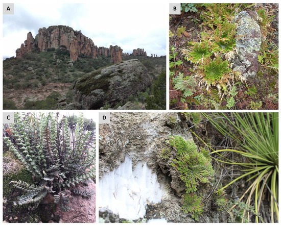
Figure 12. Common habitats for desiccation-tolerant pteridophytes. Photograph of an ecosystem with rock formations in the national park Sierra de Órganos, Mex. (A), and a close-up of a representative community of desiccation-tolerant organisms (including several ferns and mosses, Selaginella sp., lichens) growing on a rock outcrop (B). Resurrection plants can occupy rock crevices and shallow depressions where they experience periodic dryness, which represent inadequate sites for the establishment of desiccation-sensitive plants. Examples of desiccation-tolerant pteridophytes growing at these types of sites: the fern Myriopteris aurea (C) and the lycophyte Selaginella pilifera (D). All photographs were taken during the rainy season.
In contrast to bryophytes that can be rehydrated by dew or rain, desiccation-tolerant vascular plants seem to rehydrate only after receiving rain
[4][15][15,26]. Some desiccation tolerant pteridophyte species can occupy desert regions with as little as 20 to 50 cm of total precipitation per year
[16][27]. Therefore, to restore a metabolically active state when favorable conditions arise, these plants must be very efficient in water acquisition. Foliar water uptake represents a common feature shared between desiccation-tolerant pteridophytes and angiosperms. Leaf structures with a role in water uptake in ferns include trichomes, hairs, and scales
[5][16], but their function is not limited to water uptake, but also to prevent rapid water influx that could produce cellular damage
[15][26].
ForA study in the desiccation-tolerant fern
Pleopeltis polypodioides determined that scales contribute to general water management during the desiccation process
[17][28]. Besides improving water absorption during the rehydration stage, frond scales also have a role during dehydration by preventing rapid water loss. Furthermore, an
in situ one was
tudy showed that leaf irrigation is insufficient for the recovery of the entire plant. When only the leaves of the ferns
Pentagramma tirangularis and
Pellaea andromedifoli receive water, the stele and roots remain desiccated
[18][29].
ItThis wasstudy suggest
eds that in natural conditions, recovery after desiccation involves not only foliar water uptake but also root turgor and capillary action.
Apparently, there are no mandatory anatomical characteristics for VDT in pteridophytes, and these species can show contrasting anatomical and morphological characteristics. Specifically, desiccation-tolerant ferns can be divided in two groups: (1) ferns that show a well-developed cuticle and includes several families, and (2) filmy ferns, mainly represented by Hymenophyllaceae species, with a very simple frond structure composed of one or a few cell layers with a rudimentary or absent cuticle
[19][20][30,31]. Filmy ferns are generally associated with microenvironments subjected to recurrent desiccation and rewatering cycles. In contrast to ferns with well-developed cuticle, filmy ferns do not have an efficient mechanism to retain water and can lose water within minutes when exposed to dry air
[5][16]. Most of the characteristics exhibited by filmy ferns suggest that the DT ability in this group of plants is mainly conferred by a constitutive strategy, rather than the widespread inducible response observed in tracheophytes. An additional similarity of filmy ferns to bryophytes is their rapid rehydration, as some of these species fully recovered the hydrated state after one hour of soaking in water
[20][31]. Overall, the desiccation characteristics of filmy ferns appear to be more related to that of bryophytes than those of vascular plants. However, few
studies have shown that inducible mechanisms can also occur in filmy ferns. For instance,
Crepidomanes inopinatum increases the concentration of soluble sugars and activity of superoxide dismutase (SOD) in response to dehydration
[19][30].
2. Overview of Recent Insights about the Origin of VDT in Vascular Plants
The ability to tolerate desiccation is a common and widespread feature of plant reproductive structures. Although the ability to tolerate extreme dehydration in vegetative tissues of angiosperms is rare, most seeds can survive desiccation (known as orthodox seeds). Even though some angiosperms produce desiccation-sensitive seeds (recalcitrant seeds), the pollen of such species display DT
[21][32]. Spores are the reproductive structure of non-seed plants, and they also exhibit DT. Tolerance to extremely low water contents (equilibrium with ~1% relative humidity) has been reported for some fern spores, but they can display a huge variation in their longevity in the desiccated state, ranging from a few days to several months
[22][33]. Therefore, apparently all vascular plants possess the genetic potential for DT, but in most species, DT is restricted to reproductive structures.
In contrast to the constitutive or inducible protection strategies displayed in vegetative tissues, the DT of a reproductive structure such as orthodox seeds is developmentally regulated. During the last stage of seed development, denominated seed maturation, orthodox seeds acquire DT
[23][34]. The acquisition of DT during seed development is associated with multiple cellular processes including the accumulation of carbohydrates and late embryogenesis abundant (LEA)
[24][35]. The hormone abscisic acid (ABA), which mediates the response to several environmental stresses (e.g., drought, salinity, cold), also participates in the induction of dormancy and DT during seed maturation
[25][36]. The embryos of orthodox seeds generally lose their DT during germination. Interestingly, there is a short time window when the germinated seeds of some plants can re-activate DT when treated with ABA, indicating a major role of this hormone in the regulation of DT
[23][34].
The protection mechanisms exhibited in vegetative tissues of angiosperms resemble those observed in orthodox seeds. Due to such similarities, previous
ly, it wasstudies have suggested that VDT in angiosperms evolved from the re-activation of the developmental-seed program
[26][27][28][29][6,37,38,39]. Analysis of the genome of the resurrection monocot
Xerophyta viscosa supports the notion of the activation of seed pathways in VDT
[30][40]. The genome of this species has a significant expansion of some LEA families, and specifically genes encoding the LEA 4 family are accumulated during drying and rehydration. Interestingly, the promoters of LEA 4 members in
X. viscosa have a significant enrichment of the DNA binding motif for the transcription factor ABI5, an ABA-responsive transcription factor that participates in seed development. These results suggest that VDT in this resurrection angiosperm could have arisen from the reactivation of regulatory networks such as those activated during the maturation of orthodox seeds.
A rewiring event of pre-existing seed pathways has been proposed as the origin of VDT in angiosperms including the reactivation of seed development master regulators in vegetative tissues. Seed maturation is mainly controlled by a set of transcription factors named the LAFL network (LEC1, ABI3, FUS3, and LEC2) and the specific expression of some of these regulators in the reproductive structures of non-seed plants suggests a conserved role in spore development
[31][41]. Although the function of these transcription factors has been primarily associated with reproductive structures, there is evidence that some of them also participate in VDT. The moss
Physcomitrella patens can survive desiccation if incubated with ABA, but this desiccation-tolerant phenotype is dependent of the presence of ABI3 genes
[32][42]. Interestingly, a co-expression analysis in
X. viscosa determined a partial conservation of the
Arabidopsis ABI3 regulon, indicating a shared regulatory network between seeds and VDT
[30][40]. A further analysis determined that desiccation tolerant
Xerophyta species have at least four paralogs of the ABI3 gene, but only one is induced during both seed maturation and VDT
[33][43]. However, this ABI3 gene expressed in response to vegetative desiccation in
Xerophyta lacks the B3 domain required for binding to its cognate binding site in the promoters of some of its target genes. Furthermore, most of the putative target genes of ABI3 in
Xerophyta viscosa did not show the motif recognized by the B3 domain (
for i.e
xample., the RY
cis-acting element)
[33][43]. Then, the role of this specific ABI3 paralogue in VDT could likely be via the interaction with other transcription factors.
Current knowledge in resurrection angiosperms strongly suggests that VDT evolved from the reactivation of protective mechanisms of orthodox seeds. Thus, this reactivation of seed pathways in vegetative tissues have been proposed by means of conserved seed regulators including LAFL transcription factors. Comparative analysis between seed developmental stages and vegetative tissue in response to desiccation in the same species could determine if this hypothesis is correct. The characterization of the gene expression changes during seed maturation in a desiccation-tolerant plant has only been performed in the monocot
Xerophyta humilis [33][43].
IAlthough this st
wasudy also determined a significant overlap in the induced genes during seed development and during the dehydration of vegetative tissue, the canonical LAFL was only induced during seed maturation but not in vegetative tissues. Therefore, this observation indicates that the activation of DT in vegetative tissue is the result of the rewiring of the original regulatory network activating DT in
X. humilis seeds.
A similar scenario regarding DT in reproductive structures (
for i.e
xample., spores) and its reactivation in vegetative tissues of pteridophytes has also been proposed. Unfortunately, to
theour knowledge, there are no
onestudies characterizing global gene expression programs during spore maturation in resurrection pteridophyte species. Since processes that take place during spore development are similar to those observed in seeds, it has been proposed that the maturation phase shares some similarities between both reproductive structures
[34][44]. However, it is important to consider that some of the major events described as related to DT acquisition in seeds may not be present during spore development. For example, late maturation in seeds is associated with chlorophyll degradation because its retention could be detrimental to longevity in the dry state
[35][45]. In contrast, several fern species produce chlorophyllous spores, which could be associated with a rapid recovery of photosynthesis at the gametophytic stage
[36][46]. Furthe
r studies are required to determine the key protection mechanisms for DT in spores and the regulatory networks controlling such processes to gain insights into the origin of VDT in pteridophytes.
3. Intermediate DT Strategy in Pteridophytes: Constitutive and Inducible Responses
Plants can experience different types of cellular damage associated with mechanical, structural, or metabolic stresses during desiccation
[27][37][5,37]. Desiccation-tolerant species possess several mechanisms to avoid or minimize the detrimental effects of desiccation. The most common and widespread VDT mechanisms described in vascular plants include increased antioxidant levels (enzymatic and non-enzymatic antioxidants), changes in lipid composition, adjustment of carbohydrate metabolism, modifications in cell wall properties, induction of early light-induced proteins (ELIPs), accumulation of members of the LEA, and small heat shock proteins (sHSPs) families, among others
[38][39][40][41][47,48,49,50]. An extensive survey of the main findings of transcriptomic
onstudies identified a core of desiccation responses apparently conserved among all green plants, suggesting that VDT was derived from the mechanisms found in ancestral land plants
[42][51]. The transcriptional response displayed by resurrection plants showed a set of universally utilized strategies for VDT including the accumulation of LEA, ELIPs, sHSPs, compatible solutes, and antioxidants. However, the same
wa
uthors pointed out that plants have additional species-specific mechanisms to acquire VDT.
The intermediate position of pteridophytes in the land plant phylogeny correlated with their VDT strategy, which shares some features with the dehydration-inducible protection response observed in angiosperms and others with the constitutive mechanisms present in bryophytes. As discussed previously, desiccation-tolerant pteridophytes display highly diverse and even contrasting morphological adaptations. The presence of leaf structures to diminish water loss during dehydration suggest a dehydration-induced VDT strategy. On the other hand, a relatively simple leaf structure with no adaptations to retain water indicates a major constitutive component for VDT. Here, it wase compiled increasing evidence that some pteridophyte species with a predominantly inducible VDT strategy could also show a constitutive component and vice versa.
The photosynthetic apparatus is very liable to injury during water stress; therefore, resurrection plants have several mechanisms to prevent irreversible damage
[43][52]. Desiccation-tolerant plants can present a strategy to preserve the photosynthetic machinery at the desiccated state (homoiochlorophyllous), allowing for fast recovery during rehydration, or alternatively, a strategy to dismantle the photosynthetic apparatus during dehydration and resynthesize and reassemble it upon rehydration (poikilochlorophyllous)
[44][53]. In poikilochlorophyllous, degradation of chlorophyll and dismantling of chloroplast significantly reduce ROS formation, but reconstruction of the photosynthetic apparatus upon rehydration results in longer recovery times compared to the homoiochlorophyllous species. Homoiochlorophyllous plants are associated with habitats with rapid alternations of wet and dry cycles, whereas poikilochlorophyllous evolved in habitats where plants remain in a desiccated state for 8–10 months
[45][54]. Poikilochlorophylly is restricted to monocot species in which the recovery of full metabolic activity can take up to 72 h after rehydration
[46][55].
Desiccation-tolerant pteridophytes are classified as homoiochlorophyllous species
[1][11]. Therefore, their chloroplast ultrastructure (including stromal thylakoid, grana thylakoid and chloroplast membrane system) remains intact at the desiccated state
[47][56], allowing for recovery of full photosynthetic activity a few hours after rehydration. Most homoiochlorophyllous species are subjected to a partial loss of chlorophyll during desiccation, but never exceeding critical levels. Nevertheless, the fern
Pleopeltis pleopeltifolia loses more than two-thirds of its chlorophyll during desiccation and despite only recovering half of the initial chlorophyll content 24 h after rehydration, its photosynthetic activity has similar values to the fully hydrated conditions indicating rapid recovery
[48][57]. Fast recovery after desiccation is only possible in homoichlorophyllous species, but the loss of most chlorophyll in this fern contrasts such a strategy. A similar pattern was also observed in the filmy ferns
Hymenoglossum cruentum and
Hymenophyllum dentatum, which experience a significant decrease in chlorophyll pigments during a dehydration–rehydration cycle, but not complete dismantling of the photosynthetic apparatus
[20][31].
The It was authors of this last study proposed an intermediate strategy between homoiochlorophylly and poikilochlorophylly in these filmy ferns. Interestingly, desiccation-tolerant corms of
Isoetes can neither be classified as homoiochlorophyllous nor poikilochlorophyllous because its leaves (and also roots) decay and lose function after desiccation, and their corms develop completely new photosynthetic tissue upon rehydration
[11][22]. The way that these species have evolved to deal with desiccation increases the diversity of VDT strategies observed in pteridophytes. Furthermore,
I. taiwanensis also exhibits crassulacean acid metabolism (CAM)
[49][58], providing the opportunity to
knownstudy the crosstalk between a strategy to improve water-use efficiency, and the ability to tolerate extreme dehydration.
Although a significant number of pteridophytes have been described as desiccation-tolerant species, few
studies have been carried out to characterize their desiccation response. Among the desiccation-tolerant pteridophytes, the best characterized species is the lycophyte
Selaginella lepidophylla [50][59].
TheOur knowledge on this species includes physiological
onstudies to determine photosynthetic characteristics
[51][52][60,61], ultrastructural changes at the desiccated state
[53][54][62,63], early analysis of gene expression
[55][64], metabolic profiling
[56][65], and a sequenced genome
[57][66]. However, desiccation-tolerant pteridophytes display diverse responses to desiccation and the knowledge of VDT in this group is still limited. Therefore,
it w
ase present
ed an overview of the VDT mechanisms in pteridophytes as determined by physiological and biochemical approaches as well as those identified by metabolic, proteomic, and transcriptomic analysis.
Mechanisms for ROS scavenging could also differ between desiccation-tolerant species, but a general response in resurrection pteridophytes includes high levels of ROS scavenging enzymes such as superoxide dismutases, catalases, peroxidases, and glutathione reductase. Indeed, it has been proposed that some desiccation tolerant
Selaginella species are already primed for desiccation
[9][58][20,83]. These desiccation-tolerant species exhibit significantly higher levels of compounds with antioxidant capacity compared to desiccation-sensitive relatives in normal conditions. Both tolerant and sensitive plants display an increase in antioxidant compounds upon dehydration, but the levels in sensitive species are generally lower than those observed in tolerant plants. The adaptive mechanism of priming that some resurrection plants exhibit has been proposed as an important component of VDT. For example, the desiccation-tolerant grass
Sporobolus stapfianus is metabolically
[59][88] and transcriptionally
[60][89] primed for desiccation. While desiccation-sensitive species direct their metabolism to support faster growing rates, a desiccation-tolerant plant such as
S. stapfianus invests part of its metabolism to resist dehydration and associated damage with the accumulation of osmolytes in non-stressful conditions.
Resurrection pteridophyte species can also deal with oxidative stress by the induction of secondary metabolism pathways. A detailed analysis in
S. lepidophylla identified the accumulation of several of these metabolites including flavonoids (
for e
xample.g., apigenin), vanillate, and some phenylpropanoids (e.g., coniferyl alcohol)
[56][65], all considered as potent antioxidants. A significant accumulation of sugar alcohols or polyols (mainly sorbitol and xylitol) is observed in
S. lepidophylla compared to the desiccation sensitive
Selaginella moellendorffii [58][83]. Polyols have a dual function participating in redox control and osmotic adjustment
[61][90]. Due to the strong water-binding activity of polyols, they probably act to slow down water loss, providing a longer time for the induction and establishment of VDT mechanisms
[56][65]. In response to dehydration, plants also accumulate compatible solutes (
for e
xample.g., proline, mannitol, glycine betaine) to increase their cellular osmolarity and reduce water efflux from cells
[62][91]. Proline accumulation in plants is a common response to several stresses, and in addition to its role as an osmolyte, it has been proposed to act as a chaperone preventing protein aggregation and as a ROS scavenger, among other functions
[63][92]. The results of
theour survey showed that proline accumulation in response to desiccation is one of the most widespread mechanisms employed by desiccation-tolerant pteridophyte species.
Among conserved responses in resurrection plants during dehydration, accumulation of LEA proteins and their transcripts to high levels is among the most frequent responses to water loss
[38][47]. Surprisingly, LEA proteins are not restricted to plants and can be found in a wide range of organisms including animals that survive severe water stress
[64][93]. The occurrence of LEA proteins in distinct life forms suggests an ancient and ubiquitous protective role in DT. Diverse functions have been proposed for LEA proteins including roles as molecular chaperones and the protection of cellular components
[37][5]. However, characterization of the distinct LEA families showed multiple functions, interaction with proteins and nucleic acids and other activities, thus, apparently no single function is universal across LEA families
[42][51]. As discussed above, several mechanisms involved in seed DT are also present in vegetative tissues of resurrection plants. For instance,
Arabidopsis seeds acquired DT during the last stage of their development by accumulating high levels of LEA proteins and some sugars (i.e., sucrose and the raffinose family oligosaccharides)
[23][27][34,37], which also accumulate in several desiccation-tolerant species. The interaction between both LEA and sugars has been proposed as the main constituents for the formation of intracellular glasses
[65][94]. This glassy state, also known as vitrification, has been proposed as an important mechanism to relieve mechanical stress, reduce membrane fusion, and also in the inhibition of chemical reactions that could produce cellular damage
[37][5]. A genome analysis of the lycophyte
S. lepidophylla identified a total of 65 LEA genes, of which 48 showed significant expression changes during a rehydration–dehydration cycle
[57][66]. Analysis of other resurrection lycophyte of the same genus, the species
Selaginella tamariscina, also reported LEA genes as highly expressed during dehydration
[66][95]. Most LEA genes in the desiccation-sensitive relative
S. moellendorffii are also induced in response to dehydration
[66][95]. The latter observation suggests a conserved protective role of LEA proteins on both DT and in response to dehydration in sensitive plants. Maybe the protection characteristics of LEA proteins during desiccation are due to its level of expression, the induction of particular members of some LEA families, or their combined activity with other compounds (e.g., sugars) produced during desiccation.
A decrease in the photosynthetic activity is a conserved response in all desiccation-tolerant plants
[44][53]. Most of the physiological
onstudies performed in resurrection pteridophytes have also described a decline in photosynthesis during dehydration. Such a decrease is likely to be a consequence of a lower diffusion of CO
2 caused by stomata closure and downregulation in the expression of photosynthesis-related genes, among other factors. An important difference between desiccation-tolerant and sensitive plants is that the former shuts down photosynthesis at early stages of dehydration to diminish oxidative damage
[44][53]. The controlled cessation of photosynthesis also avoids the excessive production of ROS
[43][52]. Although photosynthetic activity is arrested during dehydration, several photosynthesis related proteins including those involved in the maintenance of chloroplast stability and enzymes of the Calvin cycle are upregulated upon desiccation in some lycophytes
[67][82]. Deactivation of photosynthetic activity at the proteome level has been reported in
S. tamariscina, which shows a significant reduction in the abundance of key enzymes involved in CO
2 fixation including the RuBisCO large subunit (rbcL)
[68][78]. As discussed throughout this
review, resurrection pteridophytes can show contrasting patterns for the same function. For example, the rbcL protein has been reported to remain constant and highly abundant during the desiccation process in some filmy ferns
[69][68]. Apparently, regulation of photosynthesis in homoiochlorophyllous species occurs at the physiological, transcriptomic, and proteomic level, indicating a crucial role in the proper regulation of this process in the acquisition of VDT.
Desiccation-tolerant pteridophytes also undergo morphological modifications during drying. Common mechanical responses to dehydration include leaf folding and stem curling, which has been proposed to diminish oxidative stress by reducing the area of photosynthetic tissue exposed to direct sunlight. A field experiment determined that stem curling in
S. lepidophylla is a mechanism that limits photoinhibitory and thermal damage
[70][96]. The ordered dehydration-induced morphological packing in
S. lepidophylla S. lepidophylla is produced by a differential rate of water loss between the inner and outer stems
[71][97]. A more detailed analysis showed the complexity of these mechanical processes, finding gradients of tissue density and even cell wall composition differences between the adaxial and abaxial sides of the stem
[72][98]. Additionally, desiccation leads to a significant decrease in cell volume, causing mechanical stress that could be ameliorated by cell wall folding and vacuolation
[5][5]. Structural characterization and transcriptomic analysis in
Selaginella Selaginella suggest a pronounced shift in cell wall structure and composition during desiccation
[73][74][76,84].