Your browser does not fully support modern features. Please upgrade for a smoother experience.
Please note this is a comparison between Version 2 by Vivi Li and Version 1 by Elisa Gnodi.
The use of nanoparticles (NPs) has surely grown in recent years due to their versatility, with a spectrum of applications that range from nanomedicine to the food industry. Recent research focuses on the development of NPs for the oral administration route rather than the intravenous one, placing the interactions between NPs and the intestine at the centre of the attention. This allows the NPs functionalization to exploit the different characteristics of the digestive tract, such as the different pH, the intestinal mucus layer, or the intestinal absorption capacity. On the other hand, these same characteristics can represent a problem for their complexity, also considering the potential interactions with the food matrix or the microbiota.
- nanoparticles
- nanocarriers
- insulin delivery
- inflammatory bowel diseases
- colon cancer
- food additives
1. Introduction
The development of nanotechnology in recent years has dramatically changed the approaches for drug delivery, initially improving the efficacy of the intravenous route for various drugs, such as anti-cancer ones. These results have also prompted researchers and pharma companies to look into the possibility of using nanocarriers for drug delivery through the oral route. On the other hand, in addition to pharmacological use, nanoparticles (NPs) have also been used quite extensively in the food industry, due to their ability to improve food characteristics, as well as product shelf-life. This implies that the interactions between NPs and the intestine can become quite frequent; for this reason, it is necessary to understand the pros and cons of the NPs presence in the intestine, also considering the possible interactions with the lumen components (food, acid environment, enzymes) and the different cell types.
1.1. The Intestinal Barrier
The gastrointestinal tract is a difficult environment for nanocarriers, both due to the aggressive conditions present in the lumen, as well as for the presence of the barrier separating the lumen from the rest of the body. The first challenge is a large pH gradient, ranging from pH 1–2.5 in the stomach to pH 7–8 in the colon, fact which can affect the structure of the nanocarriers or of the vehiculated drug. Moreover, the lumen enzymes, both in the stomach (ex. Pepsin) and in the duodenum (biliary and pancreatic secretions that include lipases, peptidases, and amylases) can affect the nanocarrier stability and/or their capacity to bind different substances (including food components) [1][2].
The intestine is composed of different cell types that have specific functions, and the composition changes according to the anatomical site, i.e., the small or large intestine. In the small intestine the main absorption function is performed by the enterocytes, which are also responsible for the tight junctions, the most important structure creating the intestinal barrier. The mechanisms of uptake of lumen substances could be either paracellular or transcellular, i.e., through the enterocytes. The paracellular pathway usually plays a minor role in the passage of NPs, which are usually transported through the transcellular route. This occurs through vesicle-mediated mechanisms, either endocytosis or pinocytosis; it is easy to understand that the intrinsic characteristics of the NPs can affect the ability to bind to the enterocytes and to be transported through the transcellular route. In Figure 21, the main NPs administration routes are represented, along with the main aspects related to the oral administration (Figure 21).
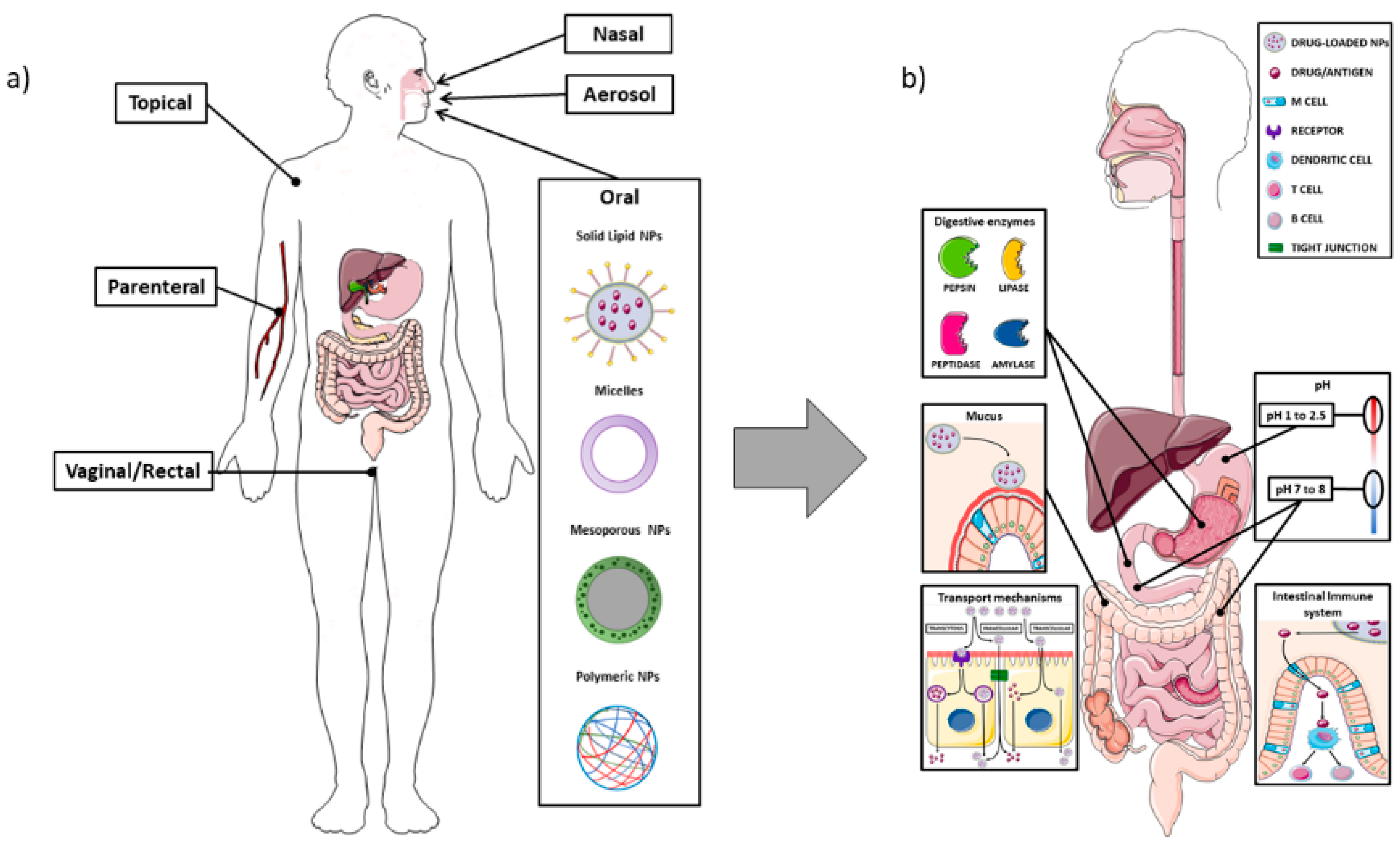
Figure 21. Schematic representation of the different routes for nanoparticle drug delivery, with attention to the oral administration and the interactions with the intestinal barrier. Panel (a): Overview of the main administration routes for nano-drug delivery; Panel (b): Different enzymes (pepsin, lipase, peptidase, and amylase) located in the gastrointestinal tract can impair nanocarriers stability and their ability to reach the target tissue. The mucus layer also plays an important role in the entrapment of NPs, which may lead to reduced uptake at cellular level. The enterocytes transport mechanisms of NPs can occur through the intestinal cells, either by transcytosis (mediated by endocytic vesicles), or through a direct apical-basolateral passage, or by the paracellular route (passing through the intercellular space). The difference in pH among the stomach, duodenum, and colon represent one of the main challenges in delivering NPs, particularly in order to avoid their premature degradation through the acidic environment. M cells, as part of the GALT (gut-associated lymphoid tissue), can detect antigens from the intestinal lumen and bring them to antigen presenting cells (APC), which, in turn, are able to present them to B or T lymphocytes located at the mucosal level. The image was created with the use of Servier Medical Art modified templates, licensed under a Creative Common Attribution 3.0 Unported License (https://smart.servier.com, accessed on 19 February 2022).
There is also another point that needs to be considered, i.e., the presence of multidrug resistance transporters (MDR) in the epithelium, fact which could dampen the total amount of the drug bound to NPs which had been taken up by the enterocytes. In addition to enterocytes, there are other cells in the small intestine, such as goblet cells (localized in the villi), as well as Paneth and stem cells (in the crypts); the former are producing the mucus which covers the intestinal epithelium, whereas Paneth cells are responsible for the production of antimicrobial peptides and immunomodulating proteins. Mucus is a complex hydrogel composed of water and different types of proteins, among which mucins are the most abundant ones. Most mucins are glycosylated, so they have a negative charge, characteristic which could lead to the adhesion of positively charged nanocarriers through electrostatic interactions. This ability of nanocarriers to bind to the mucus layer could be regarded only partially as positive, since the intestinal mucus is structured in two different layers: the first one, nearer to the intestinal lumen, is more loose whereas the layer in contact with the epithelium is firmly adhered. The firm binding of the NPs to the upper layer can, thus, lead to a prompt clearance and to a reduction in the opportunity to reach the epithelium [3].
1.2. Nanoparticles
The NPs that can interact with the intestine can be divided into different categories, mainly according to the material used to generate them.
Lipid-based nanocarriers have been quite extensively used in drug delivery because of their versatility, biocompatibility and low toxicity profile, and their use by i.v. administration has already been approved by Food and Drug Administration (FDA) and European Medicines Agency (EMA) [4] (recently reviewed by Halwani). However, the oral route presents a series of advantages, e.g., ease of administration and high patient compliance, and, thus, a large amount of research is now being undergone, aiming at developing the best lipid-based nanocarriers for oral delivery. This task also takes advantage of the fact that most oils and fats used for the development of these nanocarriers derive from dietary lipids, thus facilitating oral permeability and biodegradability. The term lipid-based nanocarrier includes liposomes, self-nano and microemulsifying drug delivery systems, nanoemulsions and nanocapsules.
Liposomes are spherical vesicles constituted by lipid bilayers and an aqueous inner core. Their basic composition is phospholipids and sterols (such as cholesterol), with the latter ones being used in order to stabilize the liposomal membrane. However, different components can be added to this simple structure, such as surfactants, bile acids, or specific ligands that could help the targeting of the liposomes to intestinal cells (see below). Moreover, due to their composition, liposomes can carry hydrophilic molecules into their inner cavity, whereas hydrophobic drugs can be inserted into the lipid bilayer [5]. Solid lipid NPs are composed by a lipid core (triglycerides, fatty acids or phospholipids) with a monolayer surfactant shell, such as lecithin or bile salt derivatives [6]. Nanoemulsions are dispersions of an oily and an aqueous phase with the addition of an appropriate surfactant, but due to the percentage of surfactant (3–10%) they are thermodynamically unstable. On the contrary, microemulsions are stabilized by surfactants added in higher concentrations (≥20%), thus making them thermodynamically stable [7]. Lipid nanocapsules are constituted by an oily phase and an aqueous one, stabilized by surfactants and a polymeric shell. Due to their nature, lipid nanocapsules can present with different biological properties, which depend on their surface characteristics. In fact, the characteristics of the polymeric shell can determine the ability of the NPs to interact with the intestinal environment, in particular with the mucus and/or the enzymes present in the lumen [8].
MNPs can interact with the intestine either because they are used as therapeutic agents or because they are ingested with food, since they can be used as food preservatives or colouring agents (such as TiO2) [9][10]. From the medical point of view, the most extensively utilized are Ag or gold (Au) NPs, but data have also been obtained on palladium, titanium, zinc, and copper ones. Due to their chemical properties, their surface can be easily functionalized to conjugate targeting agents and active biomolecules, and multiple drugs can also be loaded on the same MNP. MNPs have been mainly employed as anticancer agents [11] or to counteract infections, either bacterial or viral [12]. Due to their small size MNPs (in particular Ag and Au) can also perform a passive targeting of cancer cells, i.e., reaching them more easily due to the leakiness of the vasculature growing within the tumour mass. Moreover, MNPs, in particular Ag NPs, are extremely reactive and can interact with many cellular components through the induction of ROS, leading to mitochondrial damage and eventually apoptosis. Although this effect could be quite desirable in cancer therapy, it should be definitely avoided in the interaction with normal cells, in this case the enterocytes [13][14].
Polymeric NPs can be of synthetic origin but also made of natural substances, such as polysaccharides; in a biological setting these latter ones are obviously preferred, since they do not provoke or produce toxic effects. Among the natural polymers, the most commonly used are polysaccharides including chitosan, hyaluronic acid (HA), alginates, etc.; due to their chemical structure, they present both hydrophilic groups (necessary for the solubility in water) but also residues able to interact with biological membranes, as further discussed below [15] (Figure 32).
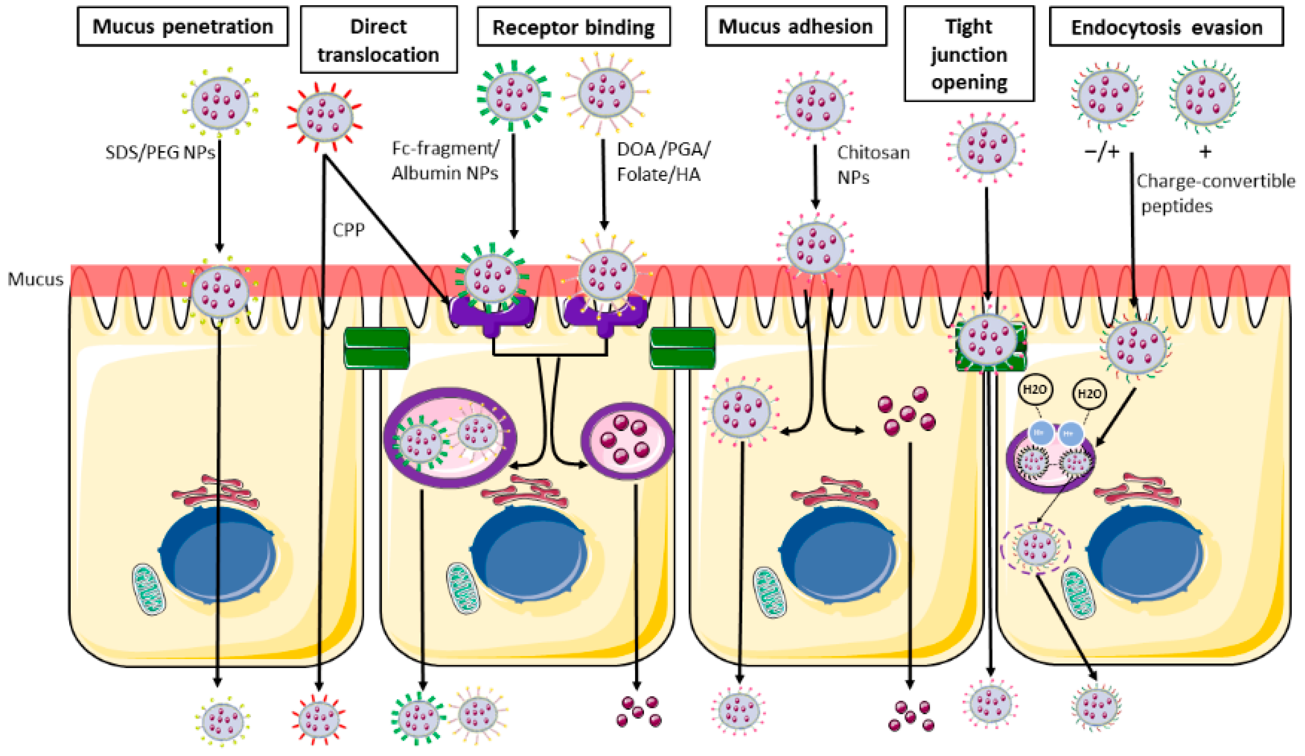
Figure 32. Main nanoparticles functionalization and their intestinal transport. From the left: Schematic representation of mucus penetrating NPs (SDS/PEG), able to penetrate the mucus layer and directly pass through the blood flow. Receptor binding NPs (DOA/PGA/folate/HA/albumin/Fc-fragment) able to bind the cell surface using the ligand-receptor binding and are then internalized in endocytic vesicles and released in the systemic circulation. CPP (cell penetrating peptides) are able to undergo both receptor binding internalization and direct translocation. Muco-adhesive NPs and tight junction opening NPs (chitosan) are able to be retained in the mucus layer, and then undergo transcellular passage or pass through the opened tight junction. Charge-convertible peptides are able to evade the lysosomal degradation using the proton sponge mechanism. SDS: sodium dodecyl sulphate; PEG: polyethylene glycol; CS: chondroitin sulphate; DOA: deoxycholic acid; PGA: poly-glutamic acid; HA: hyaluronic acid. The image was created with the use of Servier Medical Art modified templates, licensed under a Creative Common Attribution 3.0 Unported License (https://smart.servier.com, accessed on 19 February 2022).
1.3. Nanoparticles—Intestine Interaction
The gastrointestinal tract, as mentioned, represents a harsh environment for drug delivery, since the active component has to survive the low pH but also cross the intestinal barrier, i.e., the mucus layers and the enterocytes. For this reason, NPs can be functionalized, in order to prevent the attack of pH and enzymes or to favour their passage through the intestine in order to have a systemic effect.
The protection from low pH can use polymers that have been already employed in the drug industry, such as all the different formulations of the Eudragit®, which can be dissolved above specific pHs, thus allowing the drug delivery in the various regions of the gastrointestinal tract, i.e., small intestine or colon [16]. Other molecules can be used to create a shell, and among them there is alginate, which can provide resistance to low pH and, if associated with other molecules, also to enzyme digestion. Alginate is a linear anionic polymer derived from brown seaweed consisting of β-D-mannuronic acid (M) and α-L-guluronic acid (G) linked by glycosidic bonds [17]. The monomer composition can affect the general structure of alginate, making it more rigid and with larger pores (thus with a higher release of the drug) or more soft with smaller pores according to a high or low presence of G blocks, respectively [18][19]. Alginate also responds to pH, and researchers have developed specific emulsions able to swell or shrink according to the environmental pH [20]; in particular, the presence of low pH will maintain alginate in a stable hydrogel form, thus protecting the associated drug, whereas neutral pH will cause the hydrogel dissolution and the release of the active compound.
Since mucus covers the apical part of the enterocytes, the NPs need to attach to it, but also be able to cross the two different layers in order to reach the enterocytes. In order to design NPs able to deliver their load, several mucus characteristics should be kept in mind; mucins, the main mucus component, contain glycosylated section with negative charges that could bind positively charged NPs, trapping them. Moreover, some part of these proteins are hydrophobic, and this strongly reduces the transport of hydrophobic particles, such as PLGA and polystyrene (PS), which are quite used as NPs. Last but not least, mucins create a sieve-like structure, thus the size of the NPs should also be kept to a minimum.
Substances employed in NPs can interact with the mucus either increasing NPs ability to adhere to it or augmenting their penetration.
One of the most used molecules belonging to the first group is chitosan, a nontoxic, cationic polysaccharide derived from chitin (naturally obtained from marine organisms) which has been approved by FDA for biomedical applications. It is biocompatible, biodegradable, and non-toxic; in addition the presence in its sequence of positively-charged N-acetyl glucosamine units favours the binding to the mucus [15]. Chitosan is also rich in hydroxyl, amino, and carboxyl groups, which allow a series of chemical modifications that can increase, for example, its water solubility or its stability [21]. In addition to mucus adhesion, chitosan can induce the opening of the tight junctions, as demonstrated by alteration in the trans-epithelial resistance and by electron microscopy [22][23][24][25]; these opening has been demonstrated to be reversible, at least in in vitro experiments on Caco2 cells, and associated with a redistribution of claudin-4, an essential component of tight junctions [26]. This effect is mediated by a direct interaction between positively charged chitosan and negatively charged integrin aVβ3, fact which causes a conformational change of this latter proteins that aggregate along cell boundaries, reorganization of F-actin and a downregulation of claudin-4 [27].
Another molecule widely used in NPs that is able to increase mucus binding is HA, a natural linear glycosaminoglycan, biocompatible, and biodegradable through the action of the host enzymes. Its ability to bind to biological substrates is mediated by the presence of abundant COOH groups [28] and also by the molecular weight (MW), with a higher efficiency of the adhesion being observed in presence of a lower MW [29].
The most used substance which helps the passage of NPs through the mucus is polyethylene glycol (PEG) [30]; the addition of this component can change, even in an important manner, the ability of the NPs to cross the mucus layer; Xu et al. observed that, in the case of PLGA NPs, a percentage of at least 5% is necessary to reduce the interaction with mucus, and higher PEG concentrations improved the passage [30]. This could be explained by a “shielding effect” by the PEG molecules, which prevented the interactions between mucin proteins and the NPs core. These data were obtained using a 5 kDa PEG molecule and the authors used as in vivo model mouse vaginal mucosa; the situation could be totally different in the intestine, and also the PEG size could influence the mucus penetration, as demonstrated by Inchaurraga et al., who analysed the effect of different MW PEGs on the ability of NPs to reach the enterocytes [31]. Interestingly, better results were obtained using PEG 2000 or 6000, whereas the 10,000 molecule showed a worse performance. Other polymers have been developed, such as poly-N-2-hydroxypropyl methacrylamide (HPMA), a water-soluble polymer with excellent mucus-permeating properties similar to PEG. This compound can dissociate from chitosan NPs during the passage in the mucus, as demonstrated by Liu M et al., but it can also cause an opening of the tight junctions [32].
To increase the passage through the intestinal barrier there are, in theory, two possibilities, i.e., cause a loosening of the tight junction or increase the uptake of the NPs by the M cells or the enterocytes. Several compounds can actually interfere with the proteins that are forming the junctions sealing off the intestinal content, such as occludins, claudins, and integrins. Natural food compounds can have an effect on the permeability of in vitro systems, as reviewed by Kosińska et al., and, more recently, demonstrated by Haasbroek et al. that focused their attention on aloe extracts that were able to decrease trans-epithelial resistance in a trans-well Caco2 cell model and increase the passage of 4 kDa dextran [33][34]. Chen et al. developed an hydrogel able to adhere to the mucosa and, at the same time, to chelate calcium ions, which are essential for the maintenance of the junctions [35]. They tested these particles, carrying HbS antigen, in mice and were able to demonstrate a higher intestinal immune response compared to the usual vaccination route. Last, but not least, it should be remembered that bacteria causing gastrointestinal disorders are able to secrete toxins acting on the integrity of the tight junctions, causing a damage or a rearrangement of their protein components. Although this kind of intervention could cause the passage of a large quantity of the cargo drug through the barrier, it could be hazardous; for this reason synthetic peptides mimicking the effect of these toxins have been produced. In particular, AT-1002 is a hexamer peptide (FCIGRL) derived from zonula occludens toxin (ZOT) produced by Vibrio cholera [36]. This toxin is able to bind to a receptor present on the apical portion of the enterocytes, activate protein kinase C and cause a transitory disassemble of the tight junctions. It can be added to other NPs components, such as chitosan, as reported in a delivery system for insulin [37] that was able to obtain a good glycaemic control in diabetic rats. It must be kept in mind, however, that increasing the permeability of the junctions could also allow the passage of intestinal antigens; further studies on this aspect must be performed in vivo, although a paper by Sonaje et al. failed to detect an increased passage of LPS following chitosan NPs administration [38].
The passage through cells, either M or enterocytes, could be increased by adding to the NPs peptides that are able to interact with specific receptors present of the apical part of the cells. As regards M cells, studies in mice demonstrated that various types of lectins added to NPs can increase the uptake due to their ability to interact with cellular α-L-fucose moieties [39][40]; unfortunately, these specific moieties are not present on human cells, thus NPs should be functionalized with other peptides. Among them, the Gly-Arg-Gly-Asp-Ser (GRGDS) pentapeptide could be a good candidate, since it binds B1 integrins, present also on human cells. Up to now, it has only been evaluated in a human cell model (Caco2 + Raji), and demonstrated able to increase the passage through Raji cells [41]. Last but not least the route through which NPs can reach other organs can be important in order to avoid the hepatic first pass; for this reason, NPs can be designed to use the lymphatic system, and this means that intestinal absorption should occur through M cells.
As regards enterocytes, several receptors have been described on the apical surface, and known enterocyte-targeting ligands include lectins, transferrin, vitamins, oligopeptides, and monoclonal antibody fragments as summarized in Table 1. The cell entry could also occur through the action of some specific peptides, identified as cell-penetrating peptides (CPPs), that are able to allow the attachment and penetration of the NPs (such as Trans-Activator of Transcription (TAT)), or through a classical receptor-mediated endocytotic process (Table 1 and Figure 32 and Figure 43) [42].
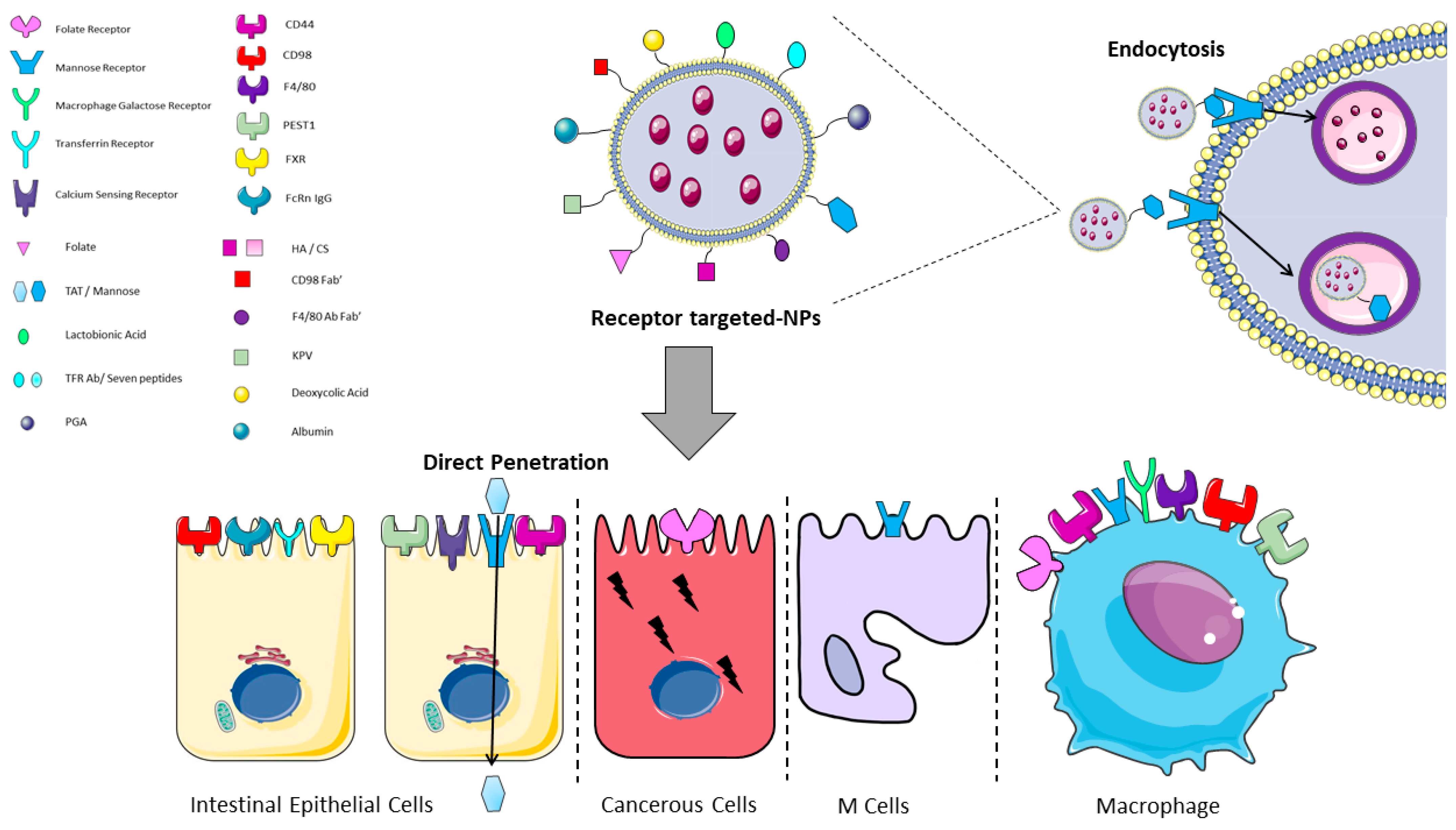
Figure 43. Graphical representation of receptor/ligand NPs interactions in the intestine. Functionalized receptor-binding NPs are able to bind the cell membrane through the binding of the NP (ligand) to the receptor on the cell surface and then to undergo the endocytotic process. Trans-Activator of Transcription (TAT) is the only mentioned ligand that undergoes direct penetration. CS: chondroitin sulphate; FXR: farnesoid X receptor; HA: hyaluronic acid; KPV: lysine-proline-valine; PEST1: peptide transporter1; PGA: polyglutamic acid; TFR: transferrin receptor. The image was created with the use of Servier Medical Art modified templates, licensed under a Creative Common Attribution 3.0 Unported License (https://smart.servier.com, accessed on 19 February 2022).
Table 1.
Main receptor-ligand interactions used for NPs functionalization in the intestine.
| Reference | Receptor | Ligand | Cell Type Expression |
Direct Penetration |
Endocytosis | |||||
|---|---|---|---|---|---|---|---|---|---|---|
| Hua S 2020 [43 | ||||||||||
| Li L 2017 | ] | [58Mannose Receptor | Mannose | Macrophages, Enterocytes, M cells |
No | Yes | ||||
| ] | Chitosan | CPP | n/a | Yes | Tian 2018 [44] |
CD44 | HA/CS | Macrophages, Intestinal Epithelial Cells |
No | Yes |
| Wu J-Z 2017 [59] | diethylene glycol dimethacrylate | n/a | phenylboronic acid | Yes | Xiao, 2018 [45] |
CD98 | CD98 Fab’/single chain CD98 Ab | |||
| Alfatama 2018 [60 | Intestinal | Epithelial Cells, Macrophages | No | ] | Alginate/Chitosan | Yes | ||||
| n/a | n/a | Yes | Peng L, 2021 [46] |
F4/80 | F4/80 Ab Fab’ | Macrophages | No | Yes | ||
| Liu W, 2018 [47] |
Macrophage Galactose Receptor | Lactobionic Acid | Macrophages | No | Yes | |||||
| Xi Z 2022, Álvarez-González, 2020 [48][49] | ||||||||||
| Czuba 2018 [61] | PLGA | SDS | n/a | Yes | Folate Receptor | Folate | ||||
| Fan 2018 [62] | Chitosan | Deoxycholic acid | Macrophages, Epithelial Cancer Cells | No | Yes | |||||
| n/a | Yes | |||||||||
| Hou 2018 [63] | Mesoporous silica nanoparticle | n/a | phenylboronic acid | Yes | Yong, 2019 | |||||
| Jamshidi 2018 [64] | [50] | Transferrin Receptor | TFR Ab/Seven peptides | ChitosanIntestinal Epithelial Cells |
No | n/a | n/a | Yes | ||
| Yes | Zhang W, 2021 [51] |
PEST1 | KPV | |||||||
| Ji N 2018 [65] | Macrophages, Intestinal | Zein + CSA | n/aEpithelial Cells | No | Yes | |||||
| n/a | n/a | Liu L, 2018 [52] |
Mannose Receptor | TAT | Intestinal Epithelial Cells, Macrophages |
Yes | No | |||
| Liu L 2018 [52] | Azevedo, 2020 [53] |
FcRn IgG | Albumin | Intestinal Epithelial Cells |
No | Yes | ||||
| Huang X, 2021 [54] |
FXR | Deoxycolic Acid | Intestinal Epithelial Cells |
No | Yes | |||||
| Urimi, 2019 [55] |
Calcium Sensing Receptor | PGA | Intestinal Epithelial Cells |
No | Yes |
CS: chondroitin sulphate; FXR: farnesoid X receptor; HA: hyaluronic acid; KPV: lysine-proline-valine; PEST1: peptide transporter1; PGA: polyglutamic acid; TAT: Trans-Activator of Transcription; TFR: transferrin receptor.
Various bacteria-derived peptides can also be used, since they are recognized by TLR4, but these peptides carry the risk of activating the intestinal immune system. Li et al. recently evaluated the possibility of employing a non-toxic form of Pseudomonas aeruginosa exotoxin A associated with alginate/chitosan particles; the presence of the exotoxin favoured the transcitosis, but the in vivo administration of these NPs to rats showed that they co-localized with CD11c+ cells, which have an important role in intestinal immune response [56]. In all cases, the NPs and their cargos should be vehiculated to the basolateral side of the cells, thus requiring transcytosis (Figure 21b and Figure 32). This step should not be regarded as trivial, since there is the risk that the fusion of endocytotic vesicles with lysosomes damages the NPs, both in its structure or inactivating the carried drug. For this reason, some researchers developed NPs associated with charge-convertible peptides [48][57]. The presence of these components allows the NPs to survive the acidic pH of late endosomes, since they can act as “sponge” for H+ ions. Last, but not least, the NPs have to cross the basolateral membranes of enterocytes and be released into the circulation; interestingly, Xi et al. observed that the addition of the charge-convertible peptides increased the interaction of the NPs with the proton-coupled oligopeptide transporter present in the basolateral membrane, thus boosting the exocytosis [48]. The interactions between NPs and the intestine could be subdivided in three main categories, i.e., the use of NPs to deliver systemic drugs, which implies the passage through the intestinal barrier and reaching the blood or lymphatic flow, NPs as carriers for drugs that should act on the intestinal mucosa or the “involuntary” interaction due to NPs used as food additives. In this entreview, wey, researchers are going to discuss some examples in each category, pointing out advantages and pitfalls.
2. Nanoparticles for Systemic Drug Delivery
The possibility to deliver drugs through the intestinal route rather than using other more invasive ways has been quite captivating for various pharma products, in particular anti-cancer drugs or vaccines. However, due to the large number of the employed molecules and the great differences among the NPs, wresearchers decided to focus on a single molecule tackling another disorder, i.e., insulin. Due to the high social impact of diabetes and the need to administer the drug few times during the day, several groups throughout the world have been involved in the development of NPs able to provide the oral delivery of recombinant insulin. The most used cores for NPs are polymers, either natural or synthetic ones; among the natural polymers there is chitosan, either alone or in combination with alginate; these NPs have some characteristics that make them suitable for insulin delivery, such as biodegradability, nontoxicity, muco-adhesiveness, and low immunogenicity, as previously described (see Table 2). Other employed natural polymers are HA, albumin, starch (amylose), zein, and lignin, as reported in Table 2.Table 2.
Summary of the different NPs and functionalization for the delivery of insulin.
| Reference | Core of the NPs | Further Functionalization for Adhesion/Passage |
Release Control | Reduces Glycaemia in Animal Model |
|---|---|---|---|---|
| Chitosan + hydrogel | ||||
| n/a | ||||
| n/a | ||||
| Yes | ||||
| Song M 2018 [66] | Cyclodextrin/chitosan | n/a | n/a | Yes |
| Tian 2018 [44] | Chitosan/hyaluronic acid | n/a | n/a | Yes |
| Wang W 2018 [67] | Polyamidoamine/polyaspartic acid/phenylboronic acid/PEG | PEG | phenylboronic acid | Yes |
| Xu Y 2018 [68] | solid lipid nanoparticle + endosomal escape agent | n/a | n/a | Yes |
| Zhang Y 2018 [69] | hydroxyapatite | PEG | n/a | Yes |
| Zhang L. 2018 [70] | PLGA + chitosan + alginate | n/a | pH dependent | Yes |
| Alsulays 2019 [71] | Solid lipid nanoparticles | CPP | n/a | Yes |
| Guo 2019 [72] | Chitosan | CPP | n/a | yes |
| Hu 2019 [73] | phospholipids | n/a | n/a | Yes |
| Jamwal 2019 [74] | dextran | n/a | Glucose oxidase | n/a |
| Ji 2019 [75] | Chitosan/zein-carboxymethylated short-chain amylose | n/a | n/a | Yes |
| Mohammadpour 2019 [76] | PLGA + chitosan | n/a | Glucose oxidase | Yes |
| Muntoni 2019 [77] | Lipid nanoparticles | n/a | n/a | Yes |
| Mudassir 2019 [78] | Methyl methacrylate/itaconic acid nanogels | n/a | pH dependent | Yes |
| Tsai 2019 [79] | Chitosan + fucoidan | n/a | pH dependent | n/a |
| Urimi 2019 [55] | Chitosan | Polyglutamic acid | n/a | Yes |
| Azevedo 2020 [53] | Albumin | n/a | n/a | Yes |
| Bai 2020 [80] | PLGA + glutamic acid conjugated amphiphilic dendrimer | n/a | n/a | Yes |
| Chai 2020 [81] | Poly (acrylamido phenylboronic acid)/sodium alginate | n/a | Cicloborate (Glucose sensing) and glucose oxidase | Yes |
| Chen Z 2020 [82] | Chitosan/Hyaluronic acid | CPP | n/a | Yes |
| Cheng 2020 [83] | Poly (n-butylcyanoacrylate) | Ratio insulin/Poly (n-butylcyanoacrylate) | Ratio insulin/Poly (n-butylcyanoacrylate) | Yes |
| Ding 2020 [84] | amphiphilic cholesterol- phosphate conjugate |
n/a | pH dependent | Yes |
| Han X 2020 [85] | Zwitterionic micelles | Betaine | n/a | Yes |
| Jana 2020 [86] | hyaluronic acid | n/a | Glucose oxidase | n/a |
| Mumuni 2020 [87] | Chitosan/mucin | n/a | n/a | yes |
| Sladek 2020 [88] | Hyaluronic acid/chitosan | Sucrose laurate | n/a | Yes |
| Sudhakar 2020 [89] | Chitosan | n/a | pH dependent | Yes |
| Tan X 2020 [90] | Mesoporous silica | PEG + CPP | n/a | Yes |
| Wang T 2020 [91] | Lipid nanoparticles | n/a | n/a | Yes |
| Zhou S 2020 [92] | Chitosan | PC6 | pH dependent | Yes |
| Zhou X 2020 [93] | Alginate | n/a | Glucose oxidase | Yes |
| Zhou Y 2020 [94] | FeCl3·6H2O + BTC | SDS | pH dependent | Yes |
| Bao X 2021 [95] | Zein/casein-dextran | Cholic acid | n/a | Yes |
| Benyettou 2021 [96] | Nanoscale imine-linked covalent organic frameworks | n/a | pH dependent | Yes |
| Cui 2021 [97] | Chitosan + Hyaluronic acid | Biotin | n/a | Yes |
| Huang X 2021 [54] | layered double hydroxide nanoparticle + hyaluronic acid | Deoxycholic acid | n/a | Yes |
| Kim WJ 2021 [98] | POSS-APBA | n/a | phenylboronic acid | n/a |
| Li H 2021 [99] | polyphosphoesters-based copolymer |
n/a | phenylboronic acid | Yes |
| Li J 2021 [100] | Alginate/chitosan | n/a | pH dependent | Yes |
| Liu X 2021 [101] | PLGA/PEG | Angiopep-2 | n/a | Yes |
| Qin 2021 [102] | Mesoporous silica + Alginate + Boronic acid Mesoporous silica + Chitosan + boronic acid |
n/a | phenylboronic acid | Yes |
| Rao 2021 [103] | Porous silicon nanoparticles | Zwitterionic dodecyl sulfobetaine | n/a | Yes |
| Volpatti 2021 [104] | Polycation | n/a | Glucose oxidase | Yes |
| Wang W 2021 [105] | PLGA | Chitosan + Cholanic acid | n/a | Yes |
| Zhang Y 2021 [106] | mesoporous silica nanoparticles | CPP | n/a | Yes |
| Fu 2022 [107] | Glycopolymer | n/a | phenylboronic acid | Yes |
| Li J 2022 [108] | PLGA-Hyd-PEG | PEG | n/a | Yes |
| Martins 2022 [109] | Lignin-encapsulated silicon | Fc fragment of IgG | pH dependent | n/a |
| Reboredo 2022 [110] | Zein | PEG | n/a | Yes |
| Rohra 2022 [111] | Gold nanoparticle-encapsulated zeolitic imidazolate framework-8 | n/a | Glucose oxidase | n/a |
| Xi Z 2022 [48] | PLGA/PEG | PEG, folate and charge-convertible tripeptide | n/a | Yes |
| Xu 2022 [112] | konjac glucomannan/concanavalin A | n/a | Glucose sensing | Yes |
Most recent articles were considered (starting from 2017). APBA: 3-Aminophenylboronic acid monohydrate; BTC; 1,3,5-Benzenetricarboxylic acid; CPP: cell-penetrating peptides; CSA: Carboxymethylated Short-Chain Amylose; PC6: poly(acrylic acid)−cysteine−6-mercaptonicotinic acid; PLGA: poly (d, l-lactic-co-glycolic acid); POSS: PSS-[2-(3,4-epoxycyclohexyl)ethyl]-heptaisobutyl substituted.
These natural components were used alone or in combination, in order to exploit the different characteristics of the various components, such as the ability to bind to the mucus, recognize specific cell receptors etc. As regards synthetic polymers, most researchers employed PLGA, due to its characteristics such as biocompatibility, biodegradability, and its common use in in the drug industry, being FDA approved [113]. Its use is also supported by the fact that, when it is broken down, it generates glycolic acid and lactic acid, which are naturally metabolized by the body. Other synthetic polymers that have been used in insulin delivery are polymath-acrylic acid (PMAA), polyacrylic acid (PAA), and polycaprolactone (PCL) (see Table 2). Apart from polymers, also liposomes have been employed for insulin delivery, using liposomes containing bile salts, such as sodium glycocholate (SGC), sodium taurocholate (STC), or sodium deoxycholate (SDC). All the described polymers and liposomes were able to generate NPs with a high rate of incorporation of insulin and to protect the drug from the degradation that could occur in an acidic environment; however, to this basic structure of the NPs, other molecules have been added to improve the adhesion to the mucus/enterocytes and the passage through the epithelial layer.
The control of insulin release has to be tightly regulated, in order to prevent hyper- or hypo-glycaemic episodes. This concept was extremely important, throughout the years, in the design of the different injectable insulin formulations; for this reason, several research groups designed NPs containing a “glucose sensor”, i.e., a chemical compound able to react to the different glucose level present in the blood. The most commonly used systems are based on glucose oxidase or phenylboronic acid; glucose oxidase catalyses the oxidation and hydrolysis of β-D-glucose into gluconic acid and hydrogen peroxide and, in turn, the production of the gluconic acid lowers the pH within the NPs [114]. The change in pH alters the structure of the NPs, favouring the release of the encapsulated drug; this system not only controls insulin release, but it can also provide a faster release if compared to the same NPs lacking the glucose oxidase and a better glycaemic control in a rat diabetic model, as shown by Chai et al. [81]. The glucose sensing by phenylboronic acid (PBA) can be mediated by two different mechanisms: in the first one, it occurs through a contraction/expansion transition in which glucose binds to PBA altering the balance between its two forms, the hydrophilic and the hydrophobic one and, as a result, the water density in the NPs increases causing the release of insulin. In the second mechanism, called competitive, glucose displaces the drug that was bound to PBA, thus causing its release [115].
Last, but not least, these NPs must be able to provide an in vivo response; although this demonstration has been provided in animal models, as reported in Table 2, trials in humans are still under way (as discussed below), thus, an accurate evaluation of these data will be necessary before these NPs can be moved to clinic.
3. Nanoparticles with Intestinal Targets
3.1. Inflammatory Bowel Diseases
IBD, which include both Crohn’s disease (CD) and ulcerative colitis (UC), are chronic inflammatory disorders characterized by mucosal immune system dysregulation, which has an impact in the small intestine and colon. In recent decades, the necessity to conceive a novel therapeutic approach to IBD treatment has led to the increased interest in nanobased drug delivery systems [116][117][118]. This is due to the many side effects caused by the commonly used drugs to treat chronic inflammatory disorders, such as IBD. In particular, 5-aminosalicylates (5-ASA), antibiotics, and corticosteroids can cause, in the long-term, several side effects, including bone damage, such as the steroid-induced necrosis of the femoral head. Genome-wide association studies have demonstrated that genetic background is only one of the factors involved in the pathogenesis of the disease together with the environmental ones. The recent advances in understanding the pathways involved in the development of IBD have allowed to provide some more therapies, but since the exact cause is not completely understood, there is currently no cure tackling the primum movens of the disease. However, even classical drugs used to treat IBD could take advantage of new-targeted delivery systems that give the possibility to load drugs, natural compounds, antibodies, and other biological compounds inside functionalized NPs able to reach the colon. On the other hand, as described by Hartwig et al. [119], wresearchers must consider that the research on NPs was usually performed considering colonic drug delivery in healthy individuals and not in a pathological condition, so the data should be regarded with caution in IBD patients. In fact, these individuals have important changes in colon microbiota composition and pH mean values, in addition to the diarrhoea that may affect the gastrointestinal transit time.3.2. Nanoparticles Loading Drugs
Drugs available to treat IBD, such as Budesonide or Prednisolone, could have better efficacy and less side effects if properly conveyed, since a targeted delivery could, in theory, allow to reduce the total amount of drug administered to the patient. Naeem et al. designed a system in which budesonide loading-PLGA NPs were covered by Eudragit®s100, thus generating microparticles (NPinMP). Their findings showed that the orally delivered NPinMP in a mouse model of DSS-induced colitis was able to reduce the number of macrophages and neutrophils assessed by immunofluorescence imaging, reduce TNFα serum levels, and cause a restoration of normal colon length. This treatment was superior to the use of NPs alone, which failed to significantly mitigate inflammation; these data can be explained by the better protection through the gastrointestinal tract provided by the double coating of the drug, which allowed a higher quantity of budesonide to be released in the colon [120]. Zhou et al. created a negatively charged Prednisolone-loading nanogel with a high affinity for the damaged colon tissue due to the positive charges located at the inflamed intestinal site. The persistence of this NPs administered by enema in the large intestine was able to provide, through the gradual release, a reduction in inflammatory parameters in a TNBS-induced colitis rat model [121]. Patients with IBD (in particular UC) can also combine the oral treatment with the enema one to achieve better effectiveness. Date et al., tested both nano-suspensions (NS) and micro-suspensions (MS) of budesonide, embedded in an inert mucus substance (Pluronic f127) for the in vivo enema treatment. The particles were tested in TNBS-induced UC mouse model, demonstrating that both formulations were able to restore the colon length and the weight loss. However, the NS showed a better efficacy in decreasing the inflammatory state of the colon, significantly lowering the number of colon-infiltrating monocytes and the levels of pro-inflammatory cytokines within the tissue [122]. In another study, the authors used different drugs (budesonide, vancomycin, and GM-CSF) loaded in NPs composed of human serum albumin covered with heparin; this second coating was chosen since, in theory, it should be able to selectively bind to the inflamed colon area thanks to the negative charges of its glycosaminoglycan molecule. The formulation was delivered by enema in DSS- induced colitis and showed that the NPs can be efficiently loaded with different drugs at the same time. The authors also observed that smaller particles were better retained in a healthy colon, whereas larger particles preferred the binding in the inflamed area, obtaining a reduction in the inflammatory parameters [123]. Lee et al. used Dexamethasone (Dexa) to create spherical polymeric nano-constructs, composed by PLGA and Dexa core and then covered with PEG, for the treatment of IBD. These NPs were injected performing an intravenous infusion to a mouse model of UC. The near infrared imaging results demonstrated the powerful anti-inflammatory action together with the rapid intracellular release of the NPs [124]. Although i.v. administration could be extremely effective, its use in everyday treatment of IBD patients results very difficult, and this kind of approach should be reserved for biologicals. Ceria NPs (Ce NPs) are defined as nanozymes since they behave as enzymes with the ability of scavenging multiple ROS types, thus providing anti-redox and anti-inflammatory activity [125]. These nanozymes can exist in both reduced (Ce3+) and oxidized (Ce4+) state, mimicking, respectively, catalase and superoxide dismutase enzyme activity. In the work of Zhao et al., PEG-loaded Ce NPs were administered in a mouse model of UC, and showed an important reduction in colonic inflammation, as demonstrated by histology and cytokine analysis [125]. In this regard, in a report by Asgharzade et al., Ce NPs were used to deliver Sulfasalazine [126]; Sulfasalazine is the drug resulting from the combination of a sulphonamide and salicylic acid, that are released after the ingestion. Its main mechanism of action includes intrinsic anti-inflammatory and anti-redox activities, and the important reduction in iNOS levels. In a preclinical mouse model of DSS-induced colitis, these particles improved the disease activity index, as well as the histopathological score, and upregulated antioxidant molecules, such as glutathione [126]. In another work performed by Ahmada et al., Sulfasalazine was encapsulated in gelatin NPs and was then coated with Eudragit®s100. The nanodrug was then orally delivered to mice affected by UC and tested in a cellular model of Caco2 treated with DSS. The major protective effect was observed in 5-ASA NPs compared to the free drug, with an improvement at the histological level, increase in colon length, and decrease in serum inflammatory markers [127]. A model of intestinal organoid has been proposed for the study of IBD, conveying PLGA NPs covered with alginate or chitosan and loaded with 5-ASA. In particular, the alginate and chitosan coating negatively or positively charged the NPs. As expected, chitosan-covered NPs were preferentially transported through the epithelium to the intestinal organoid lumen [128]. Interestingly, the possible application of drugs not currently used to treat IBD, delivered in form of NPs, could open other possibilities in the treatment of the disease. Some of these drugs have a role in the modulation of the inflammatory state, such as Isoniazid (INH), an anti-tuberculosis drug known to have important anti-inflammatory actions and a structure similar to COX II inhibitors. The agent was entrapped into an enteric polymer Eudragit®s100, that was degraded at colon pH 7. In the DSS-induced colitis in mice, the comparison of the effect of the free drug and the drug-loaded NPs, assessed by H&E staining, revealed the restorative effect of the NPs-loaded with INH as compared to the free drug. In addition, the authors also demonstrated a possible synergistic effect of the nanodrug in combination with 5-ASA [129]. Another example is Raloxifene, an anti-cancer drug that modulates the estrogen receptor; Greish et al. proved its inhibitory effect on the pathway of NF-kB, a central player able to regulate the production of inflammatory cytokines in IBD. Their report compared the use of the free drug with the drug loaded on PS co-maleic acid micelles, testing them on in vitro and in vivo IBD models. Both formulations were able to induce a protective effect downregulating the NF-kB-dependent signalling pathway, even though the NPs-associated drugs had a major inhibiting power, particularly in lowering the production of IL-6 and TNFα [130]. Cai et al. studied a pH responsive system based on the administration of Tacrolimus, a calcineurin inhibitor that regulates the expression IL-2 and T cells signalling. The drug was loaded onto chitosan NPs functionalized with tripolyphosphate (TPP), a polyanion linked together by crosslinking, HA (with high affinity for the CD44 receptors), and Eudragit®s100 as enteric coating material. In vivo, their experiments suggested that the orally delivered NPs were able to restore colon length, reduce histological damage and prevent the development of the inflammatory cascade typical of IBD [131]. Antibiotics can also have a role in IBD by changing the microbiota composition, which could be altered in this disease; moreover, a subsequent targeted supplementation could help to restore a normal microbiota diversity. One example is Rifaximin a non-systemic antibiotic with antimicrobial capacity, which was loaded on tamarind gum NPs. These NPs were able to resist the degradation of the upper intestinal tract and showed a mucus adhesive capacity in the colon, allowing a prolonged release of the loaded drug. This experiment was carried out on Wistar rats with TNBS-induced colitis showing that these NPs were able to improve the colon length and decrease the serum levels of inflammatory cytokines as compared to the not-treated rats [132].References
- Cueva, C.; Gil-Sánchez, I.; Tamargo, A.; Miralles, B.; Crespo, J.; Bartolomé, B.; Moreno-Arribas, M.V. Gastrointestinal digestion of food-use silver nanoparticles in the dynamic SIMulator of the GastroIntestinal tract (simgi®). Impact on human gut microbiota. Food Chem. Toxicol. 2019, 132, 110657.
- Amara, S.; Bourlieu, C.; Humbert, L.; Rainteau, D.; Carrière, F. Variations in gastrointestinal lipases, pH and bile acid levels with food intake, age and diseases: Possible impact on oral lipid-based drug delivery systems. Adv. Drug Deliv. Rev. 2019, 142, 3–15.
- Ensign, L.M.; Cone, R.; Hanes, J. Oral drug delivery with polymeric nanoparticles: The gastrointestinal mucus barriers. Adv. Drug Deliv. Rev. 2012, 64, 557–570.
- Halwani, A.A. Development of Pharmaceutical Nanomedicines: From the Bench to the Market. Pharmaceutics 2022, 14, 106.
- Lee, M.K. Liposomes for enhanced bioavailability of water-insoluble drugs: In vivo evidence and recent approaches. Pharmaceutics 2020, 12, 264.
- Salah, E.; Abouelfetouh, M.M.; Pan, Y.; Chen, D.; Xie, S. Solid lipid nanoparticles for enhanced oral absorption: A review. Colloids Surf. B Biointerfaces 2020, 196, 111305.
- Anton, N.; Vandamme, T.F. Nano-emulsions and micro-emulsions: Clarifications of the critical differences. Pharm. Res. 2011, 28, 978–985.
- Wu, L.; Shan, W.; Zhang, Z.; Huang, Y. Engineering nanomaterials to overcome the mucosal barrier by modulating surface properties. Adv. Drug Deliv. Rev. 2018, 124, 150–163.
- Younes, M.; Aquilina, G.; Castle, L.; Engel, K.H.; Fowler, P.; Frutos Fernandez, M.J.; Fürst, P.; Gundert-Remy, U.; Gürtler, R.; Husøy, T.; et al. Safety assessment of titanium dioxide (E171) as a food additive. EFSA J. 2021, 19, e06585.
- Medina-Reyes, E.I.; Rodríguez-Ibarra, C.; Déciga-Alcaraz, A.; Díaz-Urbina, D.; Chirino, Y.I.; Pedraza-Chaverri, J. Food additives containing nanoparticles induce gastrotoxicity, hepatotoxicity and alterations in animal behavior: The unknown role of oxidative stress. Food Chem. Toxicol. 2020, 146, 111814.
- Zhao, R.; Xiang, J.; Wang, B.; Chen, L.; Tan, S. Recent Advances in the Development of Noble Metal NPs for Cancer Therapy. Pharmacol. Ther. 2022, 2022, 2444516.
- Mubeen, B.; Ansar, A.N.; Rasool, R.; Ullah, I.; Imam, S.S.; Alshehri, S.; Ghoneim, M.M.; Alzarea, S.I.; Nadeem, M.S.; Kazmi, I. Nanotechnology as a Novel Approach in Combating Microbes Providing an Alternative to Antibiotics. Antibiotics 2021, 10, 1473.
- Huang, X.; Tang, M. Review of gut nanotoxicology in mammals: Exposure, transformation, distribution and toxicity. Sci. Total Environ. 2021, 773, 145078.
- Madni, A.; Rehman, S.; Sultan, H.; Khan, M.M.; Ahmad, F.; Raza, M.R.; Rai, N.; Parveen, F. Mechanistic Approaches of Internalization, Subcellular Trafficking, and Cytotoxicity of Nanoparticles for Targeting the Small Intestine. AAPS PharmSciTech 2021, 22, 3.
- Ways, T.M.M.; Lau, W.M.; Khutoryanskiy, V.V. Chitosan and its derivatives for application in mucoadhesive drug delivery systems. Polymers 2018, 10, 267.
- Patra, C.N.; Priya, R.; Swain, S.; Kumar Jena, G.; Panigrahi, K.C.; Ghose, D. Pharmaceutical significance of Eudragit: A review. Futur. J. Pharm. Sci. 2017, 3, 33–45.
- Szekalska, M.; Puciłowska, A.; Szymańska, E.; Ciosek, P.; Winnicka, K. Alginate: Current Use and Future Perspectives in Pharmaceutical and Biomedical Applications. Int. J. Polym. Sci. 2016, 2016, 7697031.
- Okolie, C.L.; Mason, B.; Mohan, A.; Pitts, N.; Udenigwe, C.C. Extraction technology impacts on the structure-function relationship between sodium alginate extracts and their in vitro prebiotic activity. Food Biosci. 2020, 37, 100672.
- Gómez-Mascaraque, L.G.; Martínez-Sanz, M.; Hogan, S.A.; López-Rubio, A.; Brodkorb, A. Nano- and microstructural evolution of alginate beads in simulated gastrointestinal fluids. Impact of M/G ratio, molecular weight and pH. Carbohydr. Polym. 2019, 223, 115121.
- Qin, X.S.; Luo, Z.G.; Li, X.L. An enhanced pH-sensitive carrier based on alginate-Ca-EDTA in a set-type W1/O/W2 double emulsion model stabilized with WPI-EGCG covalent conjugates for probiotics colon-targeted release. Food Hydrocoll. 2021, 113, 106460.
- Nilsen-Nygaard, J.; Strand, S.P.; Vårum, K.M.; Draget, K.I.; Nordgård, C.T. Chitosan: Gels and interfacial properties. Polymers 2015, 7, 552–579.
- Vllasaliu, D.; Exposito-Harris, R.; Heras, A.; Casettari, L.; Garnett, M.; Illum, L.; Stolnik, S. Tight junction modulation by chitosan nanoparticles: Comparison with chitosan solution. Int. J. Pharm. 2010, 400, 183–193.
- Mohammed, M.A.; Syeda, J.T.M.; Wasan, K.M.; Wasan, E.K. An overview of chitosan nanoparticles and its application in non-parenteral drug delivery. Pharmaceutics 2017, 9, 53.
- Chen, C.H.; Lin, Y.S.; Wu, S.J.; Mi, F.L. Mutlifunctional nanoparticles prepared from arginine-modified chitosan and thiolated fucoidan for oral delivery of hydrophobic and hydrophilic drugs. Carbohydr. Polym. 2018, 193, 163–172.
- Faralli, A.; Shekarforoush, E.; Ajalloueian, F.; Mendes, A.C.; Chronakis, I.S. In vitro permeability enhancement of curcumin across Caco-2 cells monolayers using electrospun xanthan-chitosan nanofibers. Carbohydr. Polym. 2019, 206, 38–47.
- Yeh, T.H.; Hsu, L.W.; Tseng, M.T.; Lee, P.L.; Sonjae, K.; Ho, Y.C.; Sung, H.W. Mechanism and consequence of chitosan-mediated reversible epithelial tight junction opening. Biomaterials 2011, 32, 6164–6173.
- Hsu, L.W.; Ho, Y.C.; Chuang, E.Y.; Chen, C.T.; Juang, J.H.; Su, F.Y.; Hwang, S.M.; Sung, H.W. Effects of pH on molecular mechanisms of chitosan-integrin interactions and resulting tight-junction disruptions. Biomaterials 2013, 34, 784–793.
- Pritchard, K.; Lansley, A.B.; Martin, G.P.; Helliwell, M.; Marriott, C.; Benedetti, L.M. Evaluation of the bioadhesive properties of hyaluronan derivatives: Detachment weight and mucociliary transport rate studies. Int. J. Pharm. 1996, 129, 137–145.
- Sandri, G.; Rossi, S.; Ferrari, F.; Bonferoni, M.C.; Zerrouk, N.; Caramella, C. Mucoadhesive and penetration enhancement properties of three grades of hyaluronic acid using porcine buccal and vaginal tissue, Caco-2 cell lines, and rat jejunum. J. Pharm. Pharmacol. 2004, 56, 1083–1090.
- Xu, Q.; Ensign, L.M.; Boylan, N.J.; Schön, A.; Gong, X.; Yang, J.C.; Lamb, N.W.; Cai, S.; Yu, T.; Freire, E.; et al. Impact of Surface Polyethylene Glycol (PEG) Density on Biodegradable Nanoparticle Transport in Mucus ex Vivo and Distribution in Vivo. ACS Nano 2015, 9, 9217–9227.
- Inchaurraga, L.; Martín-Arbella, N.; Zabaleta, V.; Quincoces, G.; Peñuelas, I.; Irache, J.M. In vivo study of the mucus-permeating properties of PEG-coated nanoparticles following oral administration. Eur. J. Pharm. Biopharm. 2015, 97, 280–289.
- Liu, M.; Zhang, J.; Zhu, X.; Shan, W.; Li, L.; Zhong, J.; Zhang, Z.; Huang, Y. Efficient mucus permeation and tight junction opening by dissociable “mucus-inert” agent coated trimethyl chitosan nanoparticles for oral insulin delivery. J. Control. Release 2016, 222, 67–77.
- Kosińska, A.; Andlauer, W. Modulation of tight junction integrity by food components. Food Res. Int. 2013, 54, 951–960.
- Haasbroek, A.; Willers, C.; Glyn, M.; Du Plessis, L.; Hamman, J. Intestinal drug absorption enhancement by Aloe vera gel and whole leaf extract: In vitro investigations into the mechanisms of action. Pharmaceutics 2019, 11, 36.
- Chen, X.Y.; Butt, A.M.; Mohd Amin, M.C.I. Molecular Evaluation of Oral Immunogenicity of Hepatitis B Antigen Delivered by Hydrogel Microparticles. Mol. Pharm. 2019, 16, 3853–3872.
- Di Pierro, M.; Lu, R.; Uzzau, S.; Wang, W.; Margaretten, K.; Pazzani, C.; Maimone, F.; Fasano, A. Zonula occludens toxin structure-function analysis: Identification of the fragment biologically active on tight junctions and of the zonulin receptor binding domain. J. Biol. Chem. 2001, 276, 19160–19165.
- Lee, J.Y.J.H.; Sahu, A.; Choi, W.I.; Lee, J.Y.J.H.; Tae, G. ZOT-derived peptide and chitosan functionalized nanocarrier for oral delivery of protein drug. Biomaterials 2016, 103, 160–169.
- Sonaje, K.; Lin, K.J.; Tseng, M.T.; Wey, S.P.; Su, F.Y.; Chuang, E.Y.; Hsu, C.W.; Chen, C.T.; Sung, H.W. Effects of chitosan-nanoparticle-mediated tight junction opening on the oral absorption of endotoxins. Biomaterials 2011, 32, 8712–8721.
- Zhang, N.; Ping, Q.N.; Huang, G.H.; Xu, W.F. Investigation of lectin-modified insulin liposomes as carriers for oral administration. Int. J. Pharm. 2005, 294, 247–259.
- Jia, Z.; Wignall, A.; Prestidge, C.; Thierry, B. An ex vivo investigation of the intestinal uptake and translocation of nanoparticles targeted to Peyer’s patches microfold cells. Int. J. Pharm. 2021, 594, 120167.
- Garinot, M.; Fiévez, V.; Pourcelle, V.; Stoffelbach, F.; des Rieux, A.; Plapied, L.; Theate, I.; Freichels, H.; Jérôme, C.; Marchand-Brynaert, J.; et al. PEGylated PLGA-based nanoparticles targeting M cells for oral vaccination. J. Control. Release 2007, 120, 195–204.
- Berillo, D.; Yeskendir, A.; Zharkinbekov, Z.; Raziyeva, K.; Saparov, A. Peptide-Based Drug Delivery Systems. Medicina 2021, 57, 1209.
- Hua, S. Advances in Oral Drug Delivery for Regional Targeting in the Gastrointestinal Tract—Influence of Physiological, Pathophysiological and Pharmaceutical Factors. Front. Pharmacol. 2020, 11, 524.
- Tian, H.; He, Z.; Sun, C.; Yang, C.; Zhao, P.; Liu, L.; Leong, K.W.; Mao, H.Q.; Liu, Z.; Chen, Y. Uniform Core–Shell Nanoparticles with Thiolated Hyaluronic Acid Coating to Enhance Oral Delivery of Insulin. Adv. Healthc. Mater. 2018, 7, 285.
- Xiao, B.; Viennois, E.; Chen, Q.; Wang, L.; Han, M.K.; Zhang, Y.; Zhang, Z.; Kang, Y.; Wan, Y.; Merlin, D. Silencing of Intestinal Glycoprotein CD98 by Orally Targeted Nanoparticles Enhances Chemosensitization of Colon Cancer. ACS Nano 2018, 12, 5253–5265.
- Liu, P.; Gao, C.; Chen, H.; Vong, C.T.; Wu, X.; Tang, X.; Wang, S.; Wang, Y. Receptor-mediated targeted drug delivery systems for treatment of inflammatory bowel disease: Opportunities and emerging strategies. Acta Pharm. Sin. B 2021, 11, 2798–2818.
- Liu, W.; Zhu, Y.; Wang, F.; Li, X.; Liu, X.; Pang, J.; Pan, W. Galactosylated chitosanfunctionalized mesoporous silica nanoparticles for efficient colon cancer cell-targeted drug delivery. R. Soc. Open Sci. 2018, 5, 181027.
- Xi, Z.; Ahmad, E.; Zhang, W.; Li, J.; Wang, A.; Faridoon; Wang, N.; Zhu, C.; Huang, W.; Xu, L.; et al. Dual-modified nanoparticles overcome sequential absorption barriers for oral insulin delivery. J. Control. Release 2022, 342, 1–13.
- Álvarez-González, B.; Rozalen, M.; Fernández-Perales, M.; Álvarez, M.A.; Sánchez-Polo, M. Methotrexate Gold Nanocarriers: Loading and Release Study: Its Activity in Colon and Lung Cancer Cells. Molecules 2020, 25, 6049.
- Yong, J.M.; Mantaj, J.; Cheng, Y.; Vllasaliu, D. Delivery of nanoparticles across the intestinal epithelium via the transferrin transport pathway. Pharmaceutics 2019, 11, 298.
- Zhang, W.; Michalowski, C.B.; Beloqui, A. Oral Delivery of Biologics in Inflammatory Bowel Disease Treatment. Front. Bioeng. Biotechnol. 2021, 9, 675194.
- Liu, L.; Zhang, Y.; Yu, S.; Yang, Z.; He, C.; Chen, X. Dual Stimuli-Responsive Nanoparticle-Incorporated Hydrogels as an Oral Insulin Carrier for Intestine-Targeted Delivery and Enhanced Paracellular Permeation. ACS Biomater. Sci. Eng. 2018, 4, 2889–2902.
- Azevedo, C.; Nilsen, J.; Grevys, A.; Nunes, R.; Andersen, J.T.; Sarmento, B. Engineered albumin-functionalized nanoparticles for improved FcRn binding enhance oral delivery of insulin. J. Control. Release 2020, 327, 161–173.
- Huang, X.; Han, S.; Chen, Z.; Zhao, L.; Wang, C.; Guo, Q.; Li, Y.; Sun, Y. Layered double hydroxide modified with deoxycholic and hyaluronic acids for efficient oral insulin absorption. Int. J. Nanomed. 2021, 16, 7861–7873.
- Urimi, D.; Agrawal, A.K.; Kushwah, V.; Jain, S. Polyglutamic Acid Functionalization of Chitosan Nanoparticles Enhances the Therapeutic Efficacy of Insulin Following Oral Administration. AAPS PharmSciTech 2019, 20, 131.
- Li, R.; Laurent, F.; Taverner, A.; Mackay, J.; De Bank, P.A.; Mrsny, R.J. Intestinal transcytosis of a protein cargo and nanoparticles mediated by a non-toxic form of Pseudomonas aeruginosa exotoxin A. Pharmaceutics 2021, 13, 1171.
- Zhang, X.; Chen, D.; Ba, S.; Zhu, J.; Zhang, J.; Hong, W.; Zhao, X.; Hu, H.; Qiao, M. Poly(l-histidine) based triblock copolymers: PH induced reassembly of copolymer micelles and mechanism underlying endolysosomal escape for intracellular delivery. Biomacromolecules 2014, 15, 4032–4045.
- Li, L.; Yang, L.; Li, M.; Zhang, L. A cell-penetrating peptide mediated chitosan nanocarriers for improving intestinal insulin delivery. Carbohydr. Polym. 2017, 174, 182–189.
- Wu, J.Z.; Williams, G.R.; Li, H.Y.; Wang, D.; Wu, H.; De Li, S.; Zhu, L.M. Glucose- and temperature-sensitive nanoparticles for insulin delivery. Int. J. Nanomed. 2017, 12, 4037–4057.
- Alfatama, M.; Lim, L.Y.; Wong, T.W. Alginate-C18 Conjugate Nanoparticles Loaded in Tripolyphosphate-Cross-Linked Chitosan-Oleic Acid Conjugate-Coated Calcium Alginate Beads as Oral Insulin Carrier. Mol. Pharm. 2018, 15, 3369–3382.
- Czuba, E.; Diop, M.; Mura, C.; Schaschkow, A.; Langlois, A.; Bietiger, W.; Neidl, R.; Virciglio, A.; Auberval, N.; Julien-David, D.; et al. Oral insulin delivery, the challenge to increase insulin bioavailability: Influence of surface charge in nanoparticle system. Int. J. Pharm. 2018, 542, 47–55.
- Fan, W.; Xia, D.; Zhu, Q.; Li, X.; He, S.; Zhu, C.; Guo, S.; Hovgaard, L.; Yang, M.; Gan, Y. Functional nanoparticles exploit the bile acid pathway to overcome multiple barriers of the intestinal epithelium for oral insulin delivery. Biomaterials 2018, 151, 13–23.
- Hou, L.; Zheng, Y.; Wang, Y.; Hu, Y.; Shi, J.; Liu, Q.; Zhang, H.; Zhang, Z. Self-Regulated Carboxyphenylboronic Acid-Modified Mesoporous Silica Nanoparticles with “touch Switch” Releasing Property for Insulin Delivery. ACS Appl. Mater. Interfaces 2018, 10, 21927–21938.
- Jamshidi, M.; Ziamajidi, N.; Khodadadi, I.; Dehghan, A.; Kalantarian, G.; Abbasalipourkabir, R. The effect of insulin-loaded trimethylchitosan nanoparticles on rats with diabetes type I. Biomed. Pharmacother. 2018, 97, 729–735.
- Ji, N.; Hong, Y.; Gu, Z.; Cheng, L.; Li, Z.; Li, C. Preparation and Characterization of Insulin-Loaded Zein/Carboxymethylated Short-Chain Amylose Complex Nanoparticles. J. Agric. Food Chem. 2018, 66, 9335–9343.
- Song, M.; Wang, H.; Chen, K.; Zhang, S.; Yu, L.; Elshazly, E.H.; Ke, L.; Gong, R. Oral insulin delivery by carboxymethyl-β-cyclodextrin-grafted chitosan nanoparticles for improving diabetic treatment. Artif. Cells Nanomed. Biotechnol. 2018, 46, S774–S782.
- Wang, W.; Liao, L.; Zhang, X.; Lei, F.; Zhang, Y.; Liu, G.; Xie, W. An intelligent nanoscale insulin delivery system. Molecules 2018, 23, 2945.
- Xu, Y.; Zheng, Y.; Wu, L.; Zhu, X.; Zhang, Z.; Huang, Y. Novel Solid Lipid Nanoparticle with Endosomal Escape Function for Oral Delivery of Insulin. ACS Appl. Mater. Interfaces 2018, 10, 9315–9324.
- Zhang, Y.; Zhang, L.; Ban, Q.; Li, J.; Li, C.H.; Guan, Y.Q. Preparation and characterization of hydroxyapatite nanoparticles carrying insulin and gallic acid for insulin oral delivery. Nanomed. Nanotechnol. Biol. Med. 2018, 14, 353–364.
- Zhang, L.; Qin, H.; Li, J.; Qiu, J.N.; Huang, J.M.; Li, M.C.; Guan, Y.Q. Preparation and characterization of layer-by-layer hypoglycemic nanoparticles with pH-sensitivity for oral insulin delivery. J. Mater. Chem. B 2018, 6, 7451–7461.
- Alsulays, B.B.; Anwer, M.K.; Soliman, G.A.; Alshehri, S.M.; Khafagy, E.S. Impact of penetratin stereochemistry on the oral bioavailability of insulin-loaded solid lipid nanoparticles. Int. J. Nanomed. 2019, 14, 9127–9138.
- Guo, F.; Ouyang, T.; Peng, T.; Zhang, X.; Xie, B.; Yang, X.; Liang, D.; Zhong, H. Enhanced oral absorption of insulin using colon-specific nanoparticles co-modified with amphiphilic chitosan derivatives and cell-penetrating peptides. Biomater. Sci. 2019, 7, 1493–1506.
- Hu, X.B.; Tang, T.T.; Li, Y.J.; Wu, J.Y.; Wang, J.M.; Liu, X.Y.; Xiang, D.X. Phospholipid complex based nanoemulsion system for oral insulin delivery: Preparation, in vitro, and in vivo evaluations. Int. J. Nanomed. 2019, 14, 3055–3067.
- Jamwal, S.; Ram, B.; Ranote, S.; Dharela, R.; Chauhan, G.S. New glucose oxidase-immobilized stimuli-responsive dextran nanoparticles for insulin delivery. Int. J. Biol. Macromol. 2019, 123, 968–978.
- Ji, N.; Hong, Y.; Gu, Z.; Cheng, L.; Li, Z.; Li, C. Chitosan coating of zein-carboxymethylated short-chain amylose nanocomposites improves oral bioavailability of insulin in vitro and in vivo. J. Control. Release 2019, 313, 1–13.
- Mohammadpour, F.; Hadizadeh, F.; Tafaghodi, M.; Sadri, K.; Mohammadpour, A.H.; Kalani, M.R.; Gholami, L.; Mahmoudi, A.; Chamani, J. Preparation, in vitro and in vivo evaluation of PLGA/Chitosan based nano-complex as a novel insulin delivery formulation. Int. J. Pharm. 2019, 572, 118710.
- Muntoni, E.; Marini, E.; Ahmadi, N.; Milla, P.; Ghè, C.; Bargoni, A.; Capucchio, M.T.; Biasibetti, E.; Battaglia, L. Lipid nanoparticles as vehicles for oral delivery of insulin and insulin analogs: Preliminary ex vivo and in vivo studies. Acta Diabetol. 2019, 56, 1283–1292.
- Mudassir, J.; Darwis, Y.; Muhamad, S.; Khan, A.A. Self-assembled insulin and nanogels polyelectrolyte complex (Ins/NGs-PEC) for oral insulin delivery: Characterization, lyophilization and in-vivo evaluation. Int. J. Nanomed. 2019, 14, 4895–4909.
- Tsai, L.C.; Chen, C.H.; Lin, C.W.; Ho, Y.C.; Mi, F.L. Development of mutlifunctional nanoparticles self-assembled from trimethyl chitosan and fucoidan for enhanced oral delivery of insulin. Int. J. Biol. Macromol. 2019, 126, 141–150.
- Bai, Y.; Zhou, R.; Wu, L.; Zheng, Y.; Liu, X.; Wu, R.; Li, X.; Huang, Y. Nanoparticles with surface features of dendritic oligopeptides as potential oral drug delivery systems. J. Mater. Chem. B 2020, 8, 2636–2649.
- Chai, Z.; Dong, H.; Sun, X.; Fan, Y.; Wang, Y.; Huang, F. Development of glucose oxidase-immobilized alginate nanoparticles for enhanced glucose-triggered insulin delivery in diabetic mice. Int. J. Biol. Macromol. 2020, 159, 640–647.
- Chen, Z.; Han, S.; Yang, X.; Xu, L.; Qi, H.; Hao, G.; Cao, J.; Liang, Y.; Ma, Q.; Zhang, G.; et al. Overcoming multiple absorption barrier for insulin oral delivery using multifunctional nanoparticles based on chitosan derivatives and hyaluronic acid. Int. J. Nanomed. 2020, 15, 4877–4898.
- Cheng, H.; Zhang, X.; Qin, L.; Huo, Y.; Cui, Z.; Liu, C.; Sun, Y.; Guan, J.; Mao, S. Design of self-polymerized insulin loaded poly(n-butylcyanoacrylate) nanoparticles for tunable oral delivery. J. Control. Release 2020, 321, 641–653.
- Ding, Y.; Wang, Q.; Liu, G.; Feng, Y.; Zhou, W. Cholesterol moieties as building blocks for assembling nanoparticles to achieve effective oral delivery of insulin. Biomater. Sci. 2020, 8, 3979–3993.
- Han, X.; Lu, Y.; Xie, J.; Zhang, E.; Zhu, H.; Du, H.; Wang, K.; Song, B.; Yang, C.; Shi, Y.; et al. Zwitterionic micelles efficiently deliver oral insulin without opening tight junctions. Nat. Nanotechnol. 2020, 15, 605–614.
- Jana, B.A.; Shinde, U.; Wadhwani, A. Synthetic enzyme-based nanoparticles act as smart catalyst for glucose responsive release of insulin. J. Biotechnol. 2020, 324, 1–6.
- Mumuni, M.A.; Kenechukwu, F.C.; Ofokansi, K.C.; Attama, A.A.; Díaz, D.D. Insulin-loaded mucoadhesive nanoparticles based on mucin-chitosan complexes for oral delivery and diabetes treatment. Carbohydr. Polym. 2020, 229, 115506.
- Sladek, S.; McCartney, F.; Eskander, M.; Dunne, D.J.; Santos-Martinez, M.J.; Benetti, F.; Tajber, L.; Brayden, D.J. An enteric-coated polyelectrolyte nanocomplex delivers insulin in rat intestinal instillations when combined with a permeation enhancer. Pharmaceutics 2020, 12, 259.
- Sudhakar, S.; Chandran, S.V.; Selvamurugan, N.; Nazeer, R.A. Biodistribution and pharmacokinetics of thiolated chitosan nanoparticles for oral delivery of insulin in vivo. Int. J. Biol. Macromol. 2020, 150, 281–288.
- Tan, X.; Yin, N.; Liu, Z.; Sun, R.; Gou, J.; Yin, T.; Zhang, Y.; He, H.; Tang, X. Hydrophilic and Electroneutral Nanoparticles to Overcome Mucus Trapping and Enhance Oral Delivery of Insulin. Mol. Pharm. 2020, 17, 3177–3191.
- Wang, T.; Shen, L.; Zhang, Y.; Li, H.; Wang, Y.; Quan, D. “Oil-Soluble” Reversed Lipid Nanoparticles for Oral Insulin Delivery. J. Nanobiotechnol. 2020, 18, 98.
- Zhou, S.; Deng, H.; Zhang, Y.; Wu, P.; He, B.; Dai, W.; Zhang, H.; Zhang, Q.; Zhao, R.; Wang, X. Thiolated Nanoparticles Overcome the Mucus Barrier and Epithelial Barrier for Oral Delivery of Insulin. Mol. Pharm. 2020, 17, 239–250.
- Zhou, X.; Wu, H.; Long, R.; Wang, S.; Huang, H.; Xia, Y.; Wang, P.; Lei, Y.; Cai, Y.; Cai, D.; et al. Oral delivery of insulin with intelligent glucose-responsive switch for blood glucose regulation. J. Nanobiotechnol. 2020, 18, 96.
- Zhou, Y.; Liu, L.; Cao, Y.; Yu, S.; He, C.; Chen, X. A Nanocomposite Vehicle Based on Metal-Organic Framework Nanoparticle Incorporated Biodegradable Microspheres for Enhanced Oral Insulin Delivery. ACS Appl. Mater. Interfaces 2020, 12, 22581–22592.
- Bao, X.; Qian, K.; Yao, P. Insulin- And cholic acid-loaded zein/casein-dextran nanoparticles enhance the oral absorption and hypoglycemic effect of insulin. J. Mater. Chem. B 2021, 9, 6234–6245.
- Benyettou, F.; Kaddour, N.; Prakasam, T.; Das, G.; Sharma, S.K.; Thomas, S.A.; Bekhti-Sari, F.; Whelan, J.; Alkhalifah, M.A.; Khair, M.; et al. In vivo oral insulin delivery via covalent organic frameworks. Chem. Sci. 2021, 12, 6037–6047.
- Cui, Z.; Qin, L.; Guo, S.; Cheng, H.; Zhang, X.; Guan, J.; Mao, S. Design of biotin decorated enterocyte targeting muco-inert nanocomplexes for enhanced oral insulin delivery. Carbohydr. Polym. 2021, 261, 117873.
- Kim, W.J.; Kwon, Y.J.; Cho, C.H.; Ye, S.K.; Kim, K.O. Insulin smart drug delivery nanoparticles of aminophenylboronic acid–POSS molecule at neutral pH. Sci. Rep. 2021, 11, 21894.
- Li, H.; Zhou, R.; He, J.; Zhang, M.; Liu, J.; Sun, X.; Ni, P. Glucose-Sensitive Core-Cross-Linked Nanoparticles Constructed with Polyphosphoester Diblock Copolymer for Controlling Insulin Delivery. Bioconjug. Chem. 2021, 32, 2095–2107.
- Li, J.; Wu, H.; Jiang, K.; Liu, Y.; Yang, L.; Park, H.J. Alginate Calcium Microbeads Containing Chitosan Nanoparticles for Controlled Insulin Release. Appl. Biochem. Biotechnol. 2021, 193, 463–478.
- Liu, X.; Wu, R.; Li, Y.; Wang, L.; Zhou, R.; Li, L.; Xiang, Y.; Wu, J.; Xing, L.; Huang, Y. Angiopep-2-functionalized nanoparticles enhance transport of protein drugs across intestinal epithelia by self-regulation of targeted receptors. Biomater. Sci. 2021, 9, 2903–2916.
- Qin, T.; Yan, L.; Wang, X.; Lin, S.; Zeng, Q. Glucose-Responsive Polyelectrolyte Complexes Based on Dendritic Mesoporous Silica for Oral Insulin Delivery. AAPS PharmSciTech 2021, 22, 226.
- Rao, R.; Liu, X.; Li, Y.; Tan, X.; Zhou, H.; Bai, X.; Yang, X.; Liu, W. Bioinspired zwitterionic polyphosphoester modified porous silicon nanoparticles for efficient oral insulin delivery. Biomater. Sci. 2021, 9, 685–699.
- Volpatti, L.R.; Burns, D.M.; Basu, A.; Langer, R.; Anderson, D.G. Engineered insulin-polycation complexes for glucose-responsive delivery with high insulin loading. J. Control. Release 2021, 338, 71–79.
- Wang, W.; Yu, C.; Zhang, F.; Li, Y.; Zhang, B.; Huang, J.; Zhang, Z.; Jin, L. Improved oral delivery of insulin by PLGA nanoparticles coated with 5β-cholanic acid conjugated glycol chitosan. Biomed. Mater. 2021, 16, 064103.
- Zhang, Y.; Xiong, M.; Ni, X.; Wang, J.; Rong, H.; Su, Y.; Yu, S.; Mohammad, I.S.; Leung, S.S.Y.; Hu, H. Virus-Mimicking Mesoporous Silica Nanoparticles with an Electrically Neutral and Hydrophilic Surface to Improve the Oral Absorption of Insulin by Breaking through Dual Barriers of the Mucus Layer and the Intestinal Epithelium. ACS Appl. Mater. Interfaces 2021, 13, 18077–18088.
- Fu, Y.; Sun, Y.; Chen, M.; Xing, W.; Xu, Y.; Qian, X.; Zhu, W. Glycopolymer Nanoparticles with On-Demand Glucose-Responsive Insulin Delivery and Low-Hypoglycemia Risks for Type 1 Diabetic Treatment. Biomacromolecules 2022, 23, 1251–1258.
- Li, J.; Qiang, H.; Yang, W.; Xu, Y.; Feng, T.; Cai, H.; Wang, S.; Liu, Z.; Zhang, Z.; Zhang, J. Oral insulin delivery by epithelium microenvironment-adaptive nanoparticles. J. Control. Release 2022, 341, 31–43.
- Martins, J.P.; Figueiredo, P.; Wang, S.; Espo, E.; Celi, E.; Martins, B.; Kemell, M.; Moslova, K.; Mäkilä, E.; Salonen, J.; et al. Neonatal Fc receptor-targeted lignin-encapsulated porous silicon nanoparticles for enhanced cellular interactions and insulin permeation across the intestinal epithelium. Bioact. Mater. 2022, 9, 299–315.
- Reboredo, C.; González-Navarro, C.J.; Martínez-López, A.L.; Martínez-Ohárriz, C.; Sarmento, B.; Irache, J.M. Zein-Based Nanoparticles as Oral Carriers for Insulin Delivery. Pharmaceutics 2022, 14, 39.
- Rohra, N.; Gaikwad, G.; Dandekar, P.; Jain, R. Microfluidic Synthesis of a Bioactive Metal–Organic Framework for Glucose-Responsive Insulin Delivery. ACS Appl. Mater. Interfaces 2022, 14, 8251–8265.
- Xu, M.; Huang, J.; Jiang, S.; He, J.; Wang, Z.; Qin, H.; Guan, Y.Q. Glucose sensitive konjac glucomannan/concanavalin A nanoparticles as oral insulin delivery system. Int. J. Biol. Macromol. 2022, 202, 296–308.
- Pandita, D.; Kumar, S.; Lather, V. Hybrid poly(lactic-co-glycolic acid) nanoparticles: Design and delivery prospectives. Drug Discov. Today 2015, 20, 95–104.
- Qi, W.; Yan, X.; Duan, L.; Cui, Y.; Yang, Y.; Li, J. Glucose-sensitive microcapsules from glutaraldehyde cross-linked hemoglobin and glucose oxidase. Biomacromolecules 2009, 10, 1212–1216.
- Ma, Q.; Zhao, X.; Shi, A.; Wu, J. Bioresponsive functional phenylboronic acid-based delivery system as an emerging platform for diabetic therapy. Int. J. Nanomed. 2021, 16, 297–314.
- Naeem, M.; Awan, U.A.; Subhan, F.; Cao, J.; Hlaing, S.P.; Lee, J.; Im, E.; Jung, Y.; Yoo, J.W. Advances in colon-targeted nano-drug delivery systems: Challenges and solutions. Arch. Pharm. Res. 2020, 43, 153–169.
- Feng, Z.; Jiao, L.; Wu, Z.; Xu, J.; Gu, P.; Xu, S.; Liu, Z.; Hu, Y.; Liu, J.; Wu, Y.; et al. A Novel Nanomedicine Ameliorates Acute Inflammatory Bowel Disease by Regulating Macrophages and T-Cells. Mol. Pharm. 2021, 18, 3484–3495.
- Nedelcu, A.; Mosteanu, O.; Pop, T.; Mocan, T.; Mocan, L. Recent advances in nanoparticle-mediated treatment of inflammatory bowel diseases. Appl. Sci. 2021, 11, 438.
- Hartwig, O.; Shetab Boushehri, M.A.; Shalaby, K.S.; Loretz, B.; Lamprecht, A.; Lehr, C.M. Drug delivery to the inflamed intestinal mucosa—Targeting technologies and human cell culture models for better therapies of IBD. Adv. Drug Deliv. Rev. 2021, 175, 113828.
- Naeem, M.; Lee, J.; Oshi, M.A.; Cao, J.; Hlaing, S.P.; Im, E.; Jung, Y.; Yoo, J.W. Colitis-targeted hybrid nanoparticles-in-microparticles system for the treatment of ulcerative colitis. Acta Biomater. 2020, 116, 368–382.
- Zhou, H.; Ikeuchi-Takahashi, Y.; Hattori, Y.; Onishi, H. Nanogels of a succinylated glycol chitosan-succinyl prednisolone conjugate: Release behavior, gastrointestinal distribution, and systemic absorption. Int. J. Mol. Sci. 2020, 21, 2376.
- Date, A.A.; Halpert, G.; Babu, T.; Ortiz, J.; Kanvinde, P.; Dimitrion, P.; Narayan, J.; Zierden, H.; Betageri, K.; Musmanno, O.; et al. Mucus-penetrating budesonide nanosuspension enema for local treatment of inflammatory bowel disease. Biomaterials 2018, 185, 97–105.
- Zhang, S.; Cho, W.J.; Jin, A.T.; Kok, L.Y.; Shi, Y.; Heller, D.E.; Lee, Y.A.L.; Zhou, Y.; Xie, X.; Korzenik, J.R.; et al. Heparin-Coated Albumin Nanoparticles for Drug Combination in Targeting Inflamed Intestine. Adv. Healthc. Mater. 2020, 9, 2000536.
- Lee, A.; De Mei, C.; Fereira, M.; Marotta, R.; Yoon, H.Y.; Kim, K.; Kwon, I.C.; Decuzzi, P. Dexamethasone-loaded polymeric nanoconstructs for monitoring and treating inflammatory bowel disease. Theranostics 2017, 7, 3653–3666.
- Zhao, J.; Gao, W.; Cai, X.; Xu, J.; Zou, D.; Li, Z.; Hu, B.; Zheng, Y. Nanozyme-mediated catalytic nanotherapy for inflammatory bowel disease. Theranostics 2019, 9, 2843–2855.
- Asgharzadeh, F.; Hashemzadeh, A.; Rahmani, F.; Yaghoubi, A.; Nazari, S.E.; Avan, A.; Mehr, S.M.H.; Soleimanpour, S.; Khazaei, M. Cerium oxide nanoparticles acts as a novel therapeutic agent for ulcerative colitis through anti-oxidative mechanism. Life Sci. 2021, 278, 119500.
- Ahmad, A.; Ansari, M.M.; Mishra, R.K.; Kumar, A.; Vyawahare, A.; Verma, R.K.; Raza, S.S.; Khan, R. Enteric-coated gelatin nanoparticles mediated oral delivery of 5-aminosalicylic acid alleviates severity of DSS-induced ulcerative colitis. Mater. Sci. Eng. C 2021, 119, 111582.
- Davoudi, Z.; Peroutka-Bigus, N.; Bellaire, B.; Jergens, A.; Wannemuehler, M.; Wang, Q. Gut organoid as a new platform to study alginate and chitosan mediated plga nanoparticles for drug delivery. Mar. Drugs 2021, 19, 282.
- Yaghoubi, A.; Davoodi, J.; Asgharzadeh, F.; Rezaie, S.; Nazari, E.; Khazaei, M.; Soleimanpour, S. Therapeutic effect of an anti-tuberculosis agent, isoniazid, and its nano-isoform in ulcerative colitis. Int. Immunopharmacol. 2021, 96, 107577.
- Greish, K.; Taha, S.; Jasim, A.; Elghany, S.A.; Sultan, A.; AlKhateeb, A.; Othman, M.; Jun, F.; Taurin, S.; Bakhiet, M. Styrene maleic acid encapsulated raloxifene micelles for management of inflammatory bowel disease. Clin. Transl. Med. 2017, 6, 28.
- Cai, X.; Wang, X.; He, M.; Wang, Y.; Lan, M.; Zhao, Y.; Gao, F. Colon-targeted delivery of tacrolimus using pH-responsive polymeric nanoparticles for murine colitis therapy. Int. J. Pharm. 2021, 606, 120836.
- Amaldoss, M.J.N.; Najar, I.A.; Kumar, J.; Sharma, A. Therapeutic efficacy of rifaximin loaded tamarind gum polysaccharide nanoparticles in TNBS induced IBD model Wistar rats. Rep. Pract. Oncol. Radiother. 2021, 26, 712–729.
More