Nowadays, there is an interest in biomedical and nanobiotechnological studies, such as studies on carotenoids as antioxidants and studies on molecular markers for cardiovascular, endocrine, and oncological diseases. AlsMoreover, interest in industrial production of microalgal biomass for biofuels and bioproducts has stimulated studies on microalgal physiology and mechanisms of synthesis and accumulation of valuable biomolecules in algal cells. Biomolecules such as neutral lipids and carotenoids are being actively explored by the biotechnology community. Raman spectroscopy (RS) has become an important tool for researchers to understand biological processes at the cellular level in medicine and biotechnology.
1. Raman Spectroscopy in Biomedical Research
Due to the non-invasive, fast, and highly sensitive advantages of RS and its modifications, there is considerable demand for its use in biomedical research, such as in studying the structure and conformation of molecules of interest and investigating the mechanisms of the drug action
[1]. In addition, RS is used in vivo and ex vivo to solve various biomedical issues, such as early cancer detection, monitoring the effects of different drugs on the skin, determining the composition of atherosclerotic plaques, and rapid identification of pathogenic microorganisms. Detailed information about the RS application can be found in . Nowadays, RS is used in vivo and ex vivo to solve various biomedical issues, such as early cancer detection, monitoring the effects of different drugs on the skin, determining the composition of atherosclerotic plaques, and rapid identification of pathogenic microorganisms. Detailed information about the RS application can be found in
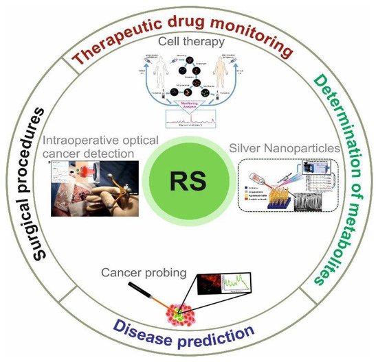
Figure 1
Schematic view of biomedical RS application. Adapted from Desroches et al.
2. Disease Prediction
Early diagnosis of diseases, such as those that are life-threatening, is essential to prevent their spread. Based on Raman light scattering, a diagnostic tool has been developed to study important molecules and events in real-time using new RS technologies with high sensitivity to biomolecular changes. In the last decades, there have been many publications showing that RS is used to define several diseases. RS is used to collect biochemical information, such as biomolecules, cells, tissues, and organs, whose biomarkers are various biological fluids, such as urine, saliva, blood, and tears. Selected biomarkers are analysed and evaluated, revealing the relationship between Raman light scattering spectral indicators and clinical condition
.
Cancer is one of the most common causes of death worldwide and RS makes it possible to diagnose undetected precancerous lesions in various organs, such as breast, skin, brain, gastrointestinal tract, heart, urinary, and reproductive tracts
investigated that confocal Raman microscopy distinguishes intestinal tumours from adenocarcinomas and normal, healthy organs. RS provides simple and immediate tissue identification during surgery, which allows for cancerous organs to be distinguished from healthy tissue. Using RS, the mechanism of malignant transformation of breast tissue has been studied with great success
.
The properties of blood vessels in a tumour mass of breast tissue were investigated by Kopeć and Abramczyk
using a combination of Raman and atomic force microscopy (AFM) imaging to determine biochemical composition. They found that individuals with breast cancer had higher concentrations of glycogen and lactic acid as well as an increase in the collagen–fibroblast network. An excellent, recent study by Winnard et al.
demonstrated the potential of RS in characterising organ-specific metastatic lesions at the molecular level to gain insight into metastatic progression. In this study, they used the combinatorial approach of RS and metabolomics. The stromal adjustments that occur in pre-metastatic lungs caused by breast cancer were analysed using RS. This work was performed with mouse lines in which mice were implanted with breast cancer cells with different metastatic potential. Changes in the extracellular matrix of the congested lungs, such as an increase in collagen and proteoglycan, were examined, and this was directly related to the metastatic potential of the breast cancer cells used
have shown that RS can be used effectively for the diagnosis of Alzheimer’s disease (AD). The novelty of this work is that it is based on the analysis of cerebrospinal fluid (CSF), while the other research has focused on different body fluids for the detection of AD. It is important to emphasise that CSF is the most relevant body fluid for detecting AD. The group of Ryzhikova et al.
suggests that early detection of AD is potentially possible using RS. It is expected that the method will be repeated on a larger subject population.
Lednev group
diagnosed early AD in saliva and serum with potential biomarkers using RS in combination with machine learning. This project aimed to use Raman hyper-spectroscopy in combination with machine learning. New methods were developed to diagnose AD based on the analysis of biological material, such as saliva. The group used biological material from saliva samples from a normal person with AD and mild cognitive impairment. In the end, it turned out that Raman hyper-spectroscopic analysis of saliva could be effective for an accurate diagnostic method in the early stages of AD. It is also possible to diagnose lung cancer with high accuracy at an early stage, as shown by the studies of Shin et al.
using a combination of SERS spectra and deep learning diseases. The advantage of this method is that tissue can be seen in the near-infrared region of the electromagnetic spectrum, which in combination with the RS instrument as well as multivariate data analysis, has become an accurately reproducible and non-invasive method for studying tissue pathology.
Barnas et al.
used Fourier transform infrared (FTIR) and RS to study endometrial hyperplasia and cancer. The study was performed on tissues from three groups of patients: normal control patients, patients with atypical hyperplasia, and patients with endometrial cancer. It has been revealed that both methods are complementary in terms of tissue examination. The results of the research suggest that the peaks of FTIR and Raman spectra and the changes in the specific peaks (absence of the peak or shift of the peak) can be used to distinguish cancer and atypical hyperplasia from normal endometrial tissue. Further studies are needed to understand whether RS is indeed a practical approach to study carcinogenesis.
SERS immunoassays have labelled/indirect or unlabelled configuration. Without labels, the Raman measurement is based on the fingerprint of the bioanalyte, and the labelled ones are identified by the spectrum of the Raman label. Therefore, labels without labels are not as complex as the labels that make up the labels on metallic nanostructures. Two systems were used to detect proteins, nucleotides, and fatty acids of lipids. The changes that occurred in the bioassay were recorded and diagnosed with infectious and non-infectious diseases
Table 1
shows some examples of bioanalytes or diseases that were detected using SERS.
Bioanalytes/diseases detected using SERS.
| Bioanalyte/Disease |
RS Substrate |
Reference |
|
| Cancer (blood plasma protein) |
|
| Ag NPs |
|
| [15] |
|
|
| Quantification of hepatitis B DNA |
|
| Ag NPs |
|
| [16] |
|
|
| Breast cancer tissue |
|
| Ag NPs |
|
| [17] |
|
|
| Sjogren’s syndrome from saliva |
|
| Cl-Ag NPs |
|
| [17] |
|
|
| Human tear uric acid |
|
| SiO2 and Au |
|
| [18] |
|
|
| Creatinine |
|
| Nano-Au |
|
| [19] |
|
|
| Mouse IgG |
|
| Au NPs |
|
| [20] |
|
|
| Single prostate cancer cells |
|
| Au NPs |
|
| [21] |
|
|
| Plasmodium falciparum DNA |
|
| Magnetic beads |
|
| [22] |
|
|
| HeLa cells |
|
| Au NPs |
|
| [23] |
|
|
| Gastritis |
|
| Au NPs |
|
| [24] |
|
In addition, RS is used to analyse the serum of patients with AD, patients with other types of dementia, and individuals from the control group. The results were analysed using multivariate statistics for differential identification of patients with AD. The study was a confirmation of the concept; this proves that RS and artificial neural network classification were able to differentiate patients with sensitivity and specificity of more than 90%, which shows that a combination test can become a blood test that can support clinical evaluation for effective and accurate differential diagnosis of AD
3. Surgical Procedures
In medicine, much attention is paid to optical instruments based on RS, which consist of intraoperative procedures in real-time. The benchmark for surgical guidance is histopathology, which also involves surgical removal of tissue followed by staining and examination under a microscope. This procedure takes a long time and in some cases, results in multiple biopsies, which causes a great deal of discomfort and suffering in patients
[13]. Therefore, a sensory system that can provide results during surgery is needed. For example, endoscopic pain analysis before surgery, delineation of the sides of the lesion during surgery, and changes of the single biopsy using RS can contribute to the absolute removal of the affected tissue and reduce the cost of secondary assessments of the disease and surgery.
Jermyn et al.
. Therefore, a sensory system that can provide results during surgery is needed. For example, endoscopic pain analysis before surgery, delineation of the sides of the lesion during surgery, and changes of the single biopsy using RS can contribute to the absolute removal of the affected tissue and reduce the cost of secondary assessments of the disease and surgery.
Motz et al. 64 have developed a small diameter Raman probe with integrated filters and a spherical lens to minimise low priority signals. In as little as one second, the probe can show the spectra of arteries and breast tissue at different stages of pathology, which is clinically useful. Jermyn et al. studied cancers of multiple human organs during surgery with 97% accuracy using a trimodal optical imaging system combining Raman. Thus, the method demonstrated that molecular imaging with high sensitivity could dramatically impact such areas of surgical and non-invasive oncological procedures for tumour detection to reduce cancer risk and improve quality of life. Kircher et al.
investigated the ternary status of magnetic resonance imaging—photoacoustic Raman imaging of nanoparticles, which revealed brain tumour boundaries and visualised tumour margins using RS. They used Raman imaging to ensure monitoring of intraoperative tumour resection, and histologic interdependence proved that Raman imaging delineated brain tumour boundaries. This latest trimodal aspect using nanoparticles can ensure the clearest visualisation of even the resection of a brain tumour.
In addition, significant steps are being taken to integrate RS with other wide-field and spectroscopic methods to provide additional data to support RS measurements. It has been shown that cancer cells can be diagnosed in less than one second using the broadband fluorescence method together with a Raman micro-spectrometer
. The trimodal optical imaging system is a combination of Raman scattering, diffuse reflectance, and intrinsic fluorescence spectroscopy, in which various cancer organs were detected during surgery with 93%, 100%, and 97% accuracy
[27] also synthesised a nanoparticle with three-component magnetic resonance imaging—photoacoustic and Raman imaging with the aim of preoperative and intraoperative separation of the sides of the leading brain tumour; the presence of this RS was used to visualise the sides of the tumour.
Nowadays, RS is increasingly used for cancer diagnosis and monitoring. As mentioned above, the improvement of algorithms for processing Raman signals, as well as the development of new methods SERS and fibre-optic probes, may make it possible to obtain results with high sensitivity and specificity and to apply RS approaches to cancer diagnosis.
4. Therapeutic Drug Monitoring
Therapeutic drug monitoring (TDM) is an important method in clinical pharmacology and clinical chemistry that aims to measure drug concentrations in human blood
also synthesised a nanoparticle with three-component magnetic resonance imaging—photoacoustic and Raman imaging with the aim of preoperative and intraoperative separation of the sides of the leading brain tumour; the presence of this RS was used to visualise the sides of the tumour.
Nowadays, RS is increasingly used for cancer diagnosis and monitoring. As mentioned above, the improvement of algorithms for processing Raman signals, as well as the development of new methods SERS and fibre-optic probes, may make it possible to obtain results with high sensitivity and specificity and to apply RS approaches to cancer diagnosis.
4. Therapeutic Drug Monitoring (TDM)
Therapeutic drug monitoring (TDM) is an important method in clinical pharmacology and clinical chemistry that aims to measure drug concentrations in human blood
. TDM has been used in medical practice since the 1960s and mainly focuses on drugs with narrow therapeutic targets
.
TDM is more commonly referred to in clinical practice as the observation of drug concentrations in biological fluids over time
. Karine is important for drugs with a limited therapeutic index, where a low dose is prescribed when the difference in dosage may lead to serious therapeutic consequences such, as drug toxicity and side effects. There is also increasing advocacy in the field of personalised medicine that will be useful for measuring the plasma concentration of a drug at individual doses. Individualised therapy planning in personalised medicine has become a great challenge for clinicians as it is very successful in improving patient care, so that each patient can reduce drug costs while receiving optimal treatment with minimal side effects.
Nowadays, there is a crucial problem of how to distinguish expired and non-expired drugs using a quick and non-invasive method. Current methods, such as HPLC, thin-layer chromatography, are time-consuming and complicated. In contrast, RS is a rapid and non-invasive method that can be used to examine expired and non-expired drugs. This is a significant problem for the medical world
The combination of AFM and RS can distinguish the characteristics of the nucleus and cytoplasm in living cells. By combining both methods, a modification of RS can be developed: tip-enhanced Raman spectroscopy (TERS). Intracellular imaging with TERS has been applied to HeLa cells. It has been shown that the regions of the nucleus and cytoplasm can be effectively distinguished using this method, for which the local information within the cell was obtained. Crucially, scientists have shown that the viability of the cell membrane is very high (around 100%) after the AFM tip penetrates the cell membrane. The method has significant potential for future use in studies where it is necessary to investigate the various organelles and biomolecules within the cell
.
The combination of AFM and RS can distinguish the characteristics of the nucleus and cytoplasm in living cells. By combining both methods, a modification of RS can be developed: tip-enhanced Raman spectroscopy (TERS). Intracellular imaging with TERS has been applied to HeLa cells. It has been shown that the regions of the nucleus and cytoplasm can be effectively distinguished using this method, for which the local information within the cell was obtained. Crucially, scientists have shown that the viability of the cell membrane is very high (about 100%) after the AFM tip penetrates the cell membrane. The method has significant potential for future use in studies where it is necessary to investigate the various organelles and biomolecules within the cell
5. Determination of Metabolites
Molecularly specific RS is well suited for profiling cellular metabolites, including neurotransmitters, amino acids, lipids, glucose, and nucleic acids, as well as in biofluids. Among all cellular metabolites, lipids are one of the most studied classes of biomolecules because they have large Raman scattering cross-sections. Lipids rich in intracellular bodies are referred to as lipid droplets, making their relationship to the physiological state of the cell increasingly apparent
. RS exploits the promise of detecting and imaging lipid droplets for quantification in cancer cells, such as HuH7 and colorectal cancer stem cells
.
RS and its modifications are widely used in cell research. A variety of microalgae
cells are studied using SERS. It has been shown that information about membrane lipids can be obtained, especially the conformation of membrane lipids and the molecular environment
.
RS has been used to study nerve myelin during excitation or the effect of neurotransmitter on nerve fibre. It has been shown that the changes in the C–C bonds of the fatty acids can be detected as well as the changes in the conformation
. This knowledge will improve our understanding of the mechanisms of lipid–lipid interactions in myelin and many processes associated with various diseases, such as multiple sclerosis, trauma and AD.
It is important to add that human skin is exposed to ultraviolet and infrared radiation, which is the cause of a number of diseases and ageing of human skin. Carotenoids are considered antioxidants that can support the antioxidant status of the human epidermis
. RS is one of the most popular methods to study carotenoids. More knowledge about the mechanisms of the photoprotective function of carotenoids is important for biomedical research and the development of commercial products. Similarly, Gellermann et al.
used resonant Raman scattering spectroscopy (RRSS) as a novel, non-invasive, in vivo optical technique to measure the concentration of the carotenoid pigments, lutein and zeaxanthin, in the human retina of adolescents and adults. Using RRSS, they found an apparent decrease in macular pigment concentration during the normal ageing process. They suggested the use of RS to measure macular carotenoids as a promising technology.
Ermakov et al.
reported that the RS technique has the potential for novel, rapid screening for carotenoid antioxidants in the largest populations at risk of vision loss due to age-related macular degeneration, an important precondition for blindness in the elderly in mature societies.
The method of RS is becoming increasingly popular in biological research. Valpapuram et al.
proposed a new technique combining optical biosensing and Raman micro spectroscopy. The particular advantage of this method is its ability to reduce the background signal and thus improve the signal-to-noise ratio. The researchers have shown that the combination of optical bio-sensing and Raman micro spectroscopy is a far more informative method than the conventional RS.
SERS has been used to spatially localise neurotransmitters on living cells and to study protein–neurotransmitter interactions
. Although it offers the best detection limits, the toxicity of metal nanoparticles in vivo limits its use
[48] have also demonstrated the usefulness of confocal RS for rapid detection of neurotransmitter predictions, but their studies were limited by in vitro spiked material. They propose real-time detection of serotonin, adenosine, and dopamine in vitro, but in addition, diffusion of dopamine in a heterogeneous base gel used as a surrogate for neural tissue. Raman mapping was performed using alpha 300 WITec confocal Raman system to obtain non-overlapping spectral data of neurotransmitters. Their work demonstrates the power of Raman spectroscopy in the biological sciences and likely provides a novel mechanism for testing the adaptability and kinetics that stimulate the brain.
A rapid, non-invasive, label-free approach to biological studies is currently essential for scientific purposes. However, RS has some limitations - it requires longer acquisition times and it is not possible to optically slice the collected signal. This makes it difficult to use RS for tissue research alone. Therefore, Marchetti et al.
have also demonstrated the usefulness of confocal RS for rapid detection of neurotransmitter predictions, but their studies were limited by in vitro spiked material. They propose real-time detection of serotonin, adenosine, and dopamine in vitro, but in addition, diffusion of dopamine in a heterogeneous base gel used as a surrogate for neural tissue. Raman mapping was performed using alpha 300 WITec confocal Raman system to obtain non-overlapping spectral data of neurotransmitters. Their work demonstrates the power of Raman spectroscopy in the biological sciences and likely provides a novel mechanism for testing the adaptability and kinetics that stimulate the brain.
A rapid, non-invasive, label-free approach to biological studies is currently essential for scientific purposes. However, RS has some limitations—it requires longer acquisition times and it is not possible to optically slice the collected signal. This makes it difficult to use RS for tissue research alone. Therefore, Marchetti et al.
combined three methods: multiphoton microscopy, fluorescence lifetime imaging microscopy, and RS to perform an efficient study of tissues ex vivo. The mentioned tailored technique is a promising approach to expand the application of RS in biological research.
The use of the RS method in medical studies is becoming more common. For example, RS is used in dentistry. The short- and long-term effects of demineralization can be studied using the RS tool. The major advantage of RS is that it is non-invasive while providing a high degree of sensitivity. In the study of Marin et al.
, quantitative information on the crystalline structure of the phosphate groups and the loss of the mineral fraction in the organic collagen matrix was discovered.
Nowadays, a precise, fast, and direct analysis tool is needed. The capillary sensor SERS, developed by the group of Arabi et al.
, is proposed as an ultrasensitive tool and used for protein analysis. Trypsin is a protein that can be used as a biomarker (in urine) for the diagnosis of pancreatitis. The idea is that this approach can be effectively used for early diagnosis of the disease. In addition, and to test the feasibility of the tool, other biological fluids such as saliva and sweat have also been measured. The microsensors are relatively quick and inexpensive to produce.
Another application of RS in biological studies is high-throughput screening Raman spectroscopy (HTS-RS)—presented in the work of Arend et al.
. This application is a customised platform for single-cell analysis. In the study in which the group of Arend et al.
examined the different types of neutrophils, both infected and uninfected, it has been shown that this type of platform can potentially help to speed up the diagnosis of pathogens. Currently, the routine for such analyses takes 1 working day.
It has been discovered that RS can be used effectively in chronic renal failure (CRF) to differentiate patients with this disease from healthy patients. The group of Chen et al.
[34] conducted a study on 47 samples from patients with CRF and 54 samples from control subjects. There is a prospect that the application used, which can be effectively utilised as a rapid diagnostic method for CRF. The plasma RS has been effectively used to study giant unilamellar vesicles (GUV)-simplified models of cellular plasma membranes conducted a study on 47 samples from patients with CRF and 54 samples from control subjects. There is a prospect that the application used, which can be effectively utilised as a rapid diagnostic method for CRF. The plasma RS has been effectively used to study giant unilamellar vesicles (GUV)—simplified models of cellular plasma membranes
Raman spectroscopy is a promising high-sense diagnostic method for assessing the oxygen transport function of erythrocytes. Haemoglobin accounts for >95% of the dry weight of erythrocytes and is a suitable subject for RS to study the conformation of globin and haeme. To assess the conformational state of the active site of haemoglobin, researchers use special Raman spectra to study the conformation of deoxyhaemoglobin (d- Hb) and oxyhaemoglobin (o- Hb), as well as the ability to release oxygen. This approach is important for monitoring changes in the ability of haemoglobin in erythrocytes to carry oxygen and, accordingly, characterising the presence or development of hypoxia in patient tissues. RS has been successfully used to analyse the properties of haemoglobin from healthy donors and patients with various cardiovascular diseases
.
Raman spectroscopy is a promising high-sense diagnostic method for assessing the oxygen transport function of erythrocytes. Haemoglobin accounts for >95% of the dry weight of erythrocytes and is a suitable subject for RS to study the conformation of globin and haeme. To assess the conformational state of the active site of haemoglobin, we use special Raman spectra to study the conformation of deoxyhaemoglobin (d- Hb) and oxyhaemoglobin (o- Hb), as well as the ability to release oxygen. This approach is important for monitoring changes in the ability of haemoglobin in erythrocytes to carry oxygen and, accordingly, characterising the presence or development of hypoxia in patient tissues. RS has been successfully used to analyse the properties of haemoglobin from healthy donors and patients with various cardiovascular diseases
[56], and astronauts after a long space flight [57], as well as for the analysis of animal models of cerebral ischemia and reperfusion, haemorrhagic shock, etc. In addition, RS has been successfully used in experiments to alter the properties of erythrocytes under in vitro conditions. A promising application of RS is the study of the molecular mechanisms of the development of pulmonary hypertension. In patients with IPAH with a typical hemodynamic picture, changes in the ability of hematoporphyrin of Hb to bind O , and astronauts after a long space flight [95], as well as for the analysis of animal models of cerebral ischemia and reperfusion, haemorrhagic shock, etc. In addition, RS has been successfully used in experiments to alter the properties of erythrocytes under in vitro conditions. A promising application of RS is the study of the molecular mechanisms of the development of pulmonary hypertension. In patients with IPAH with a typical hemodynamic picture, changes in the ability of hematoporphyrin of Hb to bind O 2 have been detected.