NowIn recent decadyes, Raman spectroscopy (RS) has been used in several studies on animal cells. Also, tThe method is popular among biophysicists and life science researchers. RS allows for the study of living cells in their natural conditions without any damage. RS is a well-known approach used in many of biomedical studies. Since biomolecules are involved, the main obstacle to the use of such methods in life science research is the low signal of Raman scattering. There are a number of modifications of RS that allow Raman scattering to be improved. There are existing approaches to detect Raman signals not only on the surface of human skin, but also inside the vasculature and various organs of patients.
1. The Principles of the Method of Raman Spectroscopy
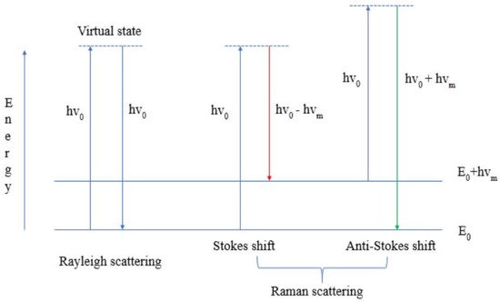
Figure 1 shows the energy transition of Rayleigh and Raman scattering. The former (Rayleigh) is based on the principle that the frequency of the absorbed and scattered photon does not change-elastic scattering. In the latter (Raman), on the other hand, there is a shift in frequency of the scattered photon (change in energy or change in wavelength) -inelastic scattering. It is also essential to note that Raman scattering is divided into Stokes and anti-Stokes shifts. The Stokes shift is more likely because it is associated with the shift of the binding maxima to the longer wavelengths (the energy and frequency of the scattered photon are correspondingly lower than those of the absorbed photon - see Figure 1). The anti-Stokes shift, on the other hand, is less likely. It results from the shift of the binding maxima to the shorter wavelengths (energy and frequency of the scattered photon are correspondingly higher than those of the absorbed photon - see
Figure 1 shows the energy transition of Rayleigh and Raman scattering. The former (Rayleigh) is based on the principle that the frequency of the absorbed and scattered photon does not change-elastic scattering. In the latter (Raman), on the other hand, there is a shift in frequency of the scattered photon (change in energy or change in wavelength) -inelastic scattering. It is also essential to note that Raman scattering is divided into Stokes and anti-Stokes shifts. The Stokes shift is more likely because it is associated with the shift of the binding maxima to the longer wavelengths (the energy and frequency of the scattered photon are correspondingly lower than those of the absorbed photon.
Energy transition of Rayleigh and Raman scattering.
In order to increase the sensitivity and resolution capability of the method, scientists use the approach based on the surface plasmon resonance effect. The electrons on the surface of the metal (silver and/or gold nanoparticles) oscillate. At a certain point, the frequency of the photon resonates with the frequency of the surface electrons. The surface plasmons substantially enhance the local electric field of the incident light (on the molecules near the surface’s vicinity of the metal nanoparticles)
[1]. In recent decades, this effect has been used to modify the method of RS to apply it in biological and medical research. . In recent decades, this effect has been used to modify the method of RS to apply it in biological and medical research.
2. Raman Spectroscopy and Its Modifications: Advantages and Use
There are many versions of RS, all of which use the phenomenon of Raman scattering in different ways. The choice of which variant to use for a particular measurement depends on inherent factors, such as the complexity of the sample and/or the concentrations of the target analyses.
The popularity of the RS method among biophysicists around the world is explained by a list of advantages of the method. RS is a non-invasive, rapid, and sensitive method for in vitro investigations
There are many variants of Raman spectroscopy, all of which use the phenomenon of Raman scattering in different ways. The choice of which variant to use for a particular measurement depends on inherent factors, such as the complexity of the sample and/or the concentrations of the target analyses.
The popularity of the RS method among biophysicists around the world is explained by a list of advantages of the method. RS is a non-invasive, rapid, and sensitive method for in vitro investigations
[4]. Usually researchers face an obstacle the biomolecules are present in the cell in very low concentrations, so the Raman scattering/signal is very low. To enhance the Raman scattering signal, the modifications of the method can be used, such as surface-enhanced Raman spectroscopy (SERS), coherent anti-Stokes Raman scattering (CARS), surface-enhanced Raman Spectroscopy (SERCS), and micro-Raman spectroscopy (see Table 1).
. Usually we face an obstacle—the biomolecules are present in the cell in very low concentrations, so the Raman scattering/signal is very low. To enhance the Raman scattering signal, the modifications of the method can be used (see Table 1), such as surface-enhanced Raman spectroscopy (SERS), coherent anti-Stokes Raman scattering (CARS), surface-enhanced Raman Spectroscopy (SERCS), and micro-Raman spectroscopy.
The list of modifications of RS with details of the objects.
| Modification of Method |
Object |
Biomolecules |
Reference/Link Number |
|---|
Coherent anti-Stokes Raman scattering (CARS) and microscopy |
|
|
microalgae |
|
|
|
lipids, carotenoids |
|
|
|
[ | 5 | ] | ,[6],[7],[8] |
|
|
|
|
Confocal Raman microscopy |
|
|
|
microalgae, algae |
|
|
|
lipids |
|
|
|
[ | 9 | ] | ,[10] |
|
|
|
|
Raman micro spectroscopy |
|
|
|
algae, animals |
|
|
|
lipids, carotenoids |
|
|
|
[ | 11 | ] | ,[12] |
|
|
|
|
Resonance Raman spectroscopy (RRS) |
|
|
|
bacteria, microalgae |
|
|
|
carotenoids |
|
|
|
[ | 10 | ] | ,[13],[14] |
|
|
|
|
Single-cell Raman spectroscopy (SCRS) |
|
|
|
microalgae |
|
|
|
lipids |
|
|
|
[ | 15 | ] | ,[16] |
|
|
|
|
Surface-enhanced Raman spectroscopy (SERS) |
|
|
|
animals, bacteria, microalgae |
|
|
|
lipids, carotenoids, proteins |
|
|
|
[ | 2 |
17 |
] | , | [ | 718],[1719],[182],[197],[20] |
|
|
As mentioned earlier, there is a special mechanism of Raman scattering (plasmon resonance) enhancement for modifications such as SERS. Moreover, each modification of the method can solve a specific task/objective.
The following sections describe the most commonly used types of RS.
As mentioned earlier, there is a special mechanism of Raman scattering (plasmon resonance) enhancement for modifications such as SERS. Moreover, each modification of the method can solve a specific task/objective.
The following sections describe the most commonly used types of RS.
2.1. Surface-Enhanced Raman Spectroscopy (SERS)
There are several clinical trials in progress with SERS. Sample types include blood
, and tears. As reported in articles from two studies, SERS had a susceptibility of 80.7% and 84.1% in detecting squamous cell carcinoma of the oral cavity by analysing blood
. Therefore, the tests show that SERS biofluid is suitable as a sample and relies on metal nanoparticles for signal amplification. The status of clinical trials is important for the understanding and future prospect of SERS and Raman spectroscopy
2.2. Coherent Anti-Stokes Raman Scattering (CARS)
Conventional Raman spectroscopy uses only a CW laser to generate spectra, while CARS and SRS use two pulsed lasers with different wavelengths to enable nonlinear optical motions. CARS microscopy can form an optical contrast of endogenous chemical structures, which is popular in various fields of biomedicine as it can provide a high-resolution image. For example, CARS microscopy has been used to visualise tissue structures, skin
[25],[26] lung, kidney, and r Consequently, CARS has been able to obtain micron-level images of brain slices, which has worked well in cancer diagnosis
Concrete prostatectomy is considered the most popular method in civilised countries for curing members of the stronger sex with clinically localised prostate cancer. In this surgery, the entire prostate is removed, but the urinary ball is reunited with the urethra
2.3. Resonance Raman Spectroscopy (RRS)
One of the drawbacks of Raman spectroscopy is the low signal intensity. This drawback can be corrected with RRS. By matching the wavelength of laser excitation to the electrical absorption maximum of a particular chemical, the Raman signal of certain bands is enhanced. The study was used for a multifaceted study of haemoglobin, and the release of cytochrome-c from mitochondria during apoptosis was also studied. Okada et al.
[30] as well as other scientists have used RRS to perform unlabelled studies of molecular dynamics in apoptotic cells. Observation of mitochondrial membrane stained with the dye JC-1 using RRS confirmed that the observed release of cytochrome was due to apoptosis.
as well as other scientists have used RRS to perform unlabelled studies of molecular dynamics in apoptotic cells. Observation of mitochondrial membrane stained with the dye JC-1 using RRS confirmed that the observed release of cytochrome was due to apoptosis.
2.4. Spatially Offset Raman Spectroscopy (SORS)
Although SORS technology may not be approved in any way for uniform diagnosis of patients, there are significant prospects for the eventual use of this technique in the clinic. Recent advances in medicine have shown SORS can be used in blood testing, such as assessing the quality of erythrocytes during blood transfusion in patients. Vardaki et al.
Although SORS technology may not be approved in any way for uniform diagnosis of patients in our time, there are significant prospects for the eventual use of this technique in the clinic. Recent advances in medicine have shown SORS can be used in blood testing, such as assessing the quality of erythrocytes during blood transfusion in patients. Vardaki et al.
have shown that SORS is able to profile changes in oxygenation when stored for 6 weeks. It is well known that in blood transfusions, the chemical composition of blood units changes differently from time to time. For this reason, a few units over the years are by no means determinative of the relationship between erythrocytes properties. Feng et al.
[32] used SORS to measure subcortical bone and biochemical changes with increasing depth in intact mouse bone.
used SORS to measure subcortical bone and biochemical changes with increasing depth in intact mouse bone.
3. Raman Spectroscopy for Photosynthetic Studies
Photosynthesis is the most basic and important process on earth. It is the natural way of synthesising carbohydrates using solar energy. Scientists from all over the world are exploring it with a number of applications, one of which is Raman spectroscopy. Findings from a number of recent studies on RS applied to photosynthetic organisms are shared below. [33] recently conducted their study on Antarctic lichens using RS. Antarctic lichens are organisms that can change their metabolism and photosynthetic activity in response to changing environmental conditions. Hydration and dehydration are the investigated triggers for the activation/deactivation of photosynthetic processes in the lichens. It has been revealed that photosynthetic activity is activated quite rapidly, which contributes to the hypothesis that the photosynthetic apparatus and carotenoids are not synthesised de novo in the early stages of photosynthesis. Another important discovery made using RS, the bands/features of the pigment scytonemin, are present in the Raman spectra of one of the lichens studied. There is a hypothesis that this pigment plays a photoprotective role in the photobionts of algae and cyanobacteriae.
recently conducted their study on Antarctic lichens using RS. Antarctic lichens are organisms that can change their metabolism and photosynthetic activity in response to changing environmental conditions. Hydration and dehydration are the investigated triggers for the activation/deactivation of photosynthetic processes in the lichens. It has been revealed that photosynthetic activity is activated quite rapidly, which contributes to the hypothesis that the photosynthetic apparatus and carotenoids are not synthesised de novo in the early stages of photosynthesis. Another important discovery made using RS, the bands/features of the pigment scytonemin, are present in the Raman spectra of one of the lichens studied. There is a hypothesis that this pigment plays a photoprotective role in the photobionts of algae and cyanobacteriae.
4. Raman Spectroscopy for Analytical Studies
There is an ongoing need for fast and accurate detection of melamine in dairy products. Melamine is a compound that can be toxic above a certain level when added to food. Therefore, it is important to propose an approach that allows accurate detection of the toxic compound in food products. Liu et al.
proposed the SERS method using silver nanoparticles (AgNPs). Not only SERS but also a colourimetric method was used for this idea. The results showed that the colourimetric method can lead to false-positives in detecting the presence of different compounds (AgNPs). The SERS method, on the other hand, can overcome this limitation. Importantly, the scientists suggested using both methods in tandem to achieve accurate and rapid detection of melamine in dairy products.
Fentanyl is one of the most commonly used opioids. However, fentanyl and its analogues caused numerous fatal drug overdose incidents. The problem raised by the group of Mirsafavi et al.
[35] is the need for novel analytical methods to effectively distinguish fentanyl from its precursors. The vibrational spectra of this family of analytes are quite similar, so it is difficult to solve the problem using conventional methods. The SERS method enables the distinguishing of fentanyl and its precursors. This approach would be an efficient and effective aid in the field of forensics.
is the need for novel analytical methods to effectively distinguish fentanyl from its precursors. The vibrational spectra of this family of analytes are quite similar, so it is difficult to solve the problem using conventional methods. The SERS method enables the distinguishing of fentanyl and its precursors. This approach would be an efficient and effective aid in the field of forensics.