Cannabis Biomolecule Effects on Cancer Cells: Comparison
Please note this is a comparison between Version 1 by Hinanit Koltai and Version 2 by Bruce Ren.

Cancer is a complex family of diseases affecting millions of people worldwide. Gliomas are primary brain tumors that account for ~80% of all malignant brain tumors. Glioblastoma multiforme (GBM) is the most common, invasive, and lethal subtype of glioma. Therapy resistance and intra-GBM tumoral heterogeneity are promoted by subpopulations of glioma stem cells (GSCs). Cannabis sativa produces hundreds of secondary metabolites, such as flavonoids, terpenes, and phytocannabinoids. Cannabis is commonly used to treat various medical conditions, and is used in palliative care of cancer patients. The anti-cancer properties of cannabis compounds include cytotoxic, anti-proliferative, and anti-migratory activities on cancer cells and cancer stem cells. Specific combinations of multiple phytocannabinoids act synergistically against cancer cells and may trigger different anti-cancer signaling pathways. Yet, due to scarcity of clinical trials, there remains no solid basis for the anti-cancer therapeutic potential of cannabis compounds.

  • cannabis
  • phytocannabinoids
  • synergy
  • cannabinoid receptors
  • cancer
  • cancer stem cells
  • cytotoxicity
  • glioma
  • glioblastoma

1. Introduction

Cancer is a complex family of diseases, in which a gradual change in the expression of multiple genes leads to genomic instability and cell death imbalance, resulting in the abnormal growth of cells [1]. Although different types of cancer present with different phenotypic clinical characteristics and different genetic modifications, there are several common molecular patterns and biological capabilities acquired during malignant transformation. The hallmarks of cancer comprise six distinctive and complementary processes essential for tumor growth and survival: sustaining proliferative signaling insensitivity to growth suppressors; disproportionately greater growth over cell death; limitless replicative potential; and the induction of angiogenesis, tissue invasion, and metastasis [2].
Cannabis sativa L. (C. sativa) is a diecious annual herb belonging to the Cannabaceae family and has been effective in treating numerous medical conditions [3][4][3,4]. The major utilization of cannabis is for recreational purposes. While many countries are legalizing cannabis production and use, cannabis remains the most widely used illegal drug globally [5]. However, the medical use of this plant has been documented in the oldest Chinese pharmacopoeia pen-ts’ao ching (compiled in 100 CE but attributed to Emperor Sheng Nung, c. 2700 BCE) for pain relief, constipation, and other ailments. In India, the plant was historically used for analgesic, tranquilizing, anesthetic, antibiotic, and anti-inflammatory functions [6][7][8][6,7,8]. Around 600 constituents have been identified in C. sativa, among them being several classes of secondary metabolites, including dozens of flavonoids, hundreds of terpenes, and more than 160 terpenophenolic compounds known as phytocannabinoids [9][10][11][12][9,10,11,12]. Among the most abundant phytocannabinoids are Δ9-tetrahydrocannabinol (THC), cannabidiol (CBD), and cannabigerol (CBG), which are all synthesized by female plants and stored mainly in epidermal glandular trichomes, which are densely concentrated in the inflorescence and bracts. Phytocannabinoids are produced as prenylated aromatic carboxylic acids and converted to neutral homologous forms by decarboxylation, which occurs to some extent within the living plant but mostly when catalyzed by heat following harvesting [9][10][11][12][9,10,11,12]. Today, several cannabis preparations or synthetic compounds have been approved by health authorities worldwide (e.g., FDA or EU) and meet the same regulatory requirements of pharmaceutical drugs in terms of safety, efficacy, and consistency. These include Nabiximols, which is a whole-plant prescription cannabinoid used in the management of patients with multiple sclerosis, chronic neuropathic pain, and cancer-related pain [13]. Another example is Dronabinol, a synthetic phytocannabinoid (THC) that is marketed as medicines in several countries and which is indicated for the treatment of anorexia and weight loss in adult patients with HIV/AIDS or cancer [14].
2. Anti-Cancer Properties of Cannabis Compounds

2.1. Pre-Clinical Studies

Studies have demonstrated that phytocannabinoids potentially possess anti-cancer properties, including the inhibition of cell migration, proliferation, and angiogenesis and the induction of apoptosis in skin, lung, breast, prostate, and glioma cancer cells [15][16][17][18][26,27,28,29]. Phytocannabinoids trigger cancer cell death via various signal transduction pathways, including oxidative stress, cell cycle arrest, endoplasmic reticulum (ER) stress, autophagy, and apoptosis [15][16][17][18][26,27,28,29].
One of the most abundant phytocannabinoids, THC, was shown to inhibit the growth of some tumors, inhibit angiogenesis, and induce apoptosis in various cancers cells in vitro and in vivo [16][17][18][19][20][27,28,29,30,31]. THC and CBD exhibited synergistic inhibition of cell proliferation in GBM cell lines [21][32]. Furthermore, CBD was found to inhibit the invasiveness of breast cancer cells and GBM cells at sub-lethal concentrations by downregulating matrix metalloproteinases (MMPs) and their inhibitors (TIMPs) [22][23][33,34]. An MMP–TIMP imbalance results in proteolysis of the matrix that may be associated with different pathological processes, including tumor invasion [24][35]. In vivo, THC and/or CBD reduced GBM tumor growth [25][36]. Furthermore, several studies have demonstrated CBG anticancer activity, including in mouse melanomas, human oral epithelioid carcinoma cells, human breast carcinomas, and colorectal cancer cells [17][28].
Recently, rwesearchers have shown that two fractions of a high-THC cannabis strain extract had a significant cytotoxic activity against Human GBM cell lines and GSCs derived from Human tumor specimens [26][37]. The two fractions were composed of different combinations of phytocannabinoids, with CBG or THC as the most abundant compound. The active fractions induced apoptosis and the expression of ER-stress-associated genes. Moreover, the fractions altered cell cytoskeletons, reduced cell invasion, and inhibited cell migration and colony formation [26][37]. Notably, the study demonstrated the therapeutic potential of combinations of cannabis compounds in exerting cytotoxic, anti-proliferative, and anti-migratory effects on human GBM cells. Furthermore, the activity of these specific combinations was higher than that of the purified primary compound in each fraction, as well as that of the crude extract [26][37]. Notably, in many cases, phytocannabinoid concentrations used in vitro do not coincide with those safely achievable in vivo, and clinical trials are needed to prove phytocannabinoid treatments’ efficacy.

2.2. A Clinical Study

One promising clinical evidence suggests effective phytocannabinoid-based treatments against GBM [27][38]. A pilot phase I clinical trial indicated that THC has a good safety profile [28][39]. The administration of THC in two of nine GBM patients in this trial led to a decrease in tumor cell proliferation [28][39].

3. Intracellular Effects of Phytocannabinoids in Glioma Cells

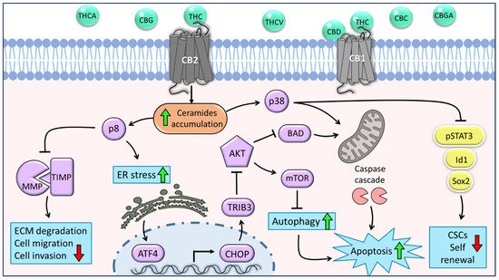
Considering the complexity and the wide distribution of the endocannabinoid system (ECS) components and their interaction with phytocannabinoids [29][30][47,49], phytocannabinoids may have the potential to impact and mediate a multitude of cancer-related signaling pathways. One common pathway activated by phytocannabinoids in different cancer types is the ER-stress pathway, which is one of the main mechanisms to induce apoptosis of glioma, astrocytoma, melanoma, and pancreatic tumor cells [31][56]. Previous studies on several models of glioma reported that CB1 receptor agonists and, more efficiently, CB2 receptor agonists stimulated the synthesis and accumulation of ceramide, a pro-apoptotic lipid second messenger which leads to the induction of stress protein p8 ([20][32][31,57]; Figure 1). Following this p8 induction, downstream ER-stress-related genes were induced (Figure 1), and as a result, the intrinsic mitochondrial pathway was activated [20][32][31,57].
Figure 1. The main molecular mechanisms underlying the anti-tumor effects of C. sativa phytocannabinoids on glioma cells and glioblastoma stem cells. Phytocannabinoids inhibit cell viability and motility through various cannabinoid receptor (CB)-mediated mechanisms. THC acts as an agonist of both CB1 and CB2 receptors; CBD may act as a CB1 antagonist. The activation of CB1 or CB2 stimulates the synthesis and accumulation of ceramides (orange shape) and, as a result, triggers the induction of p8. This leads to the inhibition of cell migration and invasion through the downregulation of MMPs. Furthermore, p8 promotes the upregulation of ER-stress-related genes ATF-4, CHOP, and TRIB-3, followed by inhibition of the Akt-mTORC1 axis and initiation of autophagy, which is upstream of apoptosis. In addition, inhibition of Akt leads to the overexpression of BAD and consequently induces apoptosis via the intrinsic mitochondrial pathway. Another signaling pathway activated by ceramides is p38-MAPK, which involves both apoptosis activation and inhibition of CSC self-renewal through the downregulation of stemness regulators, such as p-STAT3, Id1, and Sox2 (yellow shapes). Green arrows represent upregulation and red arrows represent downregulation of biological processes. Purple shapes represent genes or proteins, and blue shapes represent biological processes.
Recently, rwesearchers have shown that CBG-rich and THC-rich combinations of phytocannabinoids induced Activating transcription factor 4 (ATF4), C/EBP homologous protein (CHOP)-10 (GADD153/DDIT-3), and Tribbles homolog 3 (TRIB3) gene transcription in a CB2 activation-dependent manner ([26][37]; Figure 1), supporting the notion that phytocannabinoid treatments induce cell death via ER stress. ATF4 is a transcription factor transiently induced following treatment with ER stressors [31][56]. In turn, ATF4 induces CHOP expression, a transcription factor that regulates the expression of many pro- and anti-apoptotic genes [33][58]. Under ER-stress, CHOP activates pro-apoptotic proteins, including the B cell lymphoma-2 (BCL-2) family proteins, such as BAK and BAX, and represses anti-apoptotic BCL-2 family proteins [33][58]. TRIB3 is a pseudokinase and another protein associated with ER-stress, which was found to facilitate ER-stress-dependent apoptosis via the NF-κB pathway [34][59]. Moreover, TRIB3 has been shown to inhibit the Akt-mTORC1 axis, consequently leading to the initiation of autophagy (Figure 1), which is upstream of intrinsic mitochondrial apoptosis [35][60].
Furthermore, treatment with the cannabinoid-receptor synthetic agonist WIN-55,212-2 led to upregulation of the BCL-2 homology 3 (BH3)-only family member BAD, a pro-apoptotic protein, in response to ceramide activation and the serine/threonine kinase Akt downregulation in glioma cells ([36][61]; Figure 1). Ceramide is also an important regulator of p38 mitogen-activated protein kinase (MAPK), and previous studies on human leukemia and glioma cells reported that following THC treatment, activation of this pathway induced apoptosis partially via the CB1 and CB2 receptors ([32][37][57,62]; Figure 1).
Importantly, in contrast to malignant cells, normal brain cells, such as primary neurons and astrocytes, do not undergo apoptosis or present ceramide accumulation in response to phytocannabinoid treatments [20][31]. In addition, it has been shown in vivo that even at high doses, there is no sign of any damage or neurotoxicity to normal brain tissue following treatments with phytocannabinoids [38][63]. These findings, together with the differences in the expression of cannabinoid receptors between normal tissues and cancer cells, and the fact that cannabinoid receptors mediate the anti-cancer activities support the suggestion that cannabinoid receptors regulate cell survival and cell death signaling pathways differently in glioma cells and non-transformed cell [39][64].
Although the role of cannabis compounds in the suppression of cancer migration and invasion is elusive and poorly characterized, accumulating evidence suggests that cannabis compounds have potent anti-migrative and anti-invasive effects on GBM cells, both in vitro and in vivo. It was previously reported that treatment with THC or CBD down-regulated the expression of major proteins associated with glioma tumor migration, in particular MMP-2, MMP-9, TIMP-4, and TIMP-1 [23][40][34,65], even at low concentrations, which were insufficient to induce cell apoptosis. TIMP-1 and some MMP expression is selectively upregulated in different cancers and strictly associated with tumor malignancy and metastasis [41][66]. Interestingly, THC treatment depressed TIMP-1 and MMP-2 expression in glioma cell lines as well as in cultured human GBM primary cells. In addition, the local administration of THC down-regulated TIMP-1 and MMP-2 expression in glioma-bearing mice and in two patients with recurrent GBM [19][40][30,65]. Moreover, these effects of THC were suggested to be mediated via CB2 receptor activation and were prevented by the blockade of ceramide synthesis and by knock-down of the p8 stress protein in glioma cells ([40][65]; Figure 1).
Video Production Service