Your browser does not fully support modern features. Please upgrade for a smoother experience.
Patatin-like Phospholipase Domain-Containing Protein 6: Comparison
Please note this is a comparison between Version 1 by Doris Kretzschmar and Version 2 by Beatrix Zheng.

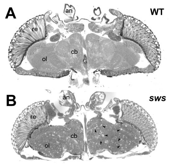
Patatin-like phospholipase domain-containing protein 6 (PNPLA6), originally called Neuropathy Target Esterase (NTE), belongs to a family of hydrolases with at least eight members in mammals. PNPLA6/NTE was first identified as a key factor in Organophosphate-induced delayed neuropathy, a degenerative syndrome that occurs after exposure to organophosphates found in pesticides and nerve agents. More recently, mutations in PNPLA6/NTE have been linked with a number of inherited diseases with diverse clinical symptoms that include spastic paraplegia, ataxia, and chorioretinal dystrophy. A conditional knockout of PNPLA6/NTE in the mouse brain results in age-related neurodegeneration, whereas a complete knockout causes lethality during embryogenesis due to defects in the development of the placenta. PNPLA6/NTE is an evolutionarily conserved protein that in Drosophila is called Swiss-Cheese (SWS). Loss of SWS in the fly also leads to locomotory defects and neuronal degeneration that progressively worsen with age. 

  • Neuropathy Target Esterase
  • Swiss-Cheese
  • organophosphate-induced delayed neuropathy
  • hereditary spastic paraplegia
  • phosphatidylcholine
  • Protein kinase A

1. Introduction

Changes in PNPLA6/NTE have now been shown to be involved in neurodegenerative symptoms in species ranging from flies to humans. That mutations in this protein can cause neurodegeneration and locomotion deficits was first shown in Drosophila by the identification of point mutations in the swiss cheese (sws) gene, the fly homolog of PNPLA6/NTE, that lead to progressive neuronal degeneration during adulthood [1]. Human PNPLA6/NTE, whose activity has long been associated with a neuropathy caused by organophosphate poisoning, was subsequently cloned and found to be homologous to fly SWS [2]. In recent years, mutations in PNPLA6/NTE have also been linked with a number of rare inherited diseases with clinical symptoms that can include spastic paraplegia, ataxia, hypogonadism, and chorioretinal dystrophy [3][4][3,4]. PNPLA6/NTE proteins contain several conserved domains: (1) a phospholipase domain that provides its catalytic activity as esterase and contains the binding sites for the organophosphates, (2) a transmembrane spanning domain that localizes the majority of the protein to the membranes of the endoplasmic reticulum, and (3) domains that have homology with the regulatory subunits of Protein Kinase A (PKA). Specifically, this domain includes three nucleotide binding sites and a region (PKA-BD) with homology to canonical regulatory subunits of PKA required for binding to the catalytic subunits of PKA [2][5][2,5]. Whereas the phospholipase function as been confirmed in several species [6][7][8][6,7,8], the PKA regulatory function has so far only been confirmed in Drosophila, although mammalian PNPLA6/NTE can bind to the fly PKA catalytic subunit [9].

2. Phenotypic Consequences Due to the Loss of PNPLA6/NTE in Model Systems

Due to the occurrence of OPIDN in humans, the first studies in animals focused on studying effects of OPs. Whereas rodents are relatively resistant to develop the clinical symptoms of OPIDN, adult hens have been established as a model early on [10] and it has been shown that they develop symptoms that are fairly similar to OPIDN in humans [11][61]. Treated hens have difficulties standing or walking and they reveal axonal degeneration and changes in myelin [12][13][62,63]. They have also been widely used to determine the effects of different OPs and to show that pre-treatment with non-neuropathic OPs can protect from the effects of neuropathic OPs [14][15][13][15,19,63].
Whereas rats do not show the severe phenotypes of chickens after OP treatment, they do reveal reduced locomotor activity and it takes them longer to cross a narrow beam [16][64]. In addition, they show axonal degeneration after administered a neuropathic OP which can also be prevented with pre-treatment with a non-neuropathic OP [17][18][65,66].

2.1. Drosophila Melanogaster

Performing a mutagenesis screen in Drosophila, Heisenberg and colleagues identified several mutants with structural brain defects, including five mutants showing severe neurodegeneration as seen by the formation of vacuoles in the brain [19][67]. A complementation analysis showed that they all affected the same gene which based on the appearance of the mutant phenotype was named swiss-cheese or sws (Figure 1). Cloning and sequencing revealed that two of the alleles contained point mutations in the sws gene [1]. Another mutation generated a stop codon early in the sequence, resulting in no protein being detectable in Western blots. This allele (sws1) therefore appears to result in a complete loss of SWS. The mutations in the remaining two alleles could not be identified. The phenotypic analyses showed that all sws alleles exhibit age-dependent neuronal degeneration due to apoptosis, glial cell death, and a reduced lifespan [1]. In addition, sws mutants develop locomotion deficits, disruptions in the ER and ER stress [20][21][31,68]. Specifically knocking down SWS in neurons also caused age-related neurodegeneration, locomotion deficits, reduced life span and a reduction of mitochondria in the CNS, whereas mitochondrial transport was reduced in the PNS [22][69]. Consistent with reduced mitochondrial function, these flies also showed an increase in reactive oxygen species, in addition to an accumulation of lipid droplets. A knockdown in all glial cells resulted in glial wrapping defects in the CNS and abnormal glial layers and fragmented glial nuclei in the PNS [23][24][54,70]. Furthermore, the pan-glial knockdown of SWS expression induced defects in neuronal transmission and locomotion, while more restricted knockdowns in particular glial subtypes revealed that SWS is specifically required in ensheathing and surface glia [24][70].
Figure 1. Loss of SWS results in neurodegeneration. (A) A horizontal head section from a 20d old wildtype Drosophila showing an intact brain. (B) In a head section from a 20d old sws1 mutant the neurodegeneration is visible by the formation of numerous sponfgiform lesions (some are indicated by the arrowheads) in all brain areas. re = retina, ol = optic lobes, cb = central brain, an = antennae.
To determine the functional importance of the different domains identified in SWS, several constructs were tested for their ability to rescue the phenotypes seen in the sws mutant. Using a wildtype form of SWS, revealed that the neuronal degeneration could be rescued by selective expression in neurons, whereas the glial phenotype could be rescued by expression in glia, showing that SWS is autonomously required in both cell types [20][31]. In addition, expression in either neurons or glia restored esterase activity and for the neuronal rescue it was also demonstrated that it reduced the abnormal increase in PC seen in sws flies. In contrast, as mentioned above, a SWS construct with a mutation in the active site serine of the phospholipase domain did not restore esterase activity in sws mutants and it only partially rescued the neurodegeneration phenotype [9][20][9,31]. Similarly, a mutation in the PKA-BD domain only partially rescued the degenerative phenotype [9], suggesting that both domains contribute equally to the neuronal function of SWS. Although the PKA-BD mutant protein has an intact phospholipase domain, it could not restore the esterase activity in sws mutants; however, it doubled the level of esterase activity when expressed in the presence of wildtype SWS. These findings support the hypothesis that the PKA-regulatory function of SWS is required for its phospholipase activity, though not directly within the same molecule. In glia, the phospholipase function seems to play a more prominent role than the PKA-regulatory function, because the mutation in the active site serine completely failed to rescue the glial phenotypes in the glia-specific knockdown of SWS [24][70]. In contrast, expression of the PKA-BD mutant resulted in a partial rescue. Lastly, to confirm the functional conservation of SWS and mammalian PNPLA6/NTE, constructs encoding either the mouse or human PNPLA6/NTE were expressed in sws mutants, and both could rescue the behavioral and degenerative phenotypes, as well as restore esterase activity [20][25][26][31,71,72].

2.2. Danio Rerio

Effects of the loss of PNPLA6/NTE have also been studied in mammalian models. A knock-down in zebrafish embryos caused various abnormalities, with a curled tail being the most prominent phenotype [27][73]. In addition, the embryos showed defects in eye development, midbrain-hindbrain boundary abnormalities, and a reduced number of motor neurons with short and abnormally branched axons. Addressing the importance of the esterase domain, the authors co-injected PNPLA6/NTE-specific morpholinos with different PNPLA6/NTE mRNAs. Whereas expression of wildtype PNPLA6/NTE rescued the curly tail and motor neuron phenotype of the knock-down, three mRNAs that contained mutations in the esterase domain, including one with a mutation in the active site serine, did not. Similar experiments by Hufnagel et al. confirmed the curly tail phenotype in the PNPLA6/NTE knockdown and also demonstrated that wildtype PNPLA6 rescued this phenotype but PNPLA6/NTE with mutations in the esterase domain did not [28][59]. In combination, these studies provide further evidence that the esterase/phospholipase activity is important for the biological function of PNPLA6.

2.3. Mus Musculus

Mice lacking PNPLA6/NTE show severe growth retardation and die around day 9 of embryonic development [29][74]. A histological analysis revealed defects in placental development (with no placental labyrinth forming), in addition to a breakdown of yolk sac circulation, leading to enlarged pericardia and dilated blood vessels in the embryo. A role early in embryonic development is also supported by findings that PNPLA6/NTE is expressed in mouse embryonic stem cells, with increased levels during differentiation, and that silencing caused changes in the expression of genes involved in neuronal development and the formation of the respiratory and vascular system [30][31][75,76]. Silencing of PNPLA6/NTE in a human pluripotent cell line induced similar changes in the transcriptome, suggesting that the function of PNPLA6/NTE during development is conserved in humans [32][33][77,78]. Interestingly, treatment with OPs did not induce these transcriptional changes, although it inhibited the esterase function, suggesting that this developmental function of PNPLA6/NTE is not dependent on its esterase activity [32][34][77,79]. Whereas the loss of PNPLA6/NTE and its developmental functions causes lethality during embryogenesis, heterozygous knock-out animals develop normally but have reduced esterase activity, increased susceptibility to certain OPs, and they appear hyperactive in open field tests [29][35][74,80]. A conditional, brain-specific knock-out did not affect development but these mice showed defects in motor coordination and neuronal degeneration when aged to 4.5 month [36][81]. Further analyses revealed a loss of Purkinje cells, disruptions of the ER, and abnormal reticular aggregates. In the spinal cord, axonal degeneration was first noticed in the distal parts of the longest spinal axons at 1 month of age, followed by axonal swelling that increased with aging in both, ascending and descending tracts [7]. These defects were accompanied by progressive hindlimb dysfunction, first detectable by clenching of the digits, and by 4 months the animals were unable to fully support their lower body when walking on a beam. Comparable axonal degeneration and progressive swelling of spinal cord axons was detected when treating animals with TOCP [7]. A glial-specific knock-out resulted in incomplete ensheathment of Remak fibers in the sciatic nerve whereas myelination was not affected [37][60], confirming that the loss of PNPLA6/NTE in glia also has functional consequences in mammals. Lastly, a selective knock-down of PNPLA6/NTE in testis demonstrated a role in the proliferation of spermatogonial stem cells and a reduction in sperm count was also observed when treating male mice with TOCP [38][82].

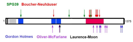
3. PNPLA6 in Human Disease

The first disease-causing mutations in PNPLA6/NTE were described in 2008 in two families with affected members showing progressive spastic paraplegia associated with distal upper and lower extremity wasting starting in childhood [39][83]. Electrophysiological studies and magnetic response imaging suggested that the locomotion defects were due to motor neuron neuropathy and spinal cord atrophy. Rainier and colleagues identified three mutations in these families, all localized within the esterase domain of PNPLA6/NTE. The mutations were recessive and whereas the patients in one family were homozygous, the affected individuals from the other family were compound heterozygotes. This inherited spastic paraplegia was consequently named NTE-related Motor Neuron Disorder or spastic paraplegia 39 (SPG39). Mutations in PNPLA6/NTE have now been identified in a few more spastic paraplegia patients and there are now also mutations outside the esterase domain [40][84] (Figure 2). In one family, the affected members also showed Parkinsonism in addition to spastic paraplegia [41][42][85,86], suggesting that the pathology can vary. In fact, it has now been shown that mutations in PNPLA6/NTE can cause a wide spectrum of neurological disorders with overlapping symptoms (Table 1), which have now been categorized as PNPLA6-related disorders [3][28][40][43][3,59,84,87].
Figure 2. Disease-causing mutations in PNPLA6/NTE. Shown are only point mutations associated with the different syndromes. The cyclic nucleotide binding sites (aa195-322, aa511-633 and aa629-749) are shown in blue. The phospholipase domain (aa981-1147) is shown in red.
Table 1. PNPLA6-related disorders. Characteristic clinical features are indicated by +. Clinical features not detected in all patients are indicated by +/−.
  Ataxia Hypogonadism/

Delay in Sexual

Development
Chorioretinal Dystrophy Trichomegaly/

Alopecia
Spastic Paraplegia Intellectual Disabilities Dwarfism/

Short Statue
SPG 39 +/−       +    
Boucher–Neuhäuser + + +/-   +/− +/−

mild
 
Gordon Holmes + +     +/− +/−

mild
 
Oliver–McFarlane +/− + +

severe
+ +/− + +
Laurence–Moon +/− + +   +/− + +
One of them is Boucher–Neuhäuser syndrome, which is characterized by delayed puberty, spinocerebellar ataxia, and in some patients, chorioretinal dystrophy. Mutations in PNPLA6/NTE as the underlying cause for Boucher–Neuhäuser syndrome were first described in 2014 and there are now more than a dozen identified pathogenic mutations [40][84]. As in the case of SPG39, the mutations are recessive and affected individuals are either homozygous for the mutation or compound heterozygotes. Disease-causing mutations in these patients are mostly, but not exclusively, found in the esterase/phospholipase domain although a few are in the cyclic nucleotide binding sites [44][45][46][47][48][49][50][51][52][53][54][88,89,90,91,92,93,94,95,96,97,98]. The onset of symptoms varies widely and although a case of a one-year-old child has been described, symptoms mostly manifest after late childhood or in adults [44][88].
Also belonging to the group of PNPLA6-related disorders is Gordon Holmes syndrome. Many of the symptoms of Gordon Holmes patients overlap with the ones described in Boucher–Neuhäuser patients, however they do not show chorioretinal dystrophy [55][56][57][58][99,100,101,102]. A prominent feature in Gordon Holmes syndrome is hypogonadotropic hypogonadism and experiments treating a mouse pituitary cell line with an OP suggest that the effects on sexual development are due to a decreased release of luteinizing hormone from the pituitary glands [55][99]. Again, the mutations associated with this syndrome are recessive, and so far, have been localized to the C-terminal half of the protein.
The list of PNPLA6-related disorders was further expanded in 2015, with the discovery that mutations in PNPLA6/NTE cause both Oliver–McFarlane syndrome and Laurence–Moon syndrome. Although symptoms in these patients can include hypogonadism and ataxia, the distinguishing feature is severe chorioretinal dystrophy/retinitis pigmentosa often occurring in early childhood [8][28][8,59]. Whereas the phenotypes in Oliver–McFarlane patients include congenital trichomegaly, this is absent in Laurence–Moon patients. Like the forementioned diseases, Oliver–McFarlane and Laurence–Moon syndrome are rare inherited recessive diseases. Although most of the mutations are localized to the esterase/phospholipase and cyclic nucleotide binding domains, others are outside these identified domains [8][28][59][60][61][8,59,103,104,105].
As described above, disease-causing mutations are not restricted to the phospholipase domain (or delete the phospholipase domain due to a frame-shift) and it is therefore still unclear whether all these diseases are due to changes in the phospholipase function and effects on lipid metabolism. However, even mutations outside the phospholipase domain may interfere with the phospholipase function of PNPLA6/NTE. This concept is supported by findings that neither point mutations in the phospholipase domain nor point mutations in the cyclic nucleotide binding sites can restore LPC levels in the sws mutant [26][72]. On the other hand, measuring the hydrolase activity in fibroblasts from patients and non-symptomatic carriers revealed that phenotypically unaffected carriers can have similar or even significantly lower esterase activity than patients [28][59], suggesting that other changes in PNPLA6/NTE may contribute to the disease.
Academic Video Service