Short-Chain Fatty Acid Receptors and Cardiovascular Function: Comparison
Please note this is a comparison between Version 1 by Anastasios Lymperopoulos and Version 2 by Jessie Wu.

Free fatty acids (FFAs) are metabolically produced and utilized as energy substrates during almost every biological process in the human body. Contrary to long- and medium-chain FFAs, which are mainly synthesized from dietary triglycerides, short-chain FFAs (SCFAs) derive from the gut microbiota-mediated fermentation of indigestible dietary fiber. Originally thought to serve only as energy sources, FFAs are now known to act as ligands for a specific group of cell surface receptors called FFA receptors (FFARs), thereby inducing intracellular signaling to exert a variety of cellular and tissue effects. All FFARs are G protein-coupled receptors (GPCRs) that play integral roles in the regulation of metabolism, immunity, inflammation, hormone/neurotransmitter secretion, etc. Four different FFAR types are known to date, with FFAR1 (formerly known as GPR40) and FFAR4 (formerly known as GPR120) mediating long- and medium-chain FFA actions, while FFAR3 (formerly GPR41) and FFAR2 (formerly GPR43) are essentially the SCFA receptors (SCFARs), responding to all SCFAs, including acetic acid, propionic acid, and butyric acid. As with various other organ systems/tissues, the important roles the SCFARs (FFAR2 and FFAR3) play in physiology and in various disorders of the cardiovascular system have been revealed over the last fifteen years.

  • cardiovascular
  • FFAR2
  • FFAR3
  • GPCR
  • Signal transduction

1. FFAR2 Signaling and Cardiovascular Function

1.1. Signaling of FFAR2

Although it has been suggested to be preferentially activated by shorter SCFAs, FFAR2 is activated by all three main SCFAs: acetate, propionate, and butyrate [1][2][60,61], albeit with species-dependent variation in agonist potency [3][62]. FFAR2 is known to couple to both Gi/o and Gq/11 proteins [4][5][1,2] (Figure 1). Therefore, via Gi/o protein activation, it inhibits AC and lowers intracellular cAMP levels, but also activates the MAPKs ERK1/2 at the same time (Figure 1). On the other hand, via Gq/11 protein activation, FFAR2 increases intracellular [Ca2+] and also promotes activation of ERKs and other MAPKs (Figure 1). Importantly, FFAR2 also activates β-arrestin2, thereby inhibiting the nuclear translocation and hence, the activation of the pro-inflammatory transcription factor nuclear factor (NF)-κB, which reduces the synthesis of pro-inflammatory cytokines, such as interleukin (IL)-1β and IL-6, in heterologous cells (Figure 1) [6][63].
Figure 1. The cardiovascular physiology relevant to FFAR2 signaling. AC: Adenylyl cyclase; DAG: Diacylglycerol; ERK: Extracellular signal-regulated (mitogen-activated protein, MAP) kinase; IP
The cardiovascular physiology relevant to FFAR2 signaling. AC: Adenylyl cyclase; DAG: Diacylglycerol; ERK: Extracellular signal-regulated (mitogen-activated protein, MAP) kinase; IP
3: Inositol 1′, 4′, 5′-trisphosphate; PLC: Phospholipase C; PKC: Protein kinase C; “.” indicates a lack of consensus (currently) for the action depicted. (See text for details and for all other molecular acronym descriptions.)
: Inositol 1′, 4′, 5′-trisphosphate; PLC: Phospholipase C; PKC: Protein kinase C; “.” indicates a lack of consensus (currently) for the action depicted. (See text for details and for all other molecular acronym descriptions.)
Ijms 23 03303 g001

1.2. Function of FFAR2 in Relation to the Cardiovascular System

The roles of FFAR2 in food allergies, cancer, arthritis/gout, and autoimmune disorders, including type 1 diabetes, have been well established [4][1]. Regarding its potential effects in the cardiovascular system, however, the current knowledge is, unfortunately, next to none. FFAR2 regulates the permeability of the blood-brain barrier (BBB) [7][64] and increases glucagon-like peptide (GLP)-1 and peptide YY (peptide tyrosine-tyrosine, PYY) synthesis [8][65]. FFAR2 agonist treatment induces PYY and GLP-1 secretions in mice via the Gi/o protein/AC inhibition signaling pathway in intestinal cells [9][66] (Figure 1). Although both of these hormones (and especially GLP-1) are known to have several beneficial actions for the heart [10][11][67,68], the extent (if any) to which FFAR2 affects cardiovascular function by regulating the production of GLP-1 and/or PYY is completely unknown at this time. In contrast, the effects of FFAR2 on GLP-1 and PYY levels shed light on the therapeutic value of FFAR2 pharmacological targeting for diabetes, obesity, and other metabolic disorders.
One of the most important functions of FFAR2 is the regulation of energy accumulation in adipose tissues and of adipogenesis, thus having significant ramifications for metabolic syndrome pathogenesis [12][69]. Indeed, FFAR2 has been shown to increase adipogenesis [13][70]. Acetate and propionate upregulate FFAR2 in murine fat tissues, leading to lower plasma FFA levels and decreased lipolysis [13][14][70,71]. The pro-adipogenic role of FFAR2 seems to be corroborated by studies in FFAR2 knockout mice fed a high fat diet (HFD), which then displayed lower body fat mass, improved glucose control, lower plasma FFA levels, increased energy expenditure and brown adipose tissue (BAT) density (“browning” of adipose tissue), as well as lower white adipose tissue (WAT) inflammation, suggesting FFAR2 as a crucial mediator in HFD-induced obesity/diabetes [15][16][28,72]. However, other studies have failed to show any effect of SCFAs on adipogenesis in vitro or in vivo, or any FFAR expression level alterations, refuting the correlation of FFAR2 with human adiposity [16][17][72,73]. Thus, the role of FFAR2 in human fat tissue homeostasis and development remains controversial and unclear at this point (Figure 1). If future studies prove a causative role for this receptor in human adipogenesis and obesity, then its pharmacological inhibition would be theoretically advantageous for heart disease, as well. A definitive answer to this conundrum however, is, at best, several years away.

2. FFAR3 Signaling and Cardiovascular Function

2.1. Signaling of FFAR3

FFAR3 was deorphanized in 2003 (it was called GPR41 until then), when it was identified as a SCFAR [1][18][60,74]. Similar to FFAR2, FFAR3 is activated by all the main SCFAs, such as propionate, butyrate, and valerate, all produced by the bacterial metabolism of otherwise indigestible dietary fiber in the gut [4][1]. However, in contrast to FFAR2, FFAR3 is minimally activated by acetate (the shortest FFA that exists in nature) and shows a preference for the longest-chain SCFAs (valerate with 5 C atoms, caproate with 6 C atoms) for activation [1][18][60,74]. Additionally, FFAR3 signaling seems to proceed exclusively via the pertussis toxin-sensitive Gi/o proteins (Figure 2), unlike FFAR2, which can couple to Gq/11 proteins, as well. Indeed, FFAR3 stimulation with SCFAs inhibits AC and lowers intracellular cAMP synthesis via Gai subunit activation, but also promotes ERK1/2 phosphorylation and activation via Gi/o-derived free Gβγ subunits [19][20][75,76] (Figure 2). Of note, although FFAR3 is not known to couple to the Gq/11 protein pathway, it can also induce the phosphoinositide hydrolysis cascade and stimulate intracellular Ca2+ signaling, like FFAR2 does, again via the Gi/o-derived free Gβγ subunit activation of PLCβ2/3 [21] (Figure 2).
Figure 2. The cardiovascular physiology relevant to FFAR3 signaling. BP: Blood pressure; ERK: Extracellular signal-regulated (mitogen-activated protein, MAP) kinase; HR: Heart rate; IP
The cardiovascular physiology relevant to FFAR3 signaling. BP: Blood pressure; ERK: Extracellular signal-regulated (mitogen-activated protein, MAP) kinase; HR: Heart rate; IP
3: Inositol 1′, 4′, 5′-trisphosphate; NE: Norepinephrine (noradrenaline); P: Phosphorylation; RGS4: Regulator of G protein signaling protein-4; SGLT2i: Sodium-glucose co-transporter type 2 inhibitor; SNS: Sympathetic nervous system; “.” indicates a signaling mechanism that is (currently) unknown. (See text for details and for all other molecular acronym descriptions.)
: Inositol 1′, 4′, 5′-trisphosphate; NE: Norepinephrine (noradrenaline); P: Phosphorylation; RGS4: Regulator of G protein signaling protein-4; SGLT2i: Sodium-glucose co-transporter type 2 inhibitor; SNS: Sympathetic nervous system; “.” indicates a signaling mechanism that is (currently) unknown. (See text for details and for all other molecular acronym descriptions.)

Ijms 23 03303 g002

2.2. Function of FFAR3 in Relation to the Cardiovascular System

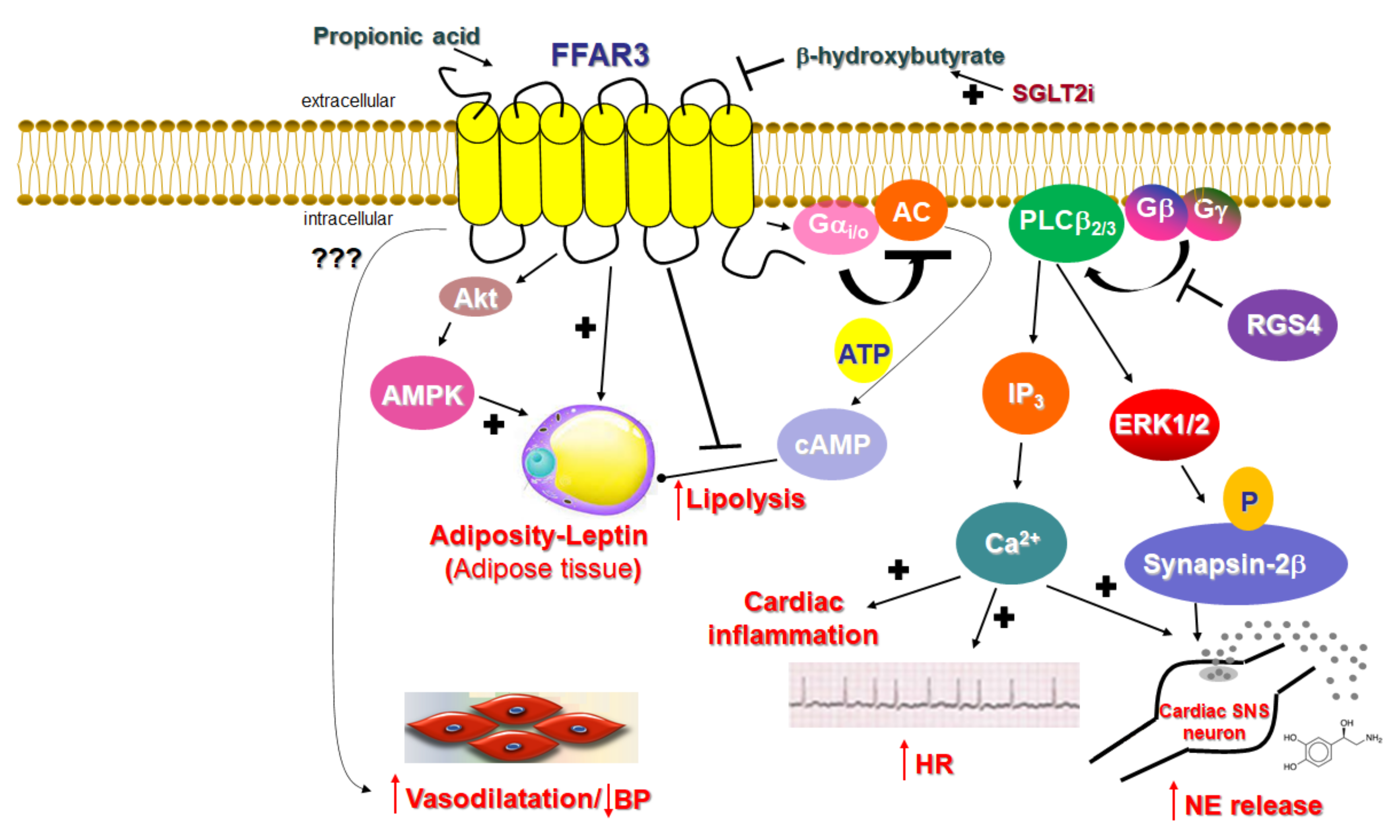
Unlike FFAR2, whose role in cardiovascular homeostasis (if any), as mentioned above, is virtually unknown, FFAR3’s involvement in cardiovascular function regulation has become increasingly clear over the past decade. The main physiological mechanism by which FFAR3 regulates cardiac function is via the effects it exerts in the sympathetic nervous system [21]. FFAR3 is robustly expressed in murine peripheral sympathetic neurons, including cardiac sympathetic nerve terminals, wherein it regulates whole body metabolic homeostasis, along with neuronal activity/firing by inducing norepinephrine release [21] (Figure 2). Although both norepinephrine and epinephrine mediate the effects of the sympathetic nervous system on all cells and tissues of the entire body, norepinephrine is the actual neurotransmitter synthesized, stored, and released from sympathetic neurons [22][23][24][77,78,79]. Epinephrine is the hormone synthesized in the adrenal medulla and secreted into the systemic circulation [22][23][24][77,78,79]. This is because sympathetic neurons lack the enzyme phenyl-ethanolamine-N-methyltransferase (PNMT), which converts norepinephrine to epinephrine [23][25][78,80]. FFAR3 is also present in portal neurons of the liver, where it regulates propionate-induced gluconeogenesis via the gut-brain axis [26][81]. FFAR3 knockout mice display significantly lower catecholamine (norepinephrine and epinephrine) synthesis, as evidenced by the downregulation of tyrosine hydroxylase, the enzyme that catalyzes the rate-limiting step of catecholamine biosynthesis [21] (Figure 2). Consistent with a lower sympathetic neuronal activity/firing rate, heart rate is also reduced in FFAR3 knockout mice [21] (Figure 2). Thus, FFAR3 clearly promotes neuronal firing and norepinephrine synthesis and release in sympathetic neurons. The signaling pathway underlying this effect of FFAR3 is the stimulation of the Gi/o-derived free Gbg subunit activation of PLCβ2/3 (see above) [21]. Gβγ-activated PLCβ2/3 activates, in turn, the MAPKs ERK1/2, which phosphorylate synapsin-2β at Ser426 to induce vesicle fusion with the neuronal plasma membrane and norepinephrine exocytosis/synaptic release from sympathetic nerve endings [27][82] (Figure 2). Notably, neither GRK2 nor β-arrestins appear to be involved in this signaling pathway [21], although GRK2 should theoretically play a role, since it interacts with free Gβγ subunits via its C-terminal pleckstrin homology (PH) domain [28][54]. In fact, this is the main mechanism for membrane targeting and the activation of GRK2 (and GRK3) [29][55]. On the other hand, RGS proteins of the B/R4 family, which inactivate Gi/o proteins, must interfere with this FFAR3-dependent signaling pathway in sympathetic neurons [30][53]. RGS4 in particular is known to directly bind Gi/o-derived free Gβγ subunits and PLCβ and to inhibit PLCβ activation independently of its RGS function [31][32][33][83,84,85]. Indeed, while studying FFAR3 signaling and function in rat cardiomyocytes, researchers have confirmed that RGS4 intervenes in FFAR3 signaling to PLCβ via the Gi/o-derived free Gβγ subunits, dampening the subsequent PLCβ-induced Ca2+ signaling from this receptor and leading to inflammation in the heart, as well as norepinephrine release and firing activity in cardiac sympathetic neurons [34][86] (Figure 2). Nevertheless, other studies have suggested that FFAR3 may actually inhibit secretion/exocytosis via N-type Ca2+ channel inhibition in enteric and vascular neurons [35][36][87,88]. More specifically, FFAR3 signaling inhibits N-type Ca2+ channels via Gβγ signaling, reducing neuronal catecholamine release in rat sympathetic neurons innervating vascular smooth muscle [35][87]. Additionally, FFAR3 modulates the cholinergic-mediated secretory response in the proximal colonic mucosal neurons of rats [36][88], and FFAR3 is a putative target for neurogenic bowel disorder treatment [37][89]. Indeed, the FFAR3 synthetic agonist AR420626 suppresses cholinergic and serotonergic-dependent colonic motility and secretions [37][89]. Therefore, the picture regarding neuronal FFAR3 effects is undoubtedly complicated, and more studies are required to provide better clarity. The decade-old study by Kimura et al. in FFAR3 knockout mice demonstrated that the FFAR3-dependent norepinephrine release from sympathetic neurons modulates energy expenditure and that the activation of FFAR3 with propionic acid elevates heart rate and increases cardiac oxygen demand/consumption [21]. It also showed that the effect of propionate/FFAR3 on heart rate was suppressed by pretreatment with a β-adrenergic receptor (AR) blocker, which indicated that FFAR3 signaling is reciprocally regulated by βARs, i.e., there is a signaling crosstalk between FFAR3 and βARs, upregulating βAR function in sympathetic ganglions [21]. Notably, in that same study, the ketone body β-hydroxybutyrate (or 3-hydroxybutyrate) was shown to block FFAR3 and antagonize its pro-sympathetic hyperactivity in neurons [21] (Figure 2). On the other hand, sodium/glucose co-transporter (SGLT)-2 inhibitors, such as dapagliflozin and empagliflozin (anti-diabetic/diuretic drugs with a plethora of beneficial cardiovascular effects that have been coming to light at an accelerating pace), increase ketone body (including β-hydroxybutyrate) production in the heart and blood vessels [38][39][90,91]. Given that dapagliflozin and empagliflozin have been shown to possess sympatholytic properties that mediate, at least in part, their beneficial effects in chronic human heart failure [40][92], it is tempting to speculate that the sympatholytic effects of SGLT2 inhibitor drugs are mediated, at least partially, by the b-hydroxybutyrate-mediated blockade of FFAR3 signaling in sympathetic neurons, which normally raises cardiac norepinephrine levels and cardiovascular sympathetic nervous system activity [39][91]. This, of course, awaits confirmation by future studies in experimental models of chronic heart failure.
FFAR3 is expressed, not only in postganglionic sympathetic and sensory neurons of the autonomic nervous system, but also in sympathetic and sensory neurons of the somatic peripheral nervous system 
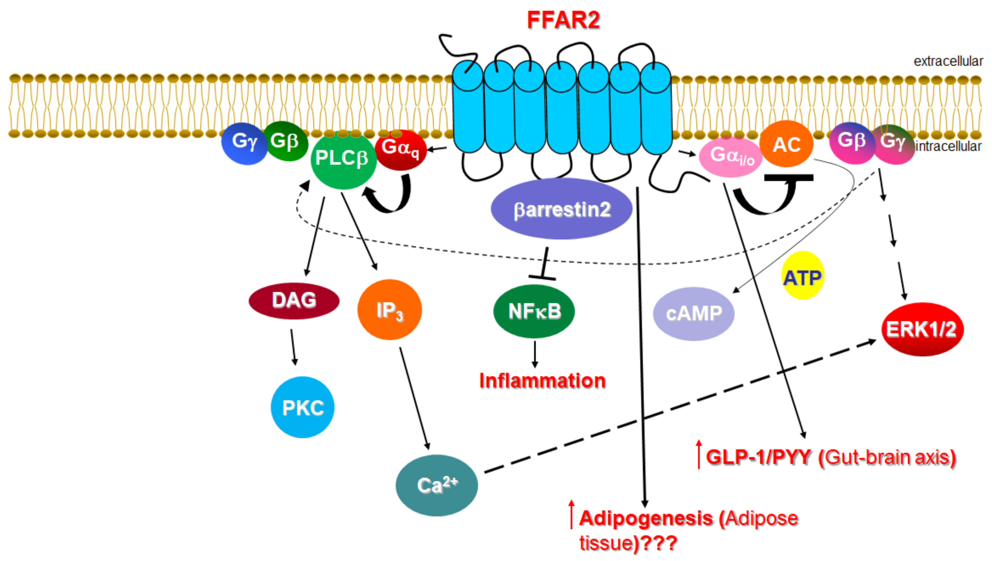
[21][35][41][21,87,93]. Thus, SCFAs exert their effects via FFAR3 not only through the enteroendocrine system, but also directly by modifying physiological reflexes integrating the peripheral nervous system and the gastrointestinal tract. Moreover, FFAR3 in submucosal, and the myenteric ganglionic plexus neurons of the small intestine regulate gut hormonal synthesis, including GLP1 and PYY synthesis, similar to FFAR2 (see above) [42][43][44][3,5,11]. Additionally, FFAR3 significantly reduces lipolysis by inhibiting hormone-sensitive lipase phosphorylation and activity via Gαi-mediated AC inhibition and cAMP lowering in peripheral adipose tissues [45][46][15,94] and increases leptin production, hepatic lipogenesis, and adipocyte growth [47][4] (Figure 2). Indeed, male FFAR3 knockout mice treated with HFD exhibit more body fat mass and higher blood glucose levels compared to wild type female littermates [48][95], and leptin synthesis is reduced in FFAR3 knockouts [49][96].
ScholarVision Creations