Your browser does not fully support modern features. Please upgrade for a smoother experience.
Please note this is a comparison between Version 2 by Yvaine Wei and Version 1 by Raffaele Parrozzani.
Neurofibromatosis type 1 (NF1) is a genetic disease affecting approximately 1 in 2500–3000 individuals. Choroidal abnormalities (CAs) have recently been introduced as one of the criteria for the diagnosis of NF1. In NF1 pediatric patients, CAs change with time, increasing both in number and dimensions, independently from the physiological growth of the eye. While the increase of the CAs number occurs particularly at an earlier age, the increase in the CAs dimensions is a slow process that remains constant during childhood.
- neurofibromatosis
- NF1
- choroidal abnormalities
1. Introduction
Neurofibromatosis type 1 (NF1) is characterized by a dominant autosomal transmission, with complete penetrance reached in 95% of cases by the age of 8, and it has a highly variable expression even in individuals of the same family [4,5][1][2].
Diagnosis can be made if two or more of the following criteria are present (one or more if a parent is affected): at least six café-au-lait macules (CALMs), axillary or inguinal freckling, at least two neurofibromas of any type or one plexiform neurofibroma, optic pathway glioma (OPG), at least two Lisch nodules (LNs) or two choroidal abnormalities (CAs) and distinctive osseous lesions [6][3]. The aforementioned criteria have been derived from a recent revision by the International Consensus Group on Neurofibromatosis Diagnostic Criteria (I-NF-DC), which introduced CAs as an ophthalmologic criterion because of its high specificity and sensitivity for the diagnosis of NF1 [6,7,8,9,10,11,12][3][4][5][6][7][8][9].
Historically, choroidal involvement in NF1 was considered a rare finding, mainly described in pathologic specimens as ovoid bodies into the choroid, consisting of hyperplastic Schwann cells, melanocytes and ganglion cells [13,14,15,16][10][11][12][13]. The in vivo detection of NF1-related choroidal abnormalities, which are completely asymptomatic and undetectable by conventional ophthalmoscopy or fluorescein angiography, was initially possible by means of indocyanine-green angiography [17][14] and, more recently, by near-infrared (NIR) reflectance imaging, a fully noninvasive tool that reveals CAs as bright patchy lesions, most often observed within the major vascular retinal arcades [18,19,20,21][15][16][17][18].
It has already been described that CAs tend to increase with patient age, both in the general NF1-population [19,22,23][16][19][20] and in the pediatric NF1-population [24][21], but none of the previous studies analyzed the same population longitudinally.

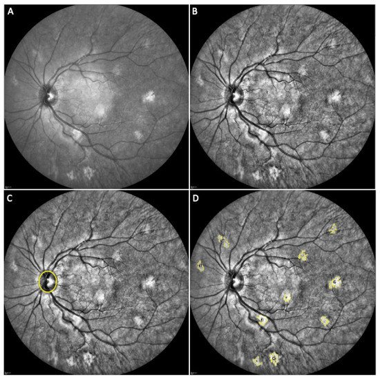
 Figure 1. Elaboration of a near-infrared (NIR) image of a neurofibromatosis type 1 (NF1) patient eye with choroidal abnormalities (CAs). (A) original NIR image; (B) application of the “Enhance Local Contrast” tool with software standard parameters; (C) identification of the optic disc margin with the “Oval” tool; (D) identification of all the visible CAs, tracing of their margin with the “Wand Tracing” tool and assignment of a reference number.
2. Elaboration of a Near-Infrared (NIR) Image
Each image was elaborated through the following steps (Figure 1): (a) the “Enhance Local Contrast” tool with software standard parameters was applied; (b) the “Oval” tool was used to manually identify the optic disc margin; three consecutive acquisitions were used to obtain the mean optic disc area (ODA) and the mean optic disc perimeter (ODP), and no significant variability was found among the measurements; (c) the “Wand Tracing” tool was used to automatically detect and trace margins of each visible CA; the area and the perimeter of each CA were automatically calculated. A reference number was assigned to each CA in each analyzed eye to allow comparison of the same lesion at each follow-up examination.
 Figure 1. Elaboration of a near-infrared (NIR) image of a neurofibromatosis type 1 (NF1) patient eye with choroidal abnormalities (CAs). (A) original NIR image; (B) application of the “Enhance Local Contrast” tool with software standard parameters; (C) identification of the optic disc margin with the “Oval” tool; (D) identification of all the visible CAs, tracing of their margin with the “Wand Tracing” tool and assignment of a reference number.
Figure 1. Elaboration of a near-infrared (NIR) image of a neurofibromatosis type 1 (NF1) patient eye with choroidal abnormalities (CAs). (A) original NIR image; (B) application of the “Enhance Local Contrast” tool with software standard parameters; (C) identification of the optic disc margin with the “Oval” tool; (D) identification of all the visible CAs, tracing of their margin with the “Wand Tracing” tool and assignment of a reference number.
3. Current Insights
It was revealed that the increase in the CAs number was positively correlated with the CAs number at baseline, highlighting that the increase is greater in children that have more CAs at the baseline evaluation. A positive correlation was also found between the CAs size progression over time and the CAs baseline dimensions, meaning that subjects presenting wider CAs at their first evaluation were those who had a greater increase in their CAs size during follow-up. The findings agree with those reported by Cassiman et al., who did not find any significant difference regarding the presence of CAs comparing two groups of NF1 patients, one with truncanting and one with non-truncanting mutations [10][7]. Two recent papers described the evolution of CAs in terms of dimensions. Chilibeck et al. reported a progression in the number and dimensions of CAs in 26 eyes from 14 children [30][22], and Touze et al. showed an increase of CAs and of their size longitudinally analyzing a pediatric NF1-population [31][23]. However, none of these works considered the physiological increase in eyeball dimension during childhood that could bias the CAs surface increase. There was not any significant correlation found between the CAs size progression and patient age, suggesting a constant increase in the CAs dimensions, at least during childhood. This finding is in contrast with the report of Touzè et al. [31][23], who described that the slope of progression was maximal between the age of 8 and 12 years. Such a spike could be explained by a growth of the eye corresponding to the puberty period [32,33,34][24][25][26], whereas herein, adjusting the CAs measures for the optic disc area and perimeter removed all the influence of the physiological growth of the eye. A possible limit could have been the use of specific follow-up timepoints (irrespective of age), rather than age-specific time points. Nevertheless, to reduce the risk of an age-related bias, the statistical correction by the patient’s age at baseline was performed. A second potential limitation was that a possible correlation was not searched between the CAs number and each NF1-related manifestation. The correlation between this quantitative changing parameter and a qualitative parameter (represented by each of the other NF-related signs) caught at a precise time-point would lack statistical and meaningful strength. Moreover, the fact that all included subjects were characterized by the presence of CAs (as an inclusion criterion) may represent a remarkable bias in correlating this sign with the other NF-related manifestations. The assessment of the CAs thickness is useful in confirming the choroidal localization of the abnormalities but it would not be precise in defining the real thickness of CAs, since their melanin component acts by blocking the underlying signal, giving a back-shadowing effect [21][18]. Moreover, a further evaluation of the behaviour of CAs in adulthood would also be interesting to investigate if a growth of the same persists even later in life.4. Conclusions
In conclusion, the natural evolution of CAs in an NF1 pediatric population is first reported on a long-term basis considering and eliminating the influence of eye growth. It was demonstrated that the CAs number increases particularly at an early age, while a slow growth in size is significant and constant during childhood.References
- Pasmant, E.; Vidaud, M.; Vidaud, D.; Wolkenstein, P. Neurofibromatosis type 1: From genotype to phenotype. J. Med. Genet. 2012, 49, 483–489.
- Clementi, M.; Barbujani, G.; Turolla, L.; Tenconi, R. Neurofibromatosis-1: A maximum likelihood estimation of mutation rate. Hum. Genet. 1990, 84, 116–118.
- Legius, E.; Messiaen, L.; Wolkenstein, P.; Pancza, P.; Avery, R.A.; Berman, Y.; Blakeley, J.; Babovic-Vuksanovic, D.; Cunha, K.S.; Ferner, R.; et al. Revised diagnostic criteria for neurofibromatosis type 1 and Legius syndrome: An international consensus recommendation. Genet. Med. 2021, 23, 1506–1513.
- Tadini, G.; Milani, D.; Menni, F.; Pezzani, L.; Sabatini, C.; Esposito, S. Is it time to change the neurofibromatosis 1 diagnostic criteria? Eur. J. Intern. Med. 2014, 25, 506–510.
- Parrozzani, R.; Clementi, M.; Frizziero, L.; Miglionico, G.; Perrini, P.; Cavarzeran, F.; Kotsafti, O.; Comacchio, F.; Trevisson, E.; Convento, E.; et al. In vivo detection of Choroidal abnormalities related to NF1: Feasibility and comparison with standard NIH diagnostic criteria in pediatric patients. Investig. Ophthalmol. Vis. Sci. 2015, 56, 6036–6042.
- Vagge, A.; Camicione, P.; Capris, C.; Sburlati, C.; Panarello, S.; Calevo, M.G.; Traverso, C.E.; Capris, P. Choroidal abnormalities in neurofibromatosis type 1 detected by near-infrared reflectance imaging in paediatric population. Acta Ophthalmol. 2015, 93, e667–e671.
- Cassiman, C.; Casteels, I.; Jacob, J.; Plasschaert, E.; Brems, H.; Dubron, K.; Keer, K.V.; Legius, E. Choroidal abnormalities in café-au-lait syndromes: A new differential diagnostic tool? Clin. Genet. 2017, 91, 529–535.
- Viola, F.; Villani, E.; Natacci, F.; Selicorni, A.; Melloni, G.; Vezzola, D.; Barteselli, G.; Mapelli, C.; Pirondini, C.; Ratiglia, R. Choroidal abnormalities detected by near-infrared reflectance imaging as a new diagnostic criterion for neurofibromatosis 1. Ophthalmology 2012, 119, 369–375.
- Parrozzani, R.; Midena, E. Author response: Choroidal abnormalities detected by near-infrared imaging (NIR) in pediatric patients with neurofibromatosis type 1 (NF1). Investig. Ophthalmol. Vis. Sci. 2016, 57, 775.
- Klein, R.M.; Glassman, L. Neurofibromatosis of the choroid. Am. J. Ophthalmol. 1985, 99, 367–368.
- Wolter, J.R. Nerve Fibrils in Ovoid Bodies: With Neurofibromatosis of the Choroid. Arch. Ophthalmol. 1965, 73, 696–699.
- Kurosawa, A.; Kurosawa, H. Ovoid Bodies in Choroidal Neurofibromatosis. Arch. Ophthalmol. 1982, 100, 1939–1941.
- Woog, J.J.; Albert, D.M.; Craft, J.; Silberman, N.; Horns, D. Choroidal ganglioneuroma in neurofibromatosis. Graefe’s Arch. Clin. Exp. Ophthalmol. 1983, 220, 25–31.
- Rescaldani, C.; Nicolini, P.; Fatigati, G.; Bottoni, F.G. Clinical application of digital indocyanine green angiography in choroidal neurofibromatosis. Ophthalmologica 1998, 212, 99–104.
- Yasunari, T.; Shiraki, K.; Hattori, H.; Miki, T. Frequency of choroidal abnormalities in neurofibromatosis type 1. Lancet 2000, 356, 988–992.
- Nakakura, S.; Shiraki, K.; Yasunari, T.; Hayashi, Y.; Ataka, S.; Kohno, T. Quantification and anatomic distribution of choroidal abnormalities in patients with type I neurofibromatosis. Graefe’s Arch. Clin. Exp. Ophthalmol. 2005, 243, 980–984.
- Ueda-Consolvo, T.; Miyakoshi, A.; Ozaki, H.; Houki, S.; Hayashi, A. Near-infrared fundus autofluorescence-visualized melanin in the choroidal abnormalities of neurofibromatosis type 1. Clin. Ophthalmol. 2012, 6, 1191–1194.
- Makino, S.; Tampo, H. Optical Coherence Tomography Imaging of Choroidal Abnormalities in Neurofibromatosis Type 1. Case Rep. Ophthalmol. Med. 2013, 2013, 292981.
- Makino, S.; Tampo, H.; Arai, Y.; Obata, H. Correlations between choroidal abnormalities, lisch nodules, and age in patients with neurofibromatosis type 1. Clin. Ophthalmol. 2014, 8, 165–168.
- Moramarco, A.; Giustini, S.; Nofroni, I.; Mallone, F.; Miraglia, E.; Iacovino, C.; Calvieri, S.; Lambiase, A. Near-infrared imaging: An in vivo, non-invasive diagnostic tool in neurofibromatosis type 1. Graefe’s Arch. Clin. Exp. Ophthalmol. 2018, 256, 307–311.
- Goktas, S.; Sakarya, Y.; Ozcimen, M.; Alpfidan, I.; Uzun, M.; Sakarya, R.; Yarbag, A. Frequency of choroidal abnormalities in pediatric patients with neurofibromatosis type 1. J. Pediatr. Ophthalmol. Strabismus 2014, 51, 204–208.
- Chilibeck, C.M.; Shah, S.; Russell, H.C.; Vincent, A.L. The presence and progression of choroidal neurofibromas in a predominantly pediatric population with neurofibromatosis type-1. Ophthalmic Genet. 2021, 42, 223–229.
- Touzé, R.; Manassero, A.; Bremond-Gignac, D.; Robert, M.P. Long-term follow-up of choroidal abnormalities in children with neurofibromatosis type 1. Clin. Exp. Ophthalmol. 2021, 49, 516–519.
- Yip, V.C.H.; Pan, C.W.; Lin, X.Y.; Lee, Y.S.; Gazzard, G.; Yong, T.Y.; Saw, S.M. The relationship between growth spurts and myopia in Singapore children. Investig. Ophthalmol. Vis. Sci. 2012, 53, 7961–7966.
- Kearney, S.; Strang, N.C.; Cagnolati, B.; Gray, L.S. Change in body height, axial length and refractive status over a four-year period in caucasian children and young adults. J. Optom. 2020, 13, 128–136.
- Ojaimi, E.; Morgan, I.G.; Robaei, D.; Rose, K.A.; Smith, W.; Rochtchina, E.; Mitchell, P. Effect of stature and other anthropometric parameters on eye size and refraction in a population-based study of Australian children. Investig. Ophthalmol. Vis. Sci. 2005, 46, 4424–4429.
More
